
Abstract
The effects of Hibiscus sabdariffa (HS) in lowering blood pressure in human and animal hypertension have been documented. This study investigated the effect of the water extract of the dried calyx of HS and Hibiscus anthocyanins (HAs) on left ventricular myocardial capillary length and surface area in spontaneously hypertensive rats (SHRs). Twelve-week-old male SHRs were divided into eight groups (six rats in each group). Three groups were given three doses; 10%, 15% and 20% of the water extract of HS in lieu of drinking water for 10 consecutive weeks (HS10, HS15 and HS20) with one group kept as control (C). Another three groups were given three doses of the HAs orally at doses of 50, 100 and 200 mg/kg for five consecutive days with one group kept as a control (C). Systolic (SBP) and diastolic (DBP) blood pressures, as well as heart rate (HR), were measured weekly. After the experimental protocols, the left ventricles (LV) of all rats were obtained. Capillary surface area density and length density were determined by unbiased sterological methods on 3 μm LV tissue samples from perfusion-fixed hearts. HS ingestion significantly reduced SBP, DBP and LV mass in a dose-dependent fashion but did not affect the HR. HS significantly increased surface area and length density of myocardial capillaries by 59%, 65% and 86%, and length density by 57%, 77% and 57%, respectively. Myocyte nuclear volume was significantly decreased in HS-treated rats. There was a decrease (although insignificant) in SBP and DBP with HA ingestion compared with controls. These changes suggest that the observed beneficial effect of HS on high BP in SHRs could be mediated through a reduction in the diffusion distance between capillaries and myocytes, as well as new vessel formation. It is proposed that these effects might be beneficial in restoring myocyte normal nutritional status compromised by the hypertrophic state of hypertension.
Introduction
Hibiscus sabdariffa (HS; botanic family: Malvaceae) is a tropical plant that grows widely all over central and West Africa, Southeast Asia and elsewhere. The calyces of the flower are consumed worldwide as a cold beverage and as a hot drink. Aqueous extracts of the plant are used in folk medicine against many complaints including high blood pressure, liver disease, fever, etc. 1–3 The red anthocyanin (AC) pigment in the calyces is used as a food coloring agent. 4 The HS extract has been documented as a hypotensive agent. Intravenous injections of the aqueous extracts to anesthetized cats 5 and rats 6 lowered blood pressure in a dose-dependent manner. Recently, the antihypertensive action was also described in rats with genetic and experimental hypertension 7,8 and in patients with moderate hypertension. 9,10 Various mechanisms were proposed to explain the hypotensive effect, 6,11 among which is inhibition of angiotensin I-converting enzyme. 12,13 Indeed, Jonadet et al. have reported an appreciable angiotensin-converting enzyme (ACE)-inhibiting activity of a crude hydroalcoholic extract of HS calyces in vitro, which was thought to be attributable to flavones. The same authors reported additional angioprotective activity of the extract which they attributed to flavones and ACs. 14 The latter work was substantiated by a clinical trial in which the obtained data confirmed that HS extract and the ACE-I captopril were similar in their hypotensive effect, antihypertensive effectiveness and tolerability. 9 In addition, a beneficial cardioreparative effect of HS extract was shown in vivo. Chronic administration of the extract to renovascular hypertensive rats at a dose of 250 mg/kg/day for up to eight weeks reduced heart weight to levels comparable to sham-operated rats. 8 It has also been shown that the dried flower powder of Hibiscus rosa sinensis has significant protective effects in ischemic heart disease. 15 The exact mechanism of its cardioprotective effect was not investigated; however, it is possible that ACE inhibition by HS improved angiogenesis in the ischemic myocardium. An antihypertensive treatment that is cardioreparative should restore myocardial structure through regression of the fibrosis and therefore normalization of the interstitium, regression of the structural coronary microvascular alterations, in addition to myocardial mass reduction. The present study was undertaken to determine the possible beneficial effects of HS, a natural product consumed by a large population, on myocardial angiogenesis in spontaneously hypertensive rats (SHRs), a well-established model for essential hypertension which in certain ways resembles hypertension in humans. 16 SHRs develop a gradual increase in systemic blood hypertension at one month of age, and have established left ventricular (LV) hypertrophy by three months. 17 To our knowledge, there are no studies using unbiased stereological methods on the effect of this extract on the myocardial structure of genetic hypertension. We used male SHRs to confirm the antihypertensive effect of HS extract and to determine by unbiased quantitative stereology if chronic administration of HS would alter the myocyte capillary bed, and if so, to specify the compartment(s) where this alteration occurred.
Materials and methods
Drugs and chemicals used were of the highest purity available, and were obtained from Sigma (St Louis, MO, USA).
HS preparation and administration
Dried HS calyxes were bought from a local market. The botanical identification of the material was checked with a taxonomist in the University Department of Biology. Five (low dose), 10 (medium dose) and 20 g (high dose) was weighed freshly every morning and placed separately in distilled water (995, 990 and 980 mL, respectively), shaken for five minutes and left overnight at room temperature. Twenty-four hours later, the water extracts of the plant were removed and given to the rats as the only drinking source (i.e. in lieu of drinking water).
AC preparation and administration
The ACs were prepared by one of the authors (GB) using methods described previously. 15,16 They were dissolved, as provided, in distilled water at different doses (50, 100 and 200 mg/kg) and given orally by gavage, daily.
Animal experiments
All procedures performed in this study were in accordance with the regulations of the Medical Research and Ethics Committee of Sultan Qaboos University. In the first part of the experiment, 12-week-old male SHRs (Taconic Farms Inc, Germantown, NY, USA) were randomly divided into four groups (6–8 each). The first (control group, C) was given access to its usual drinking water. The other three groups (HS10, HS15 and HS20) were given water extracts of 10%, 15% and 20% HS, respectively, as the only drinking source for 10 weeks.
In the second part of the experiment, 12-week-old male SHRs were divided into four groups (6 each). The first (control group, C) was given saline by gavage daily. The other three groups (AC50, AC100 and AC200) were given by daily oral gavage, 50, 100 and 200 mg/kg of AC, respectively, for five days.
All rats had free access to standard laboratory food pellets and were housed in a temperature-controlled room (23–25°C) with a 12/12-h light/dark cycle.
Body weight (BW) was measured once a week. Systolic (SBP) and diastolic blood pressures (DBP) and heart rate (HR) were measured once a week in the conscious restrained animals using a computerized tail-cuff system (Harvard apparatus; Columbus Instruments, Columbus, OH, USA).
Perfusion fixation and tissue sampling
At the end of the experimental period, each rat was anesthetized with intraperitoneal sodium pentobarbitone (40–50 mg/kg) and the abdomen was opened via a midline incision. The inferior vena cava and abdominal aorta were cannulated. Before perfusion fixation, anticoagulant sodium heparin (1000 units) was injected through the inferior vena cava, and followed one minute later by a bolus injection of 1.5 mL 2% procaine in physiological saline to arrest the heart in diastole and abolish vascular tone. The heart was then perfusion-fixed with 100 mL Karnovski's fixative injected through the aortic cannula at a pressure of 160mmHg. Thereafter, the heart was excised, and the LV free wall plus septum dissected out for the determination of its mass (LVM) and absolute volume (LVVA), and for tissue sampling. After weighing, the LVVA was determined using the Cavalieri method.
17
In brief, a series of parallel sections were cut through the LV at fixed thicknesses (T). This generated a series of slabs, which were all laid the same way up. Traditional point counting was used to estimate the area (A) of each of the cut surfaces of all slabs. Absolute volume, V
A was then calculated by the equation:

Tissue sampling was performed according to the orientator method. 18 Briefly, a slab was randomly selected from those used for evaluating V A, and processed via a series of cuts accordingly. The last cut surface was considered to be uniformly isotropic. One block of tissue was made from each ventricle, and was embedded in paraffin wax. Using a Leica microtome (Leica RM2165; Heerberg, Switzerland), three-micron-thick sections were obtained from each block and were stained with hematoxylin/eosin. A systematic uniform sample of six fields was obtained from each section and was digitally stored using a camera attached to an Olympus microscope (BH2; Olympus Optical Co, Tokyo, Japan).
Stereological analysis
Measurements of profile densities (surface area, length and volume fraction of LV capillaries) were performed using stereology software (Histometrix, MIL 6 Version 1; Kinetic Imaging Ltd, Nottingham, UK) installed on a personal computer. All microvessels without an apparent smooth muscle wall were included (Figure 1). In brief, the surface area density (S
v) of the capillaries is the surface area of capillaries/unit tissue volume of myocardium and the length density of the capillaries (L
v) is the length of capillaries/unit tissue volume of myocardium.
19
Traditional point counting was used to estimate the volume fraction, V
f, of myocardial capillaries in the LV. This parameter is expressed as a percentage of the total tissue volume that makes up the capillary network. The size of the myocyte nucleus was determined by its volume-weighted volume, which gives an indication of the size of the myocyte as a whole.
17
Although S
v is a valuable indicator of capillary exchange capacity, L
v provides the best indicator of microvascular proliferation in hearts of similar mass.
20
The total surface area (S
A) and total length (L
A) of the capillary bed were calculated by combining the absolute volume of LV (LVVA) (see above) and S
v and L
v as follows:


An unbiased counting frame with superimposed random ‘needles’ for measurement of surface area density of capillaries (c). m, myocyte. (A color version of this figure is available in the online journal)
Statistical analysis
All data are expressed as means ± SEM (standard error of the mean). After testing for normality distribution, one-way analysis of variance (ANOVA) was used to assess differences between groups. Dunnett's test was used to compare the treatment and control groups and the statistical significance was set at P > 0.05.
Results
SBP, DBP and HR
The data concerning SBP, DBP and HR at the end of the 10-week experimental protocol (from ages 12 to 22 weeks) are presented in Table 1a. At 12 weeks of age, SBP and DBP were similar in all groups. After 10 weeks, the mean SBP and DBP in all HS treatment groups showed a significant reduction when compared with the control groups (P < 0.01). No significant difference was found between SBP and DBP in the HS groups.
Mean ± (SEM) systolic blood pressure (SBP), diastolic blood pressure (DBP) and heart rate (HR) of 22-week-old spontaneously hypertensive rats (SHRs) treated with 10%, 15% and 20% Hibiscus sabdariffa (HS)
*P < 0.01 versus control; **P > 0.05 versus control
Figure 2 shows the change in SBP, DBP and HR in the HS and control groups. The mean SBP and DBP in the control groups increased significantly as expected at 22 weeks. In all three HS-treated groups, this increase was not only abolished, but significantly reversed.
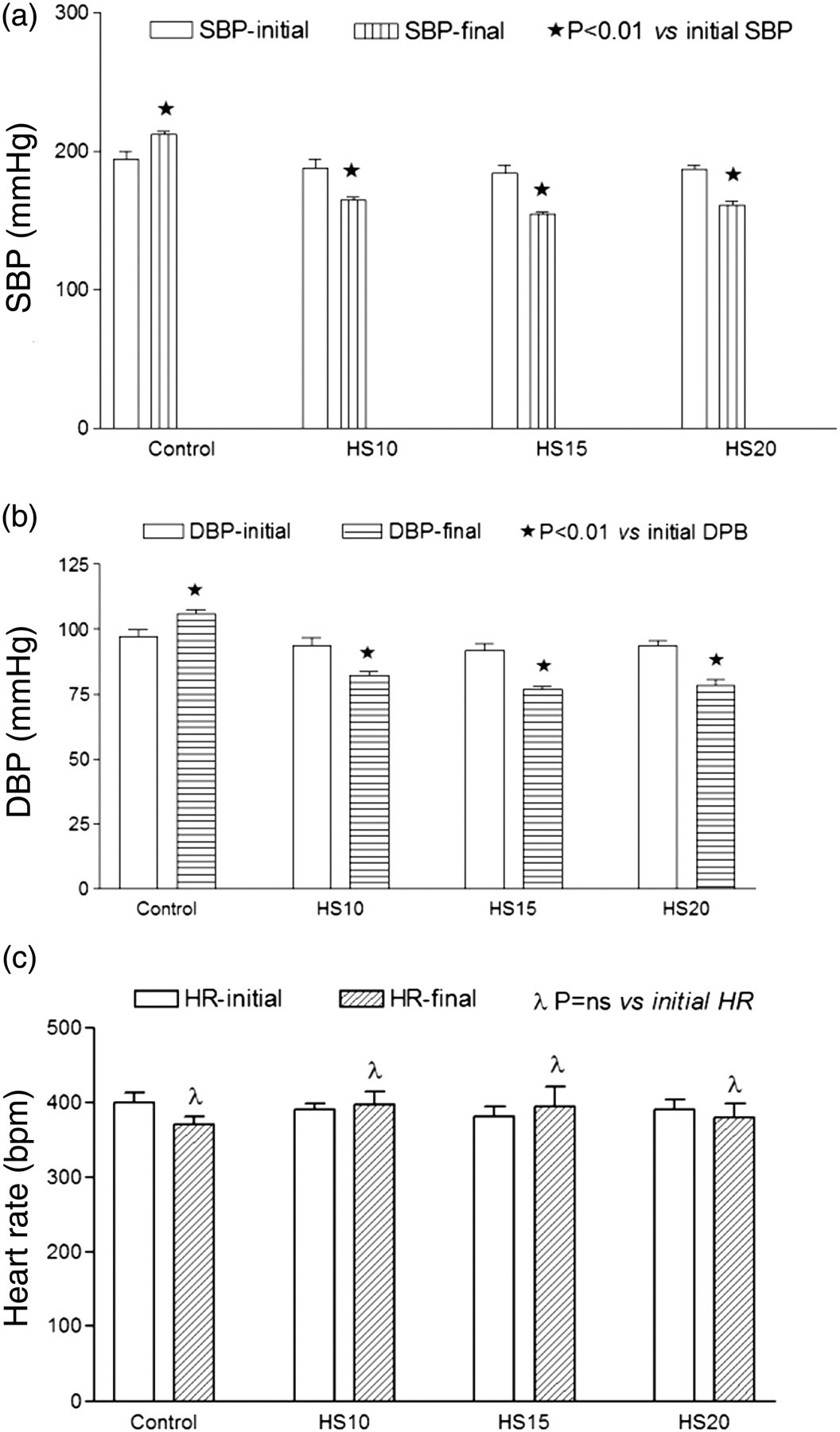
(a) Systolic blood pressure (SBP) (mmHg) of spontaneously hypertensive rats (SHRs) at the beginning and end of the treatment with Hibiscus sabdariffa (HS) n = 6. (b) Diastolic blood pressure (DBP) (mmHg) of SHRs. (c) Heart rate (HR) (bpm) of SHR
Five days of treatment with AC did not significantly reduce the SBP and DBP. However, there was a trend towards reduction of both SBP and DBP when compared with the control group. This effect was greater with the AC100 group (Table 1b). HR was not significantly different in all AC- and HS-treated groups compared with the control groups.
Mean ± (SEM) systolic blood pressure (SBP), diastolic blood pressure (DBP) and heart rate (HR) of 12-week-old spontaneously hypertensive rats (SHRs) treated with 50, 100 and 200 mg anthocyanin (AC) extract of Hibiscus sabdariff a
*P > 0.05 versus control
LVVA and total BW
The data concerning total BW, LVVA and LVVA/BW ratio after 10 weeks of experimental protocol (22 weeks of age) are shown in Table 2. Compared with the final BW in control group, there was a significant decrease by 15.7%, 24.2% and 25.8%, respectively, in the HS10, HS15 and HS20 groups (P < 0.001). Figure 3 shows the change between the initial and final BW in the HS and control groups. Significant BW gain (when compared with initial BW) occurred in the control (26.6%) and HS10 (10%) groups. In the HS15 and HS20 groups, the weight gain was 5% and −1% of the initial weight, respectively, indicating poor weight gain during the experimental period.
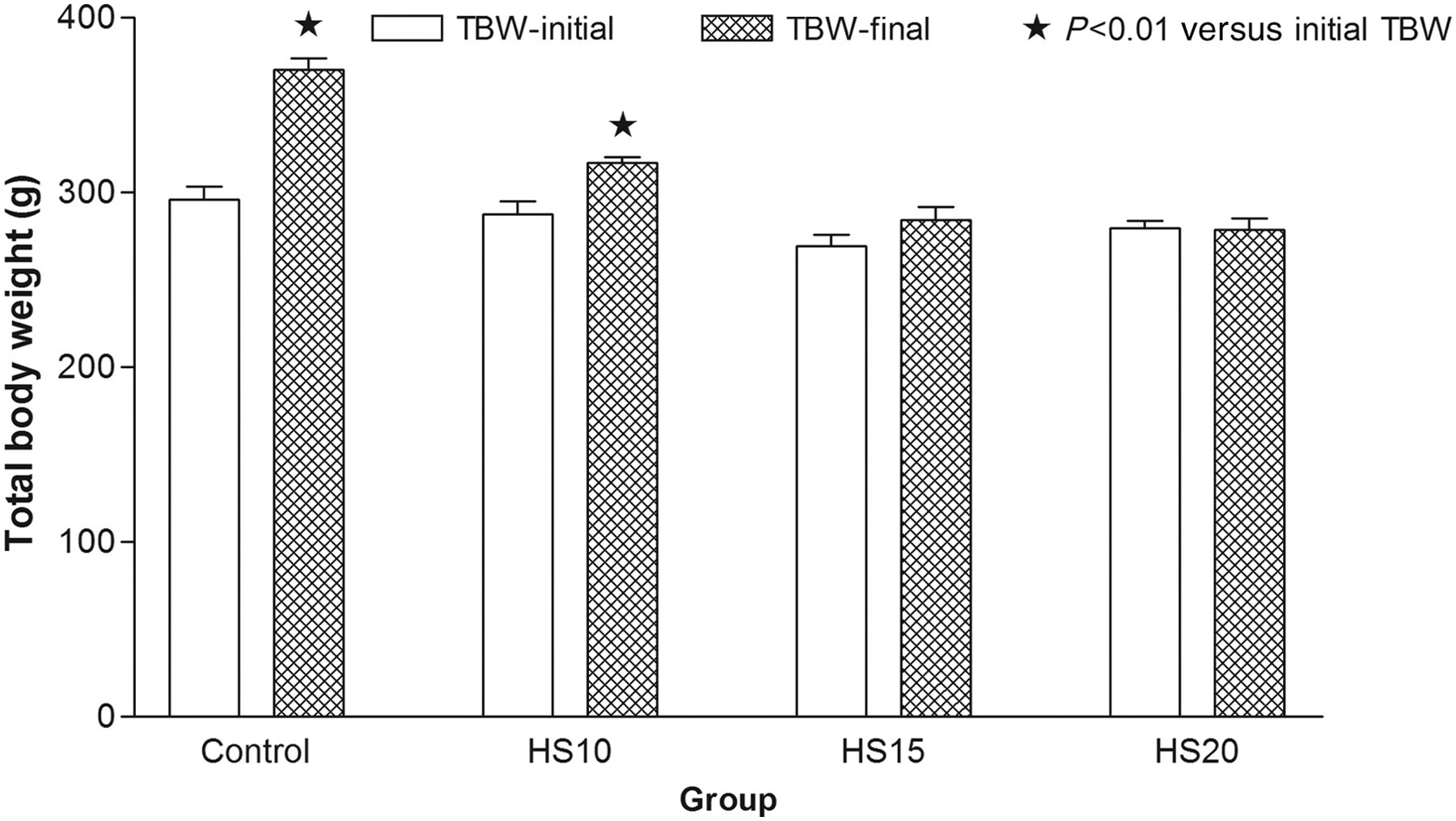
Total body weight (TBW) (g) of spontaneously hypertensive rats at the beginning and end of the treatment with Hibiscus sabdariffa (HS) (n = 6). (A color version of this figure is available in the online journal)
Total body weight (BW), left ventricular volume (LVVA) and LVVA/BW of 22-week-old spontaneously hypertensive rats (SHRs) treated with 10%, 15% and 20% Hibiscus sabdariff a (HS)
*P < 0.01 versus control; **P < 0.05 versus control ns, not significant
The LVVA was highest in the control group. Thereafter, it decreased in a dose-dependent manner by 26.5%, 27.7% and 31.3%, respectively, in the HS10-, HS15- and HS20-treated groups (P < 0.05). There was no significant difference in the LVVA/BW ratio between all HS-treated groups and the control group; however, there was a trend towards reduction of the ratio with HS treatment (Table 2).
There was no change in LVVA, total BW or LVVA/BW ratio between control and AC-treated animals.
Stereological measurement
Stereological estimation of the myocardial capillary bed showed the effects of HS treatment on myocardial capillary surface area density, total surface area, length density and total length after 10 weeks of experimental protocol (22 weeks of age).
Surface area of myocardial capillaries
The changes in surface area density, S v, of capillaries is shown in Table 3. Treatment with HS increased mean S v in a dose-dependent manner by 59%, 65.3% and 85.9% in HS10, HS15 and HS20 groups, respectively, over control values (P < 0.05). The total surface area of the capillaries in the LV was higher in the HS-treated group than in the control group; however, the difference was not significant.
Capillary surface area density, total surface area, length density, total length and myocyte volume-weighted volume of the left ventricle (LV) in 22-week old spontaneously hypertensive rats (SHRs) treated with 10%, 15% and 20% Hibiscus sabdariff a (HS)
*P < 0.01 versus control; **P < 0.05 versus control ns, not significant
Length of myocardial capillaries
Ten weeks of HS administration resulted in 57.4%, 77.6% and 56.9% increases in myocardial capillary length density, L v, in HS10, HS15 and HS20 groups, respectively, over control values. This increase was significant in the HS15 and HS20 groups (P < 0.05). Total length of capillaries in the LV was higher in the HS-treated groups than in the control group; however, the difference was not significant (Table 3).
Volume-weighted volume of myocyte nucleus
The volume density of capillaries had increased by 50–83% in HS-treated animals compared with controls (P < 0.05). The volume-weighted volume of the myocyte nucleus was decreased by 17%, 15% and 11% in the HS10, HS15 and HS20 groups, respectively, over control values (P < 0.01).
Discussion
In this study, ingestion of HS produced a marked reduction in SBP from the fourth week of treatment until the termination of the experimental period. This decrease was independent of the three dosages of HS used. A slight and insignificant tachycardia was also observed in all HS-treated groups which could well be reflex tachycardia associated with the marked decrease in blood pressure observed. Numerous previous studies have found similar results and many mechanisms explaining the results were proposed.
It was of interest to note that HS administration prevented the weight gain that was seen in control rats. Control rats gained 26.6% of their initial BW. Thereafter, there was a dose-dependent decrease both in BW and the gain in BW in the treated rats. This finding suggests that the extract may have antiobesity properties. This might be a beneficial effect, provided that more is understood about the pharmacological and toxicological properties of the extract and its mechanism of action in lowering BW. 21 Indeed, it has been reported by Alarcon-Aguilar et al. 21 that oral administration of HS aqueous extract resulted in significant BW reduction in obese rats.
The regression of LV mass after long-term treatment with ACE inhibitors has been reported in human and animal hypertension. 22–25 Similarly, Herrera-Arellano et al. have demonstrated in a clinical trial that the HS extract exerts an antihypertensive action in patients with stage I and II hypertension. It was postulated that this antihypertensive action could be through promotion of diuresis or ACE-I inhibition. 26 Indeed, Ojeda et al. isolated and extracted the ACs delphinidin and cyanidin-3-O-sambubiocides from HS and demonstrated their ACE inhibitor activity. This inhibitory activity was found to be competing with its substrate for the active site of the enzyme. 12
The reduction of myocardial hypertrophy in the HS group as evidenced by the decrease in LVV and the size of the cell nucleus might have occurred secondary to the insufficient weight gain observed in HS-treated rats or through some other mechanism that reduces myocardial hypertrophy. The mean percentage of BW decrease in all HS-treated groups was 20%, which was lower than the mean percentage of LV weight decrease of 30%. This suggests that the decrease in cardiac muscle mass was not proportional to the overall weight loss. Indeed, Lo et al. have shown that HS ACs induce apoptosis of smooth muscle cells via activation of P38 myosin-activating protein kinase and p53. 27 It could well be that this apoptotic mechanism is common to muscle tissue.
A major finding of this study was that 10 weeks of treatment of SHRs with HS produced a significant increase in S v and L v of myocardial capillaries. To our knowledge, this is the first time that such a finding has been reported. These results are comparable with previous work reporting enhanced myocardial capillarization after treatment with ACE-I inhibitors. 13,20,28,29 For instance, Unger et al. 28 reported an increase in myocardial capillary length density in SHRs treated with a low dose of the ACE inhibitor, ramipril, which did not affect blood pressure or LV mass. Similarly, Olivetti et al. 30 and Ziada et al. 20 have demonstrated an increase in myocardial capillary density associated with a decrease in LV weight in SHRs treated for three months with ACE inhibitors. In another study, Odori et al. 22 reported increased endothelial cell turnover and enlarged collateral vessels induced by another ACE inhibitor (captopril) after renal artery stenosis in the rat. Most of these previous studies interpreted the enhancement of vascularization recorded as an indicator of angiogenesis stimulated by the treatment. In this study, the mean percentage increase in capillary S v and L v induced by HS administration was far above the decrease in myocardial mass (Table 3). This implies that the enhancement in capillarization, as reflected by the increases in capillary S v and L v, was not only secondary to the HS-induced regression of myocardial mass, but also to new vessel formation. Indeed, the increase in total surface area and length of capillaries at the high dose of HS strongly suggest angiogenesis.
Previous phytochemical studies on HS have reported the presence of phenolics, organic acids, sterols, terpenoids, polysacharides and some minerals. The phenolic content consists mainly of ACs like delphinidin-3-O-glucoside, delphinidin-3-O-sambubioside and cyanidin-3-O-sambubioside. 11 By far, ACs are the major constituents in HS. 31 Employing bioassay-guided fractionation of the aqueous extract of dried calyces of HS, Ojeda et al. 12 have demonstrated inhibition of ACE I activity by the ACs delphinidin-3-O-sambubioside and cyanidin-3-O-sambubioside in vitro. We propose that the structural findings are as a result of ACE inhibitor-like effects of HS constituents. In a separate experiment, treatment with the AC extract of HS for five days did cause a reduction of blood pressure, even though not significant. This could have been due to the short duration of the treatment or insufficient dosage.
The SHRs treated with HS gained less weight than those that did not receive such treatment. This is consistent with similar previously reported observations. 32,33 It has been suggested by Bock 33 that the unpleasant taste of HS in the drinking water probably made the animals avoid sufficient food and water ingestion, thereby causing diminished weight gain. Others have demonstrated reduced fat absorbtion in rats during treatment with HS extract. 32 Interestingly, studies where SHRs were treated with ACE inhibitors were used, 22,31,34 demonstrated a similar pattern of slow weight gain. This effect was attributed either to long-lasting central ACE inhibition 31 or a strong natriuresis produced by ACE inhibition. 29
In conclusion, the results of this study demonstrate that HS ingestion was effective in enhancing myocardial capillarization in adult SHRs through structural alterations related to a reduction of myocardial mass and the stimulation of new vessel formation. The evidence for the above conclusions are: (i) HS ingestion markedly reduced LV mass and led to an increase in myocardial S v and L v; (ii) total surface area and length of capillaries were significantly increased when 15–20% of HS was used. We speculate that such changes restore the normal nutritional state of the cardiac myocytes compromised by the hypertrophic state of hypertension.
Our present findings will be substantiated by further biochemical and molecular data that may include immunostaining of the myocardium with antibodies to CD31 to detect capillary endothelial cells. Similarly, molecular markers of angiogenesis such as vascular endothelial growth factor mRNA and protein would further support our finding that HS may promote new vessel formation. 32,35–41
Footnotes
ACKNOWLEDGEMENTS
This work was supported by an internal grant from Sultan Qaboos University (SQU). We thank the staff of the Animal House of SQU for looking after the rats.