
Abstract
Cisplatin (CDDP) is one of the most active cytotoxic agents commonly used in the treatment of peritoneal carcinomatosis. The disadvantages of its clinical use are systemic side-effects, such as nephrotoxicity and myelotoxicity. Long-circulating and pH-sensitive liposomes containing CDDP (SpHL-CDDP) were developed by our research group aiming to promote the release of CDDP near the tumor as well as decreasing toxicity. The aim of this study was to evaluate the antitumor efficacy and toxicity of SpHL-CDDP after intraperitoneal administration in initial or disseminated tumor-bearing mice, at a dose of 12 mg/kg. The survival was monitored and blood samples were collected for biochemical and hematological analysis. Kidneys, liver and spleen were removed for histopathological examination. Tumor cells were evaluated for cellular viability and cell cycle. The survival of animals treated with SpHL-CDDP was higher than those treated with free CDDP. The cell death caused by treatment with SpHL-CDDP occurred through induction of apoptosis, with a cell cycle arrest at the G0/G1 phase. The treatment of mice presenting initial cancer with both formulations provoked a suppression of granulocytes. Mice treated with free CDDP also showed a decrease in platelet count, which suggests a high myelotoxicity. In an advanced cancer model, SpHL-CDDP treatment allowed an improvement of the immune response. Mice affected by cancer at an early stage and treated with free CDDP or SpHL-CDDP showed a lower urea/creatinine index compared with the saline control group. These findings indicate that both treatments were able to reduce the renal damage caused by peritoneal carcinomatosis. Microscopic analysis of kidneys from mice treated with SpHL-CDDP showed a discrete morphological alteration, while tubular necrosis was observed for free CDDP-treated mice. Concerning hepatotoxicity, no alteration in clinical chemistry parameters was observed. These findings reveal that SpHL-CDDP can improve the antitumor efficacy and decrease renal and bone marrow toxicity.
Keywords
Introduction
Peritoneal carcinomatosis is a serious concern in the treatment of abdominal tumors such as hepatic, gastric and gynecological tumors. For treatment of peritoneal carcinomatosis, intraperitoneal administration of anticancer drugs was introduced with the intention of achieving tumoricidal drug levels locally while minimizing systemic side-effects. 1 However, because most anticancer drugs are small water-soluble molecules, they are readily absorbed through the capillaries and reach the circulation system. Thus, it is difficult to sustain a high drug concentration for a long period of time in the peritoneal cavity. 2
The rationale for intraperitoneal therapy of peritoneal carcinomatosis is that the peritoneum, the predominant site of the tumor, receives sustained exposure to high concentrations of antitumor agents while normal tissues are relatively spared. 3 This procedure allows a high drug concentration to be reached at the site of the tumor, 4 without a systemic exposure to the drug for the patient. 5 Armstrong et al. 3 compared patients treated with intravenous administration of paclitaxel, followed by intravenous administration of cisplatin (CDDP) – the intravenous therapy group – and patients treated with intravenous administration of paclitaxel, followed by intraperitoneal administration of CDDP and paclitaxel – the intraperitoneal therapy group. In this study, the authors describe a statistically significant survival advantage to the intraperitoneal therapy group.
However, these patients had more toxic events than women treated with intravenous administration. The authors suggest that the toxic events may be attributed to the higher dose of CDDP in the intraperitoneal therapy group.
Thus, this fact reveals the need to find strategies to circumvent these inconveniences. The use of drug delivery systems to release CDDP locally and near the peritoneal tumor may be an attractive strategy. 6 Our research group has developed long-circulating and pH-sensitive liposomes containing CDDP (SpHL-CDDP) that were able to promote a higher concentration of the drug in the peritoneal region of Ehrlich ascitic tumor-bearing Swiss mice than the free CDDP treatment. 7 Moreover, as expected, a lower blood concentration of CDDP was observed after SpHL-CDDP treatment as compared with free CDDP treatment. In addition, a decreased renal extravasation and uptake after SpHL-CDDP administration was observed. These findings explain the absence of systemic side-effects after intraperitoneal administration of SpHL-CDDP. 8 Hematological investigation revealed neither changes in red and white blood cell (WBC) counts nor changes in the blood urea and creatinine levels following SpHL-CDDP administration in mice. 8 These liposomes are composed of dioleoylphosphatidylethanolamine (DOPE), cholesteryl hemisuccinate (CHEMS) and distearoylphosphatidylethanolaminepolyethyleneglycol2000 (DSPE-PEG2000). 9 The construction of pH-sensitive liposomes takes advantage of the polymorphic phase behavior of unsaturated phosphatidylethanolamine, which forms inverted hexagonal phases (HII) rather than bilayers. Liposome stabilization within bilayers can be achieved by using a titratable acid lipid, such as CHEMS, which is negatively charged at neutral pH. This lipid, homogeneously distributed among DOPE molecules, provides electrostatic repulsions, which decrease DOPE intermolecular interactions, thus preventing HII phase formation under physiological conditions. The protonation of CHEMS molecules in an acidic medium, such as in that within a tumor site, provokes the destabilization of liposomes and the release of CDDP. Therefore, it can be suggested that the release of CDDP in this specific site is responsible for the elimination of side-effects and improvement in therapeutic efficacy. Thus, the aim of this study was to investigate the mechanisms of cellular death involved after intraperitoneal administration of SpHL-CDDP in Ehrlich ascitic tumor-bearing mice.
Materials and methods
Materials
CDDP was purchased from Quiral Química do Brasil S.A (Juiz de Fora, Brazil). DSPE-PEG2000 and DOPE were supplied by Lipoid GmbH (Ludwigshafen, Germany). CHEMS was purchased from the Sigma Chemical Company (St Louis, MO, USA). Sodium chloride was purchased from Merck (Rio de Janeiro, Brazil). The solvents were of analytical grade and all other chemicals were commercially available.
Liposome preparation
SpHL-CDDP liposomes were prepared by reverse-phase evaporation as described by Carvalho Júnior et al.
9
Briefly, chloroform aliquots of DOPE, CHEMS and DSPE-PEG2000 (lipid concentration 40 mmol/L; molar ratio 5.7:3.8:0.5, respectively) were transferred to a round-bottom flask and submitted to evaporation. The lipid film obtained was dissolved in ethyl ether and then added to the 2 mg/mL CDDP solution prepared in the 0.9% w/v NaCl solution. The ratio of aqueous and ether phases was equal to 1:3. The resulting mixture was submitted to fast vortex agitation to produce a water/oil emulsion. Next, ethyl ether was evaporated with the resulting formation of liposomes. These liposomes were submitted to filtration through polycarbonate membranes with porous sizes of 0.4 and 0.2 μm (five cycles for each). Finally, the unencapsulated CDDP was eliminated by ultracentrifugation (Ultracentrifuge SORVALL Ultra 80; Thermo Scientific, Waltham, MA, USA) at 150,000 ×
Liposome characterization
The liposomes were characterized by their encapsulation percentage (EP), size and zeta potential (ζ). The encapsulation percentage of CDDP into liposomes was determined by the quantification of gamma radiation in non-purified and purified liposomes by means of the following equation:

The radioactivity was measured by an automatic scintillation apparatus covering an energy window of 50–150 keV (ANSR, Abbott, Chicago, IL, USA). The counting time was four minutes and the employed sample volume was in agreement with the optimum volume of the equipment (1.5 mL). The mean diameter of SpHL-CDDP was determined by quasi-elastic light scattering at 25°C and at a 90° angle, using the unimodal analysis. The zeta potential was evaluated by the electrophoretic mobility determination at a 90° angle. All samples were diluted using a 0.9% w/v NaCl solution and the size and zeta potential measurements were performed in triplicate using the 3000HS Zetasizer equipment (Malvern Instruments, Worcestershire, UK).
Ehrlich ascitic tumor model
Ehrlich tumors were grown in female Swiss mice (26 ± 5 g), eight weeks old. The animals were kept in an area maintained in a standardized light/dark cycle and had free access to food and water (Centro de Pesquisas René Rachou, FIOCRUZ, Belo Horizonte, Brazil). A total of 2.5 × 10⁶ Ehrlich tumor cells were transplanted intraperitoneally into the mice. The development of ascites was monitored by measuring the waist circumference in each animal, using an ordinary tape measure. All protocols were approved by the Ethics Committee for Animal Experiments at the Universidade Federal de Minas Gerais and CEUA – FIOCRUZ – and were in compliance with the guide for the care and use of laboratory animals recommended by the Institute of Laboratory Animal Resources.
Treatment schedule of animals
Twenty animals per group were submitted to different treatments on the third (TR3) or seventh (TR7) day after the inoculation of tumor cells. These treatment schedules were chosen to verify the treatment efficacy using initial and disseminated peritoneal carcinomatosis models, respectively. The free CDDP and SpHL-CDDP were injected into the mice immediately after their preparation at a dose of 12 mg/kg. The control groups received 0.9% w/v NaCl solution or empty liposomes (the lipid dose was equal to that administered for SpHL-CDDP treatment).
Evaluation of the viability, apoptosis and cell cycle from Ehrlich tumor cells
The cells undergoing apoptosis were determined by quantification of phosphatidylserine exposure on the cell surface using staining with fluorescein isothiocyanate (FITC)-conjugated annexin V and propidium iodide (PI) (Sigma, Steinheim, Germany). One million cells were stained with 2.5 μL of FITC-conjugated annexin V plus 5 μL of PI (10 μg/mL) for 10 min at room temperature in the dark. Cells were analyzed using a flow cytometer (FacsCalibur®; BD, San Diego, CA, USA). The analysis of apoptotic cells was performed according to the strategy proposed by Lecoeur et al., 10 with apoptotic cells being annexin V-positive/PI-negative or annexin V-positive/PI-positive. Finally, for the determination of cell cycle phase distribution, 3 × 10⁵ cells of the ascitic fluid were diluted in 0.3 mL of hypotonic fluorochrome solution and incubated for four hours at 4°C in the dark. 11 A total of 20,000 events were acquired, and the analysis of flow cytometric data was performed using FlowJo software (Tree Star Inc., Ashland, OR, USA). A histogram of DNA content (x-axis, PI fluorescence) versus counts (y-axis) was displayed. Animals that did not have ascites underwent peritoneal lavage using 1 mL of 0.9% w/v NaCl solution in order to capture tumor cells present in the abdominal cavity for the analyses.
Hematological investigations and clinical chemistry
Hematological and clinical chemistry examinations were performed on all surviving animals after seven days of treatment with free CDDP, SpHL-CDDP, saline solution or SpHL. The mice were anesthetized with a mixture of xylazine (7.5 mg/kg) and ketamine (60 mg/kg) and blood samples were collected in tubes containing 10% w/v ethylenediaminetetraacetic acid solution as well as in tubes without anticoagulants. The hematology parameters evaluated included red blood cell counts, hemoglobin, hematocrit, total WBC and platelet counts. All analyses were performed using an automated analyzer (Abacus Junior Vet, Diatron Messtechnick GmbH, Vienna, Austria). Liver function and nephrotoxicity were assessed using clinical chemistry by measuring alanine aminotransferase (ALT, IU/L), urea (mg/dL) and creatinine (mg/dL). The value of total plasma proteins (g/dL) was estimated with a refractometer (Ningbo Utech International, Ningbo, China) and fractionated protein profile (albumin, alpha-, beta- and gammaglobulin, g/dL) was determined by agarose gel electrophoresis.
Histopathological examination
The kidneys, liver and spleen were removed, washed with 0.9% w/v NaCl solution and fixed in 10% (v/v) buffered formalin. All tissues were embedded in paraffin blocks, sectioned into 5 μm in thickness and placed onto glass slides. After hematoxylin–eosin staining, the slides were observed and photos were taken using an optical microscope (Carl Zeiss Axiovert®, Göttingen, Germany) containing AxioVision 4.8 software (Carl Zeiss). The analysis of the pathology slides was blind to the pathologist.
Survival and clinical signs study
The Ehrlich ascitic tumous-bearing Swiss mice were submitted to the different treatments (free CDDP, SpHL-CDDP, SpHL or 0.9% w/v NaCl solution) and were kept for 30 days with follow-up of three times per week. During this time period, the animals were weighed and behavioral changes, signs of toxicity or deaths were recorded.
Statistical analyses
First, statistical analysis was performed to test three hypotheses: independence, normality and variance of the data groups (Minitab 15.1.1 version; Minitab Inc., San Diego, CA, USA). Data presented as true to the three hypotheses were considered parametric. Next, the difference among experimental groups was tested using the one-way analysis of variance test followed by Tukey's test. The remaining samples were considered non-parametric and the tests used were the Kruskal–Wallis test followed by Dunns multiple comparisons. The statistical program used was GraphPad Prism (5.0 version; Graphpad Software, San Diego, CA, USA). Differences were considered statistically significant when P values were lower than 0.05.
Results
Physico-chemical characterization of SpHL-CDDP
The average diameter and polydispersity index of the SpHL-CDDP vesicles were equal to 178 ± 16 nm and 0.09 ± 0.03, respectively, indicating that this formulation was monodisperse. SpHL-CDDP exhibited a zeta potential value near neutrality (−2.6 ± 1.0 mV). SpHL showed a mean diameter, polydispersity index and zeta potential values equal to those obtained for SpHL-CDDP (data not shown). These findings are similar to those described previously. 12,13
Mortality and clinical symptoms
Figure 1 shows the results of mortality after treatment of female mice with free CDDP, SpHL-CDDP and controls (saline; SpHL, administered at lipid doses present in SpHL-CDDP treatment injected at a dose of 12 mg/kg). When treatment occurred in the early stages of tumor development (TR3, Figure 1a), the intraperitoneal administration of free CDDP showed a median survival of 26.5 days and led to 70% mortality in 28 days. The intraperitoneal administration of liposomal CDDP showed an undefined median survival because only 20% of the animals died within 28 days of the study. The intraperitoneal administration of free CDDP at a dose of 12 mg/kg in female mice on the seventh day after tumor inoculation (TR7, Figure 1b) showed a median survival of 14 days, lower than the other three treatment groups. The administration of free CDDP led to a high mortality (100%) in only 16 days, while this fact (100% mortality) was observed in approximately 28 days for SpHL-CDDP treatment at the same dosage. The animals from the control groups (treated with saline or SpHL) had an increased abdominal circumference in a similar way (data not shown). The animals treated with free CDDP showed a decreased abdominal circumference, diarrhea, ataxia and weakness, clinical signs that may be related to drug toxicity. In contrast, the animals treated with SpHL-CDDP had a decreased abdominal circumference, but without the clinical signs seen in the free CDDP group animals.
Survival of initial (a) or disseminated (b) Ehrlich ascitic tumor-bearing Swiss mice treated with free CDDP (•) or SpHL-CDDP (▴). The dose of CDDP in each formulation was 12 mg/kg. The survival of the following control groups was also evaluated: SpHL (Δ) or 0.9% (w/v) NaCl solution (○)
Clinical aspects related to tumor development
Profile of treated animals on the third day (TR3) or seventh day after tumor inoculation (TR7), comparing four different groups of treatment – 0.9% w/v NaCl solution (saline), free CDDP (12 mg/kg), SpHL (lipid dosage is the same administered for the treatment with SpHL-CDDP) or SpHL-CDDP (12 mg/kg)
Staining with annexin-V/PI
In the next step, we first investigated the phosphatidylserine exposure using FITC-annexin V and PI label by flow cytometry. The animals treated with SpHL-CDDP on TR3 treatment had higher numbers of double-positive cells in late apoptosis (P = 0.0242) and lower numbers of viable, double-negative cells (P = 0.0277) when compared with the group treated with SpHL. The animals treated with SpHL-CDDP on TR7 treatment had a larger number of PI-positive cells in necrosis when compared with the groups treated with saline (P = 0.0040) or SpHL (P = 0.0028). In the SpHL-CDDP group, the cells evoluted to the necrosis process. On the other hand, the animals treated with free CDDP on TR7 treatment had a larger number of double-positive cells in late apoptosis when compared with the group treated with saline (P = 0.0259). In addition, the analysis of the number of viable, double-negative cells showed that mice treated with free CDDP or SpHL-CDDP had a lower number of viable cells when compared with the saline or SpHL-treated groups for TR7 treatment, confirming their cytotoxic effect on tumor cells. These results suggest that in an advanced tumor experimental model (TR7), both formulations containing CDDP are able to reduce cell proliferation, probably inducing apoptosis and/or necrosis.
Effect of SpHL-CDDP on cell cycle progression in Ehrlich tumor cells
An analysis of the phosphatidylserine evaluation suggested that in the ex vivo context, SpHL-CDDP treatment reduces the cell viability, inducing apoptotic mechanisms. To confirm our observations, we next investigated the DNA content of the peritoneal cells after the different treatments. We used the protocol suggested by Nicoletti et al. (1991)
14
which is based on the principle that apoptotic cells, among other typical features, are characterized by DNA fragmentation and, consequently, loss of nuclear DNA content. The use of a fluorochrome, such as PI, which is capable of binding and labelling DNA, makes it possible to obtain a rapid and precise evaluation of cellular DNA content by flow cytometric analysis and subsequent identification of hypodiploid cells.
14
As demonstrated in Figure 2, the treatment with saline and SpHL (control groups) showed that most of the cells were viable, with a typical distribution in the Sub-G0/G1, G0/G1, S and G2/M phases in flow cytometry. By contrast, after treatment of the mice with free CDDP or SpHL-CDDP, the majority of the cells were in the Sub-G0/G1 or G0/G1 phases of the cell cycle. In Figure 3, it is possible to observe that after a three-day treatment with SpHL-CDDP (TR3), a significant change in the cell cycle was observed compared with the control groups, with an increase in the percentage of cells in the G0/G1 phase (73.3%), and a simultaneous reduction of cells in the S and G2/M phases (1.9% and 6.2%, respectively). No change was observed in the subdiploid DNA content (fragmented DNA), when compared with the group treated with saline or SpHL at this time. For the group treated with free CDDP, in the TR3 schedule, among the eight surviving animals, seven animals presented no ascites and tumor cells were not recovered after peritoneal lavage; therefore, it was not possible to evaluate the cell cycle profile. As previously described, a total reduction in abdominal circumference was observed in those animals, without a sufficient number of cells to perform the analysis. For the TR7 treatment schedule, mice treated with SpHL-CDDP also presented cell cycle arrest, with 82.4% of the cells in the G0/G1 phase and a total abolition of the S and G2/M phases (Figure 3). After administration of free CDDP in mice, it was also observed that tumor cells were principally in the Sub-G0/G1 or G0/G1 phases of the cell cycle. Moreover, a significant increase on subdiploid DNA content (fragmented DNA, 34.3%) was found in the cells when compared with saline and SpHL controls (4.6% and 6.7%, respectively), suggesting apoptosis induction.
Analysis of cell cycle phase distribution in Ehrlich tumor cells after treatment with 0.9% w/v NaCl solution (a), free CDDP (12 mg/kg, b), SpHL (lipid dose is the same as that administered for the treatment with SpHL-CDDP, c), or SpHL-CDDP (12 mg/kg, d), representative of the TR7 schedule. Ehrlich tumor cells were collected and re-suspended with hypotonic fluorochrome solution, containing propidium iodide (PI). A histogram display of DNA content (x-axis, PI fluorescence) versus counts (y-axis) has been shown. The results are expressed as percentage of positive events for each phase of the cell cycle. Each plot represents the analysis of only one animal from each group mentioned above Cell cycle phases percentage in Ehrlich tumor cells after treatment with 0.9% w/v NaCl solution (□), free CDDP (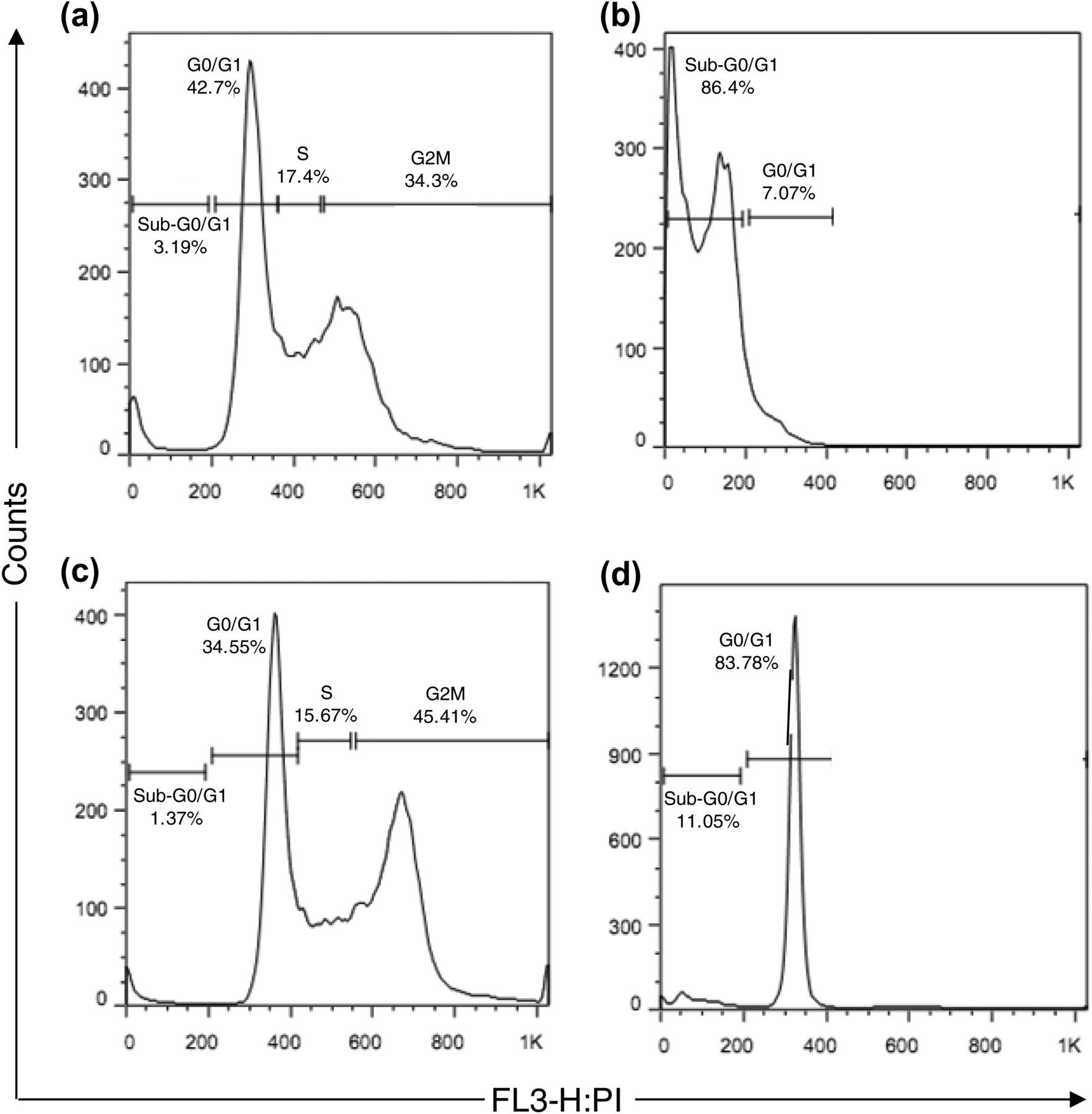
 , 12 mg/kg, B), SpHL (
 , lipid dose is the same as that administered for the treatment with SpHL-CDDP), or SpHL-CDDP (
 , 12 mg/kg), in the TR3 and TR7 schedule. aRepresents significant difference between noted phase and the same phase of the 0.9% w/v NaCl solution group. bRepresents significant difference between noted phase and the same phase of the free CDDP group. cRepresents significant difference between noted phase and the same phase of SpHL group. ND indicates that the cell cycle phases percentage was not determined from the free CDDP group due to the presence of ascites in only one animal. Data are expressed as mean ± standard error of mean
Toxicological evaluation
Hematological investigations
Hematological parameters of Ehrlich ascite tumor-bearing female Swiss mice and treated three (TR3) or seven (TR7) days after intraperitoneal treatment with 0.9% w/v NaCl solution, free CDDP, SpHL or SpHL-CDDP
Data are expressed as mean ± standard error of mean
P values less than 0.05 were set as the level of significance
*Represents significant difference between noted group and SpHL
†Represents significant difference between noted group and saline treatment
‡Represents significant difference between noted group and SpHL-CDDP
Clinical chemistry
Urea and creatinine levels of Ehrlich ascitic tumor-bearing female Swiss mice treated three (TR3) or seven (TR7) days after tumor inoculation with 0.9% w/v NaCl solution, free CDDP, SpHL or SpHL-CDDP
Data are expressed as mean ± standard error of mean
P values less than 0.05 were set as the level of significance
*Represents significant difference between group treated with SpHL-CDDP and its respective control group
Values of the urea/creatinine index of Ehrlich ascitic tumor-bearing female Swiss mice treated three (TR3) or seven (TR7) days after tumor inoculation with 0.9% w/v NaCl solution, free CDDP, SpHL or SpHL-CDDP
Data are expressed as mean ± standard error of mean
P values less than 0.05 were set as the level of significance
*Represents significant difference between noted group and the same for TR3 treatment schedule
†Represents significant difference between free CDDP, SpHL-CDDP treatments and their respective control groups
Biochemical parameters indicative of liver toxicity in Ehrlich ascite tumor-bearing female Swiss mice treated by intraperitoneal route with NaCl 0.9% (w/v), free CDDP, SpHL, or SpHL-CDDP, three (TR3) or seven (TR7) days after tumor cell inoculation
ALT, alanine aminotransferase
Data are expressed as mean ± standard error
P values less than 0.05 were set as the level of significance
*Represents significant difference between noted group and SpHL-CDDP treatment
Histopathological examination
In this study, only the histological photomicrographs of TR7 treatments were shown because both treatment schedules (TR7 and TR3) presented the same pathological profile. The microscopic examination of the kidneys from female mice after different treatments (free CDDP, saline, SpHL and SpHL-CDDP) is shown in Figure 4. Mice treated with 12 mg/kg of free CDDP revealed severe toxic tubular necrosis (Figure 4a) characterized by degeneration of tubular cells with a flattening of the lining cells of the renal cortex, coupled with proteinuria in the medullar tubules. This finding is diffuse, which indicates the toxic action of the drug. By contrast, for mice treated with SpHL-CDDP at the same dose, only a discrete presence of protein inside of the tubular lumen was observed, indicating a reduction in renal toxicity induced by CDDP in its liposomal form (Figure 4b). Concerning hepatic toxicity, we observed the degeneration of hepatocytes in the periportal regions. The degeneration of the liver is common in cases of neoplastic cachexia and may be caused by toxic agents. It is characterized by cytoplasmic vacuolization with a displacement of the nucleus to the periphery. In Figure 4c and d, only the histological photomicrographs of mice treated with 0.9% w/v NaCl solution or SpHL-CDDP were shown because the same pathological changes were observed for treatments with SpHL and free CDDP. Pratically, the degeneration in the liver of animals treated with 0.9% w/v NaCl solution or SpHL was not observed (Figure 4c) and, in contrast, severe degeneration was found in the liver of animals treated with free CDDP or SpHL-CDDP (Figure 4d). Furthermore, microscopic examination of the liver from female mice after treatments showed that tumor cells had adhered to the organ capsule (Figure 4e) and may sometimes be surrounded by inflammatory cells. This result indicates the spread of tumor cells in the peritoneal region resulting in the peritoneal carcinomatosis. The histological evaluation of the spleen showed tumor cells adhered to the organ capsule for the mice group treated with 0.9% w/v NaCl solution or SpHL. In contrast, animals treated with free CDDP or SpHL-CDDP showed no or few adhered tumor cells (data not shown). This finding shows the ability of both formulations containing CDDP to reduce the spreading of tumor cells in the abdominal region. Interestingly, vacuoles were observed in the spleen of animals treated with SpHL or SpHL-CDDP (Figure 4f), which may be related to lipid components of liposomes. It is known that nanosystems are uptaken by the organs of the mononuclear phagocyte system, such as the liver and spleen.
Photomicrographs of renal tissue from female Ehrlich ascitic tumor-bearing Swiss mice treated with free CDDP (a) or SpHL-CDDP (b). Both treatments were administered at a dose of 12 mg/kg. The arrows show the presence of protein inside the tubular lumen. Panels c and d show the profiles of the liver of mice after treatment with saline (also observed in mice treated with SpHL) and CDDP (also observed in mice treated with SpHL-CDDP), respectively. In panel e, the arrows show tumor cells adhered to the liver capsule. Panel f indicates the profile of the spleen of mice after treatment with SpHL-CDDP. The arrows show the presence of vacuoles. Hematoxylin and eosin, original magnification ×200. (A color version of this figure is available in the online journal)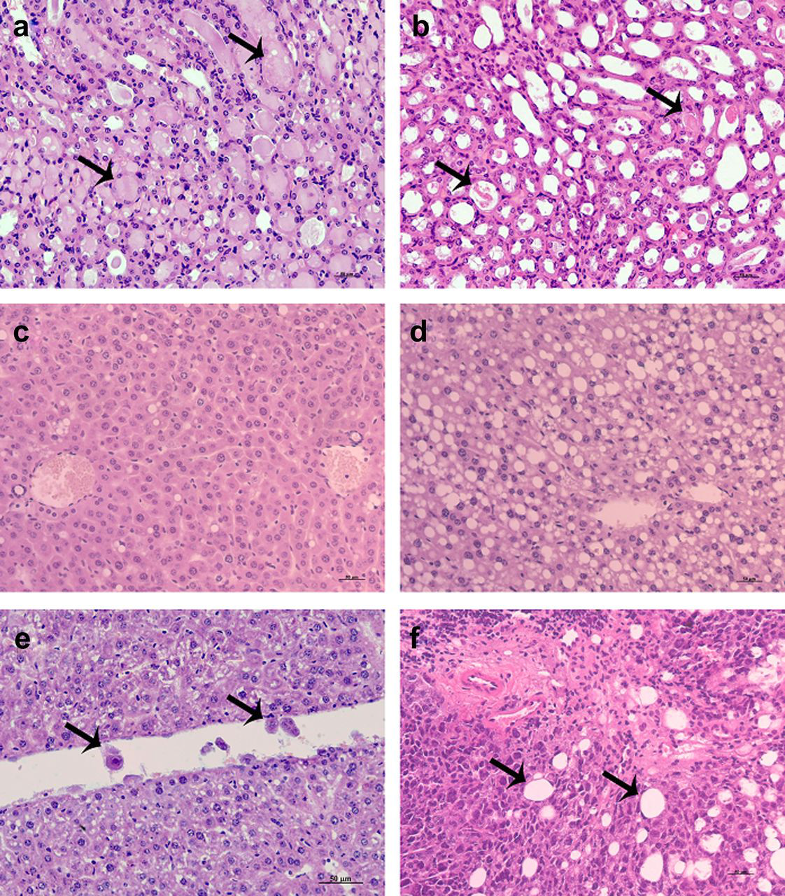
Discussion
The main objective of cancer treatment is the eradication of the disease. However, in some situations in which a cure is impossible, the objective becomes the improvement of the symptoms and quality of life associated with the increased survival of patients. 17 Currently, intraperitoneal chemotherapy for the treatment of peritoneal carcinomatosis using CDDP as an anticancer drug is limited by its absorption through the peritoneal lining. Consequently, the appearance of systemic toxicity must be observed, especially nephrotoxicity, a primary dose-limiting factor in CDDP therapy that reduces a patient's quality of life and restricts treatment protocols. 18 The development of analog drugs and new formulations are current strategies for increasing the effectiveness and safety of use of this drug. In an attempt to control the disadvantages caused by treatment with free CDDP, a strategy adopted is related to the pharmacotechnical modifications, such as the encapsulation of CDDP in drug delivery systems. 1,2,19–22 This new chemotherapeutic approach may contribute to the reduction or elimination of systemic side-effects. In this study, the antitumor effectiveness, as well as the toxicity of a novel nanostructured CDDP carrier, developed by our research group (SpHL-CDDP), was assessed using experimental animal models presenting initial or disseminated peritoneal carcinomatosis. 8,23 The treatment of initial-stage Ehrlich tumor-bearing mice with SpHL-CDDP showed a better therapeutical efficacy than that observed for free CDDP treatment. This fact could be demonstrated by higher survival of mice treated with SpHL-CDDP than with free CDDP. Moreover, SpHL-CDDP treatment avoided the formation of ascites for the TR3 treatment schedule. Probably, this finding is due to the higher concentration of the drug in the peritoneal region after SpHL-CDDP administration than free CDDP injection, as has been demonstrated previously in biodistribution studies. 7 Thus, the encapsulation of CDDP into long-circulating and pH-sensitive liposomes does not influence its antitumor activity. The evaluation of the cell cycle profile of Ehrlich tumor cells after SpHL-CDDP administration confirmed the maintenance of the antitumor activity of this formulation. The majority of Ehrlich tumor cells were arrested in the G0/G1 phase, thus stopping the progression of the cell cycle. The arrest in the G0/G1 is thought to give the cells time to repair critical damage before DNA replication occurs, thereby avoiding the propagation of genetic lesions to progeny cells and activating the apoptotic pathway. It has been reported that the conformational changes caused by the binding of CDDP to the DNA molecule induce DNA damage, affecting DNA replication and transcription, causing a twisting of the DNA strand that hinders the action of the repair enzymes and chromatin re-modeling and leads to the death of the tumor cell by apoptosis. 20 We can suggest that the interaction between CDDP and DNA occurred in our study, since the analysis of the cell cycle profile revealed that the treatment with SpHL-CDDP resulted in significant increases in the G0/G1-arrested cells, which is characteristic in cells undergoing DNA damage. In the case of treatment of disseminated peritoneal carcinomatosis with SpHL-CDDP, an improvement of antitumoral effectiveness was also achieved compared with free CDDP treatment. The median survival of mice treated with SpHL-CDDP was 26 days, whereas those treated with free CDDP presented a median survival of 14 days. In addition, SpHL-CDDP stopped cell cycle progression from the G0/G1 into S-phase. Concerning toxicity spectra of SpHL-CDDP as compared with free CDDP, it is possible to highlight the beneficial effects of these liposomes as CDDP carriers. It is worth noting the lower mortality rate for mice that received SpHL-CDDP treatment as compared with those treated with free CDDP in experimental animal models of initial or advanced peritoneal carcinomatosis. The majority of the hematological parameters investigated after administration of free CDDP or SpHL-CDDP in initial or advanced Ehrlich tumor-bearing mice showed an almost similar profile. The treatment of mice presenting initial cancer with both formulations provoked a suppression of granulocytes, suggesting a hematopoietic suppression, which is well known for CDDP based-treatments. 24,25 In addition, free CDDP treatment seems to induce a great myelotoxicity since a reduction in the platelet count can also be observed. Healthy mice treated with free CDDP at a similar dose (10 mg/kg) also showed a decrease of neutrophil count compared with the saline group. 8 By contrast, the administration of SpHL-CDDP at a dose of 12 mg/kg in healthy mice caused no alteration of neutrophil count. This finding may be due to the presence of the tumor that leads to a weakening of the health status of mice and compromises their immune response. On the other hand, disseminated tumor-bearing mice showed a significant decrease of WBCs compared with initial stage Ehrlich tumor-bearing mice. However, the SpHL-CDDP administration contributed to the improvement of the immune response of mice, which can be seen by the increase in leukocyte count, when compared with the saline control group. This finding may be explained by higher antitumor effectiveness associated with the lower bone marrow toxicity induced by SpHL-CDDP treatment when compared with the free CDDP treatment. With regards to renal toxicity, it could be demonstrated by the determination of the urea/creatinine index that only the presence of peritoneal carcinomatosis in an initial stage or widely spread is sufficient to induce renal damage. The high blood urea/creatinine index values reflect the reduction in renal perfusion as well as in the glomerular filtration rate, which consequently prevents an increase in the blood creatinine concentration. However, it is important to note that treatment of mice with free CDDP or CDDP-SpHL in an experimental animal model of peritoneal carcinomatosis in the initial stage was able to reduce the renal failure. This fact could be evidenced by the reduction in the blood urea/creatinine ratio for mice treated with these formulations. This finding demonstrates the antitumor effect of both CDDP formulations. However, when the tumor is widely disseminated in the peritoneal region, it was not possible to improve renal function after treatment of mice with free CDDP or SpHL-CDDP, since high values of the urea/creatinine index were obtained. Moreover, the histological analysis revealed that SpHL-CDDP treatment led to reduced renal toxicity in both experimental animal models. Most likely, the reduction of the renal tissue alteration observed for SpHL-CDDP-treated mice is due to the ability of liposomes to modify the biodistribution of the entrapped drug. Biodistribution studies regarding free CDDP and SpHL-CDDP in Ehrlich ascitic tumor-bearing mice, performed by our research group, demonstrated that the kidney–blood partition ratio for free CDDP treatment was higher than SpHL-CDDP treatment. 7 This finding indicates that a great extravasation and uptake of the free CDDP occurs between the renal tissue and the vascular space, which can, in turn, induce nephrotoxicity. Finally, despite the high SpHL-CDDP uptake by organs of the mononuclear phagocyte system as shown in previous studies 7 and severe hepatic tissue degeneration observed by histopathological analysis, no liver toxicity was detected in the initial or advanced stage Ehrlich tumor-bearing mice from the analysis of clinical chemistry parameters.
Conclusion
In conclusion, the treatment of initial or disseminated stages of Ehrlich ascitic tumor-bearing Swiss mice with SpHL-CDDP proved to be able to improve the antitumor efficacy and decrease renal and bone marrow toxicity of CDDP-based therapy. Moreover, it was observed that the interaction of CDDP and DNA was not altered due to its encapsulation in liposomes, since the induction of cell death by apoptosis was observed. Therefore, these results open the possibility of future use of SpHL-CDDP in chemotherapy of peritoneal carcinomatosis. New studies are underway in our research group to investigate the signaling pathways of cell death, as well as the use of high doses of SpHL-CDDP for the treatment of peritoneal carcinomatosis.
Footnotes
ACKNOWLEDGEMENTS
The authors would like to thank Fundação Oswaldo Cruz, Fundação de Amparo à Pesquisa do Estado de Minas Gerais, and Conselho Nacional de Desenvolvimento Científico e Tecnológico for their financial support. The authors also thank the Program for Technological Development of Health Products (PDTIS) for technological development in tools for health and the Oswaldo Cruz Foundation (FIOCRUZ) for use of its facilities. MCdO, GDC, OAM-F and AT-C thank the National Council for Scientific and Technological Development (CNPq) for fellowships. LdCM thanks Centro de Pesquisas René Rachou for fellowships. AT-C thanks the US Food and Drug Administration for fellowships.