
Abstract
This study was designed to examine the survival and biological behavior of adipose-derived mesenchymal stem cells (ADMSCs) under an intervertebral disc (IVD)-like acidic environment. Human ADMSCs isolated from two age groups were cultured under four different pH levels (pH 7.4, 7.1, 6.8 and 6.5) which mimicked the standard condition and the normal, mildly degenerated and severely degenerated IVD. Cell viability was measured by fluorescein isothiocyanate-Annexin-V/propidium iodide staining, and cell proliferation was measured by MTT (3-[4,5-dimethylthiazol-2-yl]-2,5-diphenyl tetrazolium bromide) assay. The expression of aggrecan, collagen-I, collagen-II, matrix metalloproteinase-2 (MMP-2), tissue inhibitor of metalloproteinase-3 (TIMP-3), p53 and caspase-3 at the mRNA level was examined by realtime quantitative polymerase chain reaction, and the expression of aggrecan, collagen-I, collagen-II, MMP-2 and TIMP-3 at the protein level was measured by enzyme-linked immunosorbent assay. Acidic pH inhibited the viability and proliferation, and the expression of aggrecan, collagen-I and collagen-II of ADMSCs from both age groups. ADMSCs harvested from young and mature donors exhibited similar responses to the acidic pH, although cells from young donors appeared less sensitive to the low pH levels. The results demonstrated that acidic pH in IVD may be an important deleterious factor for ADMSC-based IVD regeneration. ADMSCs harvested from young donors may be more suitable to be utilized for the implantation into degenerated IVD, and the implantations may be more effective at an early stage of IVD degeneration when the pH of matrix acidity is higher than 6.8.
Introduction
Low back pain is a public health problem that has significant socioeconomic impact. 1,2 It is reported that low back pain affects up to 85% of people and leads to a cost of approximately $100 billion in the United States every year. 3 Although the etiology of low back pain remains elusive, intervertebral disc (IVD) degeneration has been proposed as one of the major causes. 4
The disc cells synthesize the matrix components, which play an important role in the regulation of the biochemical and biomechanical functions of IVD. 5 IVD degeneration is accompanied by a decrease in the number of disc cells that leads to the loss of disc matrix. 6 Cell-based therapy has emerged as a promising approach for the treatment of IVD disease. 7 Because stem cells have the ability to self-renew and differentiate into specialized cells, 8 they have gained more attention as the sources of cells for IVD regeneration. Adipose-derived mesenchymal stem cells (ADMSCs) are attractive candidates for stem cell-based therapy because of their ease of isolation and their comparative abundance. 9,10 ADMSC transplantation into degenerated IVD has been proven as an effective interventional technique in many animal models. 5,11,12 In a canine disc injury model, Ganey et al. 5 found that autologous ADMSCs were effective in promoting disc regeneration, as evidenced by disc matrix production and overall disc morphology. In a rat IVD degeneration model, Jeong et al. 12 found that the rats which received ADMSC transplantation showed a significantly smaller reduction in disc heights when compared with the rats in the control group, and exhibited a restoration of magnetic resonance imaging signal intensity.
However, ADMSC transplantation for disc degeneration faces multiple challenges, the primary of which is that stem cells must remain viable and maintain normal function in the harsh microenvironment of the degenerated disc, which is characterized by low pH level, low oxygen concentrations and limited nutrition. 7 During IVD degeneration, the supply of glucose and oxygen decreases as the permeability of the endplate reduces, rendering these already harsh conditions even worse, and the pH level even lower. 13
Wuertz et al. 14 suggested that the most challenging chemical condition of the IVD microenvironment is matrix acidity, which has profound effects on cell viability and matrix turnover. The pH level in the normal IVD ranges between 7.0 and 7.2. 13,15,16 However, in serious degenerative discs, the pH usually reduces to 6.5, although pH levels as low as 5.5–5.6 have been reported in degenerative human IVD tissue removed in surgery. 17,18 The acidic condition in IVD has been shown to be a detrimental factor for cell survival and matrix production of disc cells, 13,19 as well as for the viability and function of bone marrow mesenchymal stem cells (BMMSCs). 14 However, the effects of IVD-like acidic environments on the survival and biological behavior of ADMSCs remain largely uninvestigated. Therefore, this study aimed to examine the survival and biological behavior of ADMSCs under IVD-like acidic environments, and to determine if there is a certain pH threshold under which the viability and proliferation and other biological behavior of ADMSCs will be affected significantly by the acidic microenvironment.
Materials and methods
Cell isolation and culture
The isolation of ADMSCs was performed following the approval of the Ethics Committee of Zhejiang University with informed consent from the patients. For cell culture, all reagents were purchased from Invitrogen (Carlsbad, CA, USA). Human ADMSCs were isolated from subcutaneous adipose tissues obtained from young (aged 8–12 years, n = 6) and mature (aged 33–42 years, n = 6) male donors undergoing elective surgical procedures. Approximately 1.5 g of adipose tissues were washed with phosphate-buffered saline (PBS) and finely minced, and were then digested with 0.15% collagenase type I (Sigma, St Louis, MO, USA) at 37°C for 30 min in a water-bath shaker (200 rpm). The collagenase was inactivated by the addition of Dulbecco's modified Eagle's medium (DMEM) supplemented with 10% fetal calf serum, penicillin (50 U/mL) and streptoymcin (50 μg/mL). The ADMSC-containing cell suspension was centrifuged at 600 Characterization of isolated human ADMSCs by osteogenic and adipogenic differentiation. (a) Human ADMSCs were expanded in monolayer culture. (b) ADMSCs were cultured in osteogenic media for 21 days and stained positive with alizarin red. Red staining marked mineral deposition in their newly formed extracellular matrix. (c) ADMSCs were cultured in adipogenic media for 14 days and stained positive with Oil red O staining. The lipid drop appeared red under a fluorescence microscope. ADMSC, adipose-derived mesenchymal stem cell. (A color version of this figure is available in the online journal)
Culture media with different pH levels were prepared by adding an appropriate amount of sterilized HCl (1 mol/L) and NaOH (1 mol/L) into DMEM and monitoring using a commercial pH microelectrode (Lazarlab, Los Angeles, CA, USA) (sensitive to 0.01 pH unit). Media with four pH levels, including 7.4 (standard condition), 7.1 (normal IVD), 6.8 (mildly degenerated IVD) and 6.5 (severely degenerated IVD) were obtained. The culture media were kept at 37°C with 5% CO2 for three days to allow pH equilibrium (CO2-dependent). ADMSCs in passage 2 were cultured either in 24-well plates for cell proliferation assay or in 25 cm2 cell culture flasks for cell viability and gene and protein expression analysis. The cells were re-fed with fresh medium on day 3 and harvested for analysis on day 6.
Flow cytometry analysis
After six days, the cells were harvested and incubated with 0.25 mg/mL fluorescein isothiocyanate (FITC)-conjugated Annexin-V and 10 mg/mL propidium idodide (PI) for 30 min in the dark at 4°C. Then viable, apoptotic, secondary necrotic cells were quantified by FACS analysis with a FACS Calibur cytometer (FACScan, Becton Dickinson, Mountain View, CA, USA) equipped with CellQuest software (BD Biosciences, San Jose, CA, USA). When green fluorescence (FITC) was plotted against red fluorescence (PI), three distinct cell populations could be detected in a dot–plot: viable cells (FITC−/PI−), apoptotic cells (FITC+/PI−) and secondary necrotic cells (FITC+/PI+).
MTT assay
After three and six days of exposure to an altered pH, cell proliferation was analyzed by MTT (3-[4,5-dimethylthiazol-2-yl]-2,5-diphenyl tetrazolium bromide) assay. Briefly, the media were replaced with 500 μL MTT solution (250 μg in DMEM) and the cells were cultured for another four hours. Then formazan was dissolved by adding 200 μL dimethyl sulfoxide (DMSO), and the absorbance was measured at 570 nm by using a Spectra MAX microplate reader (Molecular Devices, Sunnyvale, CA, USA).
Realtime quantitative polymerase chain reaction analysis
Total RNA was isolated from ADMSCs using TRIzol (Invitrogen) according to the manufacturer's instructions. Next, 2 μg of total RNA was reverse-transcribed using 1 μL of oligo-dT18 primer (Invitrogen), 25 units of RNase inhibitor, 1 μL of dNTPs and 200 U of reverse-transcriptase (Superscript II, Gibco BRL, Carlsbad, CA, USA) at 37°C for one hour. The reaction was stopped by incubation at 70°C for 10 min. The quantification of RNA expression levels for aggrecan, collagen-I, collagen-II, matrix metalloproteinase-2 (MMP-2), tissue inhibitor of metalloproteinase-3 (TIMP-3), p53, caspase-3 and 18S-RNA was carried out on an iCycler system (Bio-Rad Laboratories, Hercules, CA, USA). iQ™SYBR Green super mix PCR kit (Bio-Rad) was used for realtime monitoring of amplification (5 ng of template cDNA; 45 cycles: 95°C/10 s, 62°C/25 s) with appropriate primers. Human 18 S-RNA was used as the internal control. The cycle threshold (CT) values for each gene were corrected using the mean CT value. Realtime quantitative polymerase chain reaction (RT-PCR) data were quantified using the ΔCT method with the formula:
The primers used were aggrecan, forward: 5′ GGCATTTCAGCGGTTCCTTCTCCA 3′ and reverse: 5′ ACGATCCACTCCTCCACACCAGA 3′ (81 bp); collagen-I, forward: 5′ GGTGTTGTGCGATGACGTGATCTGT 3′ and reverse: 5′ CTTGGTCGGTGGGTGACTCTG 3′ (112 bp); collagen-II, forward: 5′ GGAGCAGCAAGAGCAAGGAGAAGA 3′ and reverse: 5′ GCAGTGTTGGGAGCCAGATTGTCA 3′ (103 bp); MMP-2, forward: 5′ CCAAGAACTTCCGTCTGTCCCA 3′ and reverse: 5′ GCCAAGGTCAATGTCAGGAGAG 3′ (80 bp); TIMP-3, forward: 5′ TCACCATCTCCCAGACCCTCTT 3′ and reverse: 5′ GCTTGCTGCCTTTGACTGATCCT 3′ (114 bp); p53, forward: 5′ GCCCATCCTCACCATCATCACACT 3′ and reverse: 5′ GGCACAAACACGCACCTCAAAGC 3′ (82 bp); caspase-3, forward: 5′ GACAGACAGTGGTGTTGATGATGAC 3′ and reverse: 5′ GCATGGCACAAAGCGACTGGAT 3′ (148 bp); 18S-RNA, forward: 5′ GACTCAACACGGGAAACCTCAC 3′ and reverse: 5′ CCAGACAAATCGCTCCACCAAC 3′ (122 bp).
Enzyme-linked immunosorbent assay
After six days of culture, the concentrations of aggrecan, collagen-I, collagen-II, MMP-2, and TIMP-3 in the conditioned media of ADMSCs were determined by enzyme-linked immunosorbent assay (ELISA) using commercially available kits (Uscn Life Science Inc., Wuhan, China) following the manufacturer's instructions.
Statistical analysis
ADMSCs cultured under pH 7.4 (standard condition) served as the control group. Statistical analyses were performed with SPSS 17.0 for Windows (SPSS Inc., Chicago, IL, USA). Evaluation of the effects of pH level and age was tested by two-way analysis of variance. Post hoc comparisons were performed using Turkey's test to evaluate the effects of pH level. The difference was considered significant when P was less than 0.05.
Results
Acidic pH impairs the viability of ADMSCs
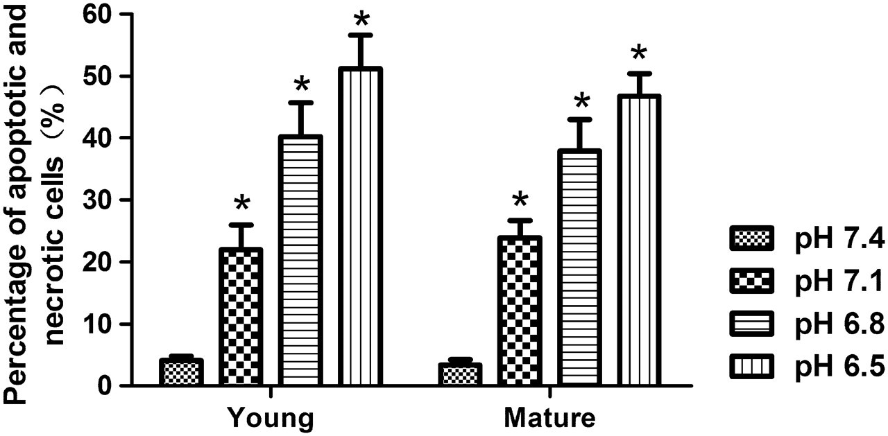
Flow cytometry analysis showed that the viability of ADMSCs decreased along with the acidity from pH 7.4 to 6.5 (Figures 2 and 3). The percentage of apoptosis and necrotic cells increased at pH 7.1 (young, 21.98%; mature, 23.85%, both P < 0.0001), pH 6.8 (young, 40.24%; mature, 37.92%, both P < 0.0001) and pH 6.5 (young, 51.24%; mature, 46.70%, both P < 0.0001) in both age groups. In addition, it was observed that pH 6.8 was a threshold under which the percentage of apoptosis and necrotic cells increased significantly. However, the age of donors of ADMSCs had no influence on cell vitality under every specific condition.
Effects of IVD-like pH levels on the viability of ADMSCs. The numbers of viable, apoptotic and secondary necrotic cells were determined by Annexin V-FITC staining. The results showed that cell vitality decreased along with acidity from pH 7.4 to 6.5 in both age groups. ADMSC, adipose-derived mesenchymal stem cell; IVD, intervertebral disc; FITC, fluorescein isothiocyanate. (A color version of this figure is available in the online journal) Percentage of apoptotic and necrotic cells of ADMSCs under IVD-like pH levels. The percentage of apoptosis and necrotic cells increased significantly at pH 7.1, 6.8 and 6.5 in both age groups. Data are presented as mean ± SD with P < 0.05 for n = 6 (young males) and n = 6 (mature males). *P < 0.05 versus pH 7.4. ADMSC, adipose-derived mesenchymal stem cell; IVD, intervertebral disc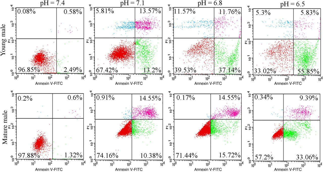
Acidic pH impairs the proliferation of ADMSCs
The MTT assay showed that the acidity had a significant effect on the proliferation of ADMSCs. Although the proliferation was not significantly affected at pH 7.1, it was inhibited at pH 6.8 and 6.5 (Figure 4). On day 3, the proliferation was not affected at pH 7.1 (young, 98.85%; mature, 97.10%), but was significantly inhibited at pH 6.8 (young, 76.23%; mature, 67.08%, both P < 0.0001) and pH 6.5 (young, 32.59%; mature, 28.64%, both P < 0.0001) in both age groups. Similar results were observed on day 6 as the proliferation was not affected at pH 7.1 (young, 99.10%; mature, 97.15%), but was significantly inhibited at pH 6.8 (young, 66.52%; mature, 57.25%, both P < 0.0001) and pH 6.5 (young, 24.61%; mature, 21.16%, both P < 0.0001) in both age groups. Neither interaction between pH and the age of donors nor a significant influence of age alone was found. In addition, a significant inhibition of the proliferation of ADMSCs from both age groups was observed when the pH level was no more than 6.8, indicating pH 6.8 as a threshold.
Effects of IVD-like pH levels on the proliferation of ADMSCs. MTT assay of ADMSCs cultured for three days or six days under IVD-like pH levels. Data are presented as mean ± SD with P < 0.05 for n = 6 (young males) and n = 6 (mature males). *P < 0.05 versus pH 7.4. ADMSC, adipose-derived mesenchymal stem cell; IVD, intervertebral disc; MTT, 3-[4,5-dimethylthiazol-2-yl]-2,5-diphenyl tetrazolium bromide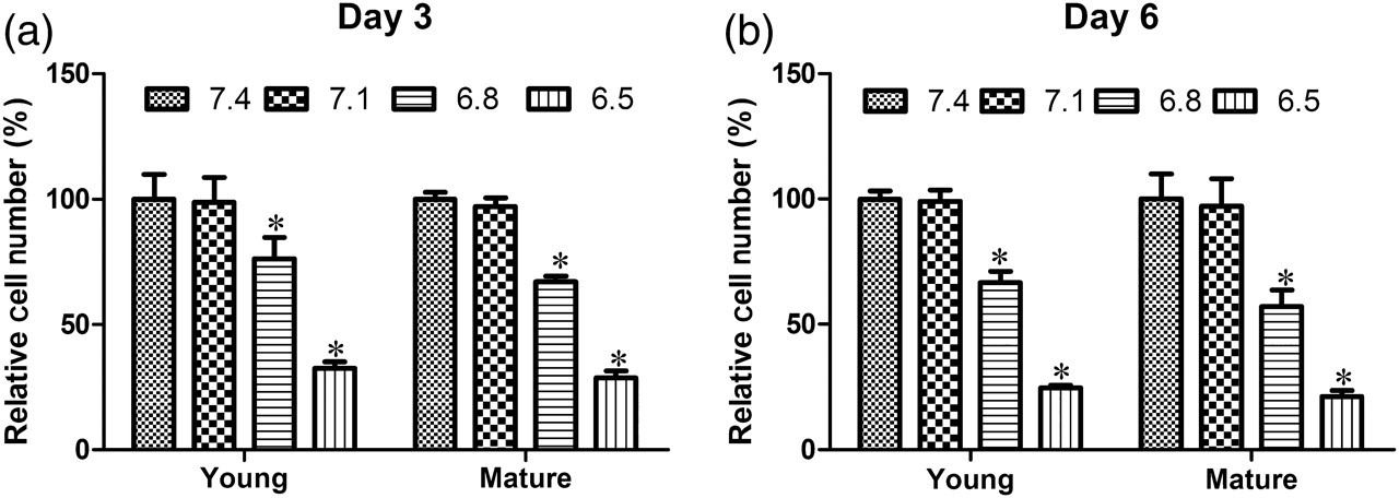
Acidic pH affects the mRNA expression of extracellular matrix components in ADMSCs
The acidic pH had a significant effect on the gene expression of extracellular matrix (ECM) components in ADMSCs in both age groups (Figure 5). The expression of aggrecan at the mRNA level was inhibited at pH 7.1 (young, 61.99%; mature, 74.78%, both P < 0.0001), pH 6.8 (young, 29.32%; mature, 15.24%, both P < 0.0001) and pH 6.5 (young, 5.05%; mature, 4.97%, both P < 0.0001) in both age groups. The expression of collagen-I at the mRNA level was inhibited at pH 7.1 in the mature group (72.40%, P = 0.0003), at pH 6.8 in both age groups (young, 69.70%, P = 0.0001; mature, 62.43%, P < 0.0001) and at pH 6.5 in both age groups (young, 35.09%; mature, 15.81%, both P < 0.0001). Similarly, the expression of collagen-II at the mRNA level was inhibited at pH 6.8 in the mature group (64.03%, P < 0.0001), at pH 7.1 in both age groups (young, 89.30%, P = 0.01; mature, 76.27%, P < 0.0001) and at pH 6.5 in both age groups (young, 29.48%; mature, 21.93%, both P < 0.0001). These data suggest that the expression of aggrecan, collagen-I and collagen-II at the mRNA level in ADMSCs is inhibited under the IVD degeneration condition.
Effects of IVD-like pH levels on the mRNA expression of aggrecan, collagen-I, collagen-II, MMP-2, TIMP-3, p53 and caspase-3 in ADMSCs. * P < 0.05 versus pH 7.4. ADMSC, adipose-derived mesenchymal stem cell; IVD, intervertebral disc; MMP-2, matrix metalloproteinase-2; TIMP-3, tissue inhibitor of metalloproteinase-3
Surprisingly, the acidic pH condition up-regulated MMP-2 mRNA expression at pH 6.8 (157.31%, P < 0.0001) in the young group, but down-regulated MMP-2 mRNA expression at pH 6.5 (31.01%, P < 0.0001) in the mature group. A significant inhibition of TIMP-3 mRNA expression was found at pH 7.1 in the mature group (59.02%, P = 0.01), at pH 6.8 in both age groups (young, 60.52%, P < 0.0001; mature, 52.79%, P = 0.0004) and at pH 6.5 in both age groups (young, 35.61%; mature, 20.64%, both P < 0.0001). As the controls, the acidic pH conditions had no significant effects on the expression of p53 and caspase-3 in both age groups as a whole, although the expression of p53 was inhibited in the mature group under pH 6.5 (68.54%, P = 0.01). Some age differences were noted with regards to the mRNA expression of aggrecan, collagen-II, MMP-2 and TIMP-3, indicating that ADMSCs isolated from young donors appeared less sensitive to the inhibitory effects of low pH on the biosynthesis of matrix (Figure 5).
Acidic pH affects the protein expression of ECM components in ADMSCs
As shown in Figure 6, the ELISA assay showed that the expression of aggrecan protein decreased significantly at pH 7.1 (young, 74.69%; mature, 74.34%, both P < 0.0001), pH 6.8 (young, 36.30%; mature, 31.96%, both P < 0.0001) and pH 6.5 (young, 15.64%; mature, 16.43%, both P < 0.0001) in both age groups. The expression of collagen-I protein decreased significantly at pH 7.1 (young, 40.63%; mature, 41.61%, both P < 0.0001), pH 6.8 (young, 53.67%; mature, 46.63%, both P < 0.0001) and pH 6.5 (young, 19.83%; mature, 20.60%, both P < 0.0001) in both age groups. Similarly, the expression of collagen-II decreased significantly at pH 7.1 (young, 80.83%; mature, 73.81%, both P < 0.0001), pH 6.8 (young, 82.05%; mature, 69.00%, both P < 0.0001) and pH 6.5 (young, 24.87%; mature, 19.55%, both P < 0.0001) in both age groups. The acidic pH up-regulated MMP-2 protein expression at pH 6.8 (young, 352.32%, P < 0.0001; mature, 169.36%, P = 0.0002), but down-regulated MMP-2 expression at pH 6.5 (young, 71.95%, P = 0.0002; mature, 44.02%, P < 0.0001) in both age groups. A significant inhibition of TIMP-3 protein expression was observed at pH 7.1 and 6.8 in the mature group (71.20%, 73.61%, both P = 0.003), and at pH 6.5 for both age groups (young, 38.38%, P = 0.001; mature, 37.07%, P < 0.0001). Some age differences were noted with regards to the expression of collagen-II, MMP-2 and TIMP-3 at protein level. These results were consistent with the findings of RT-PCR analysis.
Effects of IVD-like pH levels on the protein expression of aggrecan, collagen-I, collagen-II, MMP-2 and TIMP-3 in ADMSCs. Shown are representative blots from six independent experiments with similar results. β-Actin served as the loading control. * P < 0.05 versus pH 7.4. ADMSC, adipose-derived mesenchymal stem cell; IVD, intervertebral disc; MMP-2, matrix metalloproteinase-2; TIMP-3, tissue inhibitor of metalloproteinase-3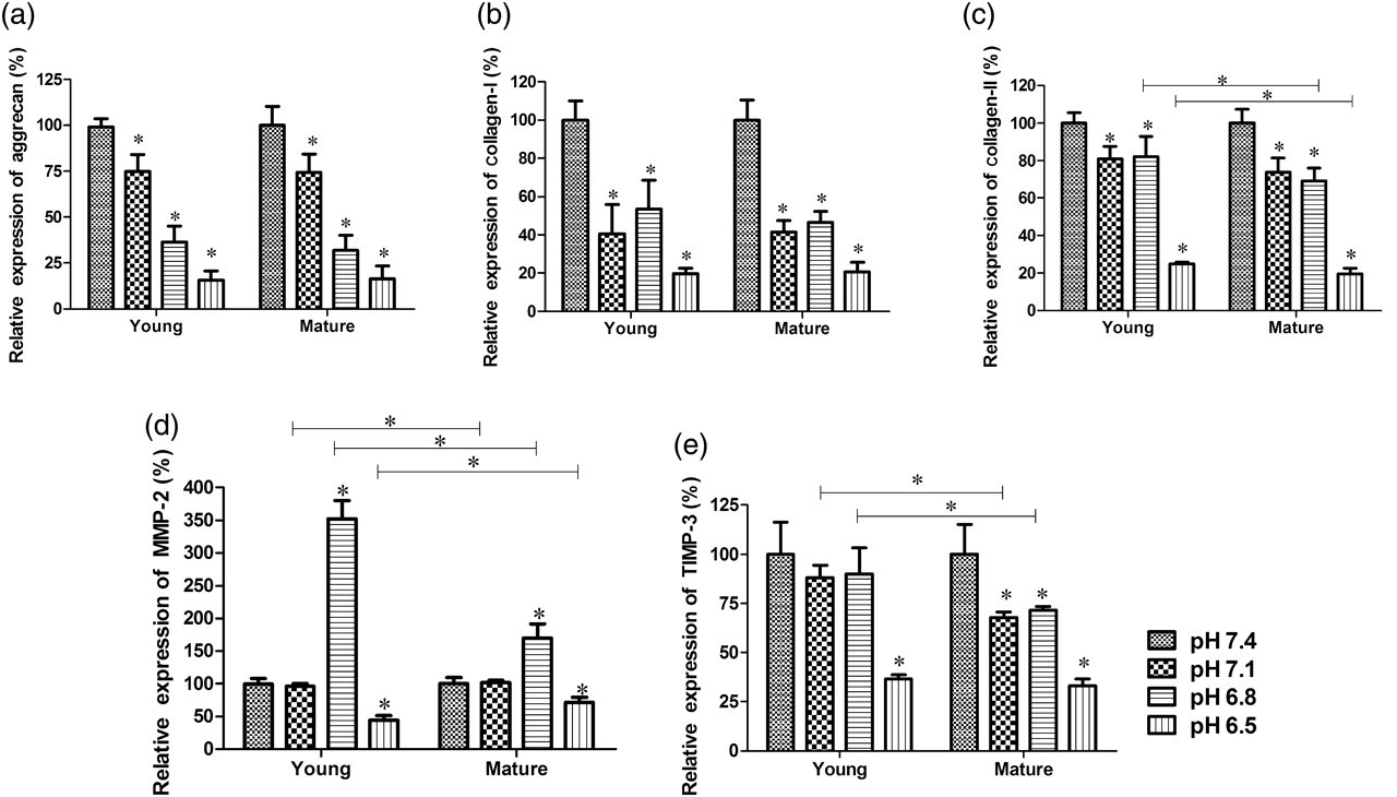
Discussion
Low back pain is an extremely common symptom, affecting nearly 85% of the population. Given that IVD degeneration is the primary cause of low back pain, effective approaches to prevent and reverse disc degeneration have been considered as promising. 20 Currently, there is an increasing interest in the potential use of stem cells in tissue engineering strategies to regenerate the degenerated IVD. 11 Both BMMSCs and ADMSCs are known to be capable of chondrogenic differentiation and recent studies have demonstrated their repair potential in IVD regeneration. 12,21
The chemical microenvironment in IVD is characterized by an acidic pH, especially during the process of disc degeneration.
13
Two main factors contribute to the acidic pH in IVD. First, the disc is the largest avascular tissue in the body with a relatively low O2 level, where the metabolism is largely anaerobic and the disc cells produce lactic acid at significant rates.
22
Second, aggrecan carries a highly negative charge due to the glycosaminoglycan chains. In order to maintain charge equilibrium, the negative charge attracts high levels of free cations such as H
Extracellular acidity is an important determinant for disc matrix turnover and may limit the success of ADMSC-based IVD regeneration. 19 To our knowledge, this is the first study to examine the survival and biological behaviors of ADMSCs under IVD-like acidic environments. We observed a significant increase in the number of apoptotic and necrotic cells under the acidic conditions. However, the apoptotic mechanisms remain elusive and may not be mediated through the up-regulation of p53 and caspase-3 because acidic pH condition did not up-regulate the expression of p53 and caspase-3 in both age groups. Surprisingly, the expression of p53 was inhibited in the mature group under pH 6.5. We currently do not know the underlying mechanism and further studies are necessary to provide the explanation. Interestingly, a previous study reported that a pH-dependent endonuclease, DNase II, could be activated at low pH level, leading to apoptosis. 23 Thus, we speculate that this endonuclease may play a role in acidic pH-induced apoptosis of ADMSCs, although this hypothesis needs to be tested in future studies.
Notably, the MTT assay showed that the proliferation of ADMSCs was inhibited at pH 6.8 and pH 6.5 in both age groups, and no difference was observed between the cells from young and mature donors. The underlying mechanism responsible for the inhibition of cell proliferation by acidic pH is not yet fully understood. Wuertz et al. 14 proposed that the changes in cytoskeleton and cell adhesion molecules are involved. Moreover, it was reported that acidification arrested cell cycle at the G1 phase. 24 Further investigations are necessary to confirm these mechanisms in ADMSCs.
The main function of IVD is to transmit load and provide flexibility of the spinal column. This function depends on the structure and biochemical composition of the IVD matrix, which consists mainly of proteoglycans and collagens. 25 Aggrecan is a large aggregated proteoglycan that carries a highly negative charge. Collagen is the major structural macromolecule of the disc with the major fibrils of the annulus and nucleus formed from the fibrillar collagens. In normal IVD, at least seven different types of collagen are present, such as I, II, III, V, VI, IX and XI, although types I and II are the most abundant. 26 The collagen network anchors the IVD to the vertebral body and allows the spine to bend and flex. IVD degeneration is accompanied by a reduction in the synthesis of ECM components such as aggrecan and collagen.
In the present study, the relative expression levels of aggrecan, collagen-I and collagen-II were used as an indicator of matrix biosynthesis. Our results showed that a decrease in pH level caused a significant fall in the rate of matrix biosynthesis (Figure 5 and 6). The expression of aggrecan appeared more significantly affected by the low pH level than the expression of collagen, consistent with a previous report. 19 In addition, below pH 6.8, the matrix expression decreased steeply, indicating that pH 6.8 was a threshold for the response of ADMSCs to acidic pH.
The disc cells also produce degrading enzymes including MMPs and the corresponding inhibitors TIMPs. 13 Consequently, the composition of IVD is determined by the balance between matrix biosynthesis and matrix degradation. Interestingly, our results revealed that the acidic environment had a strong impact on the matrix degrading enzymes. The production of TIMPs fell steeply with the decrease of pH levels. In contrast, the production of MMP-2 appeared to be not affected by low pH levels and showed a biphasic response, with the production maximal at pH 6.8 followed by a decrease at pH 6.5. These results further suggest that extracellular pH is a detrimental factor to ADMSC-based IVD regeneration. In addition, the acidic pH had a relatively less effect on the expression of proteinases in ADMSCs derived from young donors, indicating that ADMSCs harvested from young donors may be more suitable for ADMSC-based IVD regeneration.
Compared with disc cells, acidity change appears to be more crucial to ADMSCs. In a similar culture system, Ohshima et al. 19 found that the synthesis rate of human IVD increased 40–50% when the pH decreased from 7.4 to 7.0. Ichimura et al. 16 found that the rate of proteoglycan synthesis in rat nucleus pulposus cells was similar between pH 6.6 and 7.6. These differences may be attributed to disc cells possessing effective buffering and acid equivalent transport pathways to regulate intracellular pH through the operation of the Na+ × H+ exchanger and an H+-ATPase. 22
Conclusion
Acidic pH in IVD may be an important deleterious factor for ADMSC-based IVD regeneration. ADMSCs harvested from young donors may be more suitable to be utilized for the implantation into degenerated IVD and the implantation may be more effective at an early stage of IVD degeneration when the pH of matrix acidity is higher than 6.8.
Footnotes
ACKNOWLEDGEMENTS
This work was supported by grants from the National Nature Science Foundation of China (No. 81171756), the Science and Technology Planning Project of Zhejiang Province (2009C33093) and the Educational and Scientific Research Project of Zhejiang Province (No. Y201017857).