
Abstract
This study describes an innovative experimentally induced model of intervertebral disc degeneration. This innovative approach is based on the induction of extracellular matrix disorders in the intervertebral disc (IVD) using a diode laser. For this study, 15 one-year-old and five 30-month-old New Zealand White rabbits were used. Two procedures were tested to trigger IVD degeneration: needle aspiration (reference technique) and a laser approach. The IVD degeneration process was assessed 20, 40, 60, 90 and 120 days after surgery by X-ray radiography (IVD height), magnetic resonance imaging (MRI) (T2 intensity of IVD signal) and histological analysis using modified Boos’ scoring. Our data indicate that a marked IVD degeneration was found compared with sham-operated animals regardless of the procedure tested. A significant decrease in disc height on X-ray radiographs was first demonstrated. In addition, MRI disc signals were significantly reduced in both groups. Finally, a statistically significant increase in Boos’ scoring was found in both laser and aspiration-induced IVD degeneration. Interestingly, IVD degeneration induced by laser treatment was more progressive compared with aspiration. Moreover, the histological results indicated that laser-induced disc degeneration was quite similar to that obtained during the natural aging process as observed in 30-month-old rabbits. Our study describes the consistency of this innovative experimentally-induced animal model of IVD degeneration. The radiological, MRI and histological data confirm its relevance. The histological examination indicates that IVD degeneration induced by laser treatment is comparable to the degenerative process observed during the onset of spontaneous IVD degeneration. This model could be a useful tool to help us validate biomaterial-assisted, cell-based, regenerative medicine strategies for the prevention and treatment of IVD degeneration.
Introduction
The intervertebral disc (IVD) is a fibro-cartilaginous tissue composed of a peripheral network of collagen fibers (Annulus fibrosus: AF), which includes an inner, highly hydrated gel (Nucleus pulposus: NP).1,2 The IVD is the site of an inevitable age-related degeneration, which originates in the NP and is accompanied by a gradual loss of its role in spinal kinematics.2,3 During the course of IVD degeneration, the structural characteristics of the NP progressively switch from a highly hydrated gel towards a desiccated tissue. In addition, the multilayered AF gradually develops internal fissures that slowly extend radially outwards to the periphery. At the cellular level, the density dramatically reduces during aging and persistent cells are unable to ensure extracellular matrix (ECM) homeostasis, resulting in a net loss of proteoglycans and a shift in the expression of disc collagens.1,4,5 Interestingly, this age-related progressive loss of proteoglycans leads to a diminished water-binding capacity that may affect the biomechanical properties of the IVD.
This IVD degeneration is one of the major causes of low back pain, which now affects a significant proportion of the population. 6 Unfortunately, the pharmacological and surgical therapies to treat disc-related low back pain focus primarily on pain relief rather than the regeneration of the affected tissues. 7 In this context, IVD tissue engineering and regenerative medicine based on the transplantation of reparative cells with scaffolding biomaterials have recently been contemplated. 8 However, the use of biomaterials and autologous cells in cell-based therapy is still at a preliminary stage, with many issues to be addressed before the real potential of this emerging concept can be assessed. Particularly, preclinical experiments must be undertaken in relevant animal models that closely mimic human IVD degeneration.9–11
The animal models of IVD degeneration most widely used to evaluate tissue engineering strategies are based on NP aspiration with a needle or intra-NP injections of proteo-lytic enzymes.10,11 These acute models probably do not reflect the complexity of the onset of IVD degeneration during natural aging, that also involves an early dehydration of the NP extracellular matrix. In this context, we developed an innovative model of IVD degeneration by using the energy transmitted by the light beam of a diode laser (light amplification by stimulated emission of radiation) in the NP.
In the present study, laser-induced IVD degeneration was followed for a four-month period of time in one-year-old rabbits and compared with aspiration-induced IVD degeneration as well as with the IVD of a 30-month-old rabbit. IVD degeneration was proven by the measurement of discal height (X-ray radiographies), T2-weighted signal intensity (magnetic resonance imaging [MRI] pictures) and Boos’ scoring (histological analysis).
Materials and methods
Decalcifier II® was purchased from Surgipath (Richmond, VA, USA). Ketamine was Imalgene 1000® from Merial (Lyon, France) and Xylazine was 2% Rompun® from Bayer (Puteaux, France). Alcian blue and Weigert's hematoxylin were purchased from Sigma-Aldrich (St Louis, MO, USA). Histosol® was obtained from Shandom (Brussels, Belgium) and Decalcifier® from Surgipath (Richmond, VA, USA). A KaVo GENTLEray 980® diode laser was graciously lent by KaVo Dental GmbH (Biberach, Germany).
Animals and surgical procedures
All animal handling and surgical procedures were conducted according to the European Community guidelines for the care and use of laboratory animals (DE 86/609/ CEE). The ethics committee at the National Veterinary School of Nantes (ENVN-ONIRIS) approved the study protocol.
A total of 20 mature, female, New Zealand White rabbits (weighing approximately 4–5 kg each and one-year-old) were used. In parallel, five other female rabbits aged from 30 months were used in order to investigate the spontaneous aging process. Each rabbit was tranquilized by intramuscular injection of dexmedetomidine and ketamine. Each rabbit was put under general anesthesia. A longitudinal middle-line skin incision was made from the xyphoid to the pelvic rim. The anterior side of the vertebral column from L1 to L6 was exposed by the trans-peritoneal approach. Disc levels were identified under fluoroscopic control. Five IVDs (T12–L1 to L5–L6) were used for the experiment, and the lesions were spread according to the following protocol: (1) one as the control group; (2) a second as the sham-operated group that consisted of IVD puncture by the fiber of the laser (off); (3) a third received NP aspiration (reference technique) and (4) a fourth IVD was treated by laser (on). A randomization of the IVD level was performed on the different rabbits.
For NP aspiration, penetration depth was controlled by a locking forceps clamped 5 mm from the needle tip according to the technique of Sobajima et al. 12 NP aspiration consists of three successive NP aspirations for 10-s NP aspirations using a 10-ml syringe and a 21-Gauge needle. 13
Laser was performed using a KaVo GENTLEray 980® diode laser. After AF puncture with a 25-Gauge needle to allow for the introduction of the 300-μm laser fiber, the end of the fiber was introduced into the NP at a depth of 5 mm. Preliminary experiments were performed to address the optimal setting of the diode laser in order to induce a localized alteration of the NP. To perform these ex vivo preliminary experiments, the lumbar spines of rabbits were isolated, and the laser was used as described above. Several parameters were measured for different types of laser treatment in terms of duration (40–60 s) and power (1–2.4 W). The localized alteration of the extracellular matrix in the NP was macroscopically evaluated, and allowed us to define the optimal parameters to be used for all subsequent experiments (Figure 1). The parameters used were pulse sequence mode with 1.6 W pulse output, pulse length: 200 ms, pulse spacing: 200 ms, duration: 40 s. Abdominal white line and skin were closed in layers with resorbable sutures.
Gross appearance of isolated lumbar intervertebral discs (IVDs) of a one-year-old rabbit untreated (a) or exposed to a laser treatment (b–e). (a) Untreated IVD; (b) 1 W and 40 s; (c) 1.6 W and 40 s; (d) 1.6 W and 60 s; (e) 2.4 W and 40 s. (A color version of this figure is available in the online journal)
X-ray radiographs
Plain radiographs of the spines were taken using a radiograph machine (Convix 80 generator and Univesix 120 table) from Picker International (Uniontown, OH, USA). Rabbits (n = 20) were tranquilized by intramuscular injection of dexmedetomidine (100 μg/kg) and ketamine (15–20 mg/kg). Coronal and sagittal plain radiographs of the spines were taken with a collimator-to-film distance of 100 cm, exposure of 100 mAs and penetration power of 48 kVp. The image data were analyzed with Osirix software (Osirix Foundation, Geneva, Switzerland). The height of the IVD was measured and corrected by the mean antero-posterior length diameter. Data were analyzed with Osirix software 3.9 for Mac. X-ray radiographs were performed at pre- and postsurgery time and at 20, 40, 60, 90 and 120 days postsurgery. Results were expressed as relative IVD height decrease.
Magnetic resonance imaging
MRI scans were obtained using a 1 T clinical magnet Magnetom Harmony/Syngo® 1-T (Siemens, Munich, Germany). As for X-ray radiographs, rabbits (n = 20, the same as those used for the X-ray radiograph analysis) were tranquilized and placed in a supine posture within the magnet, with region centering. A 2.5 mm midsagittal section image was obtained, using a T2-weighted imaging sequence (time of repetition [TR], 5000 ms; time of echo [TE], 111 ms). The images were analyzed with Osirix software. The T2-weighted midsagittal images of the discs were analyzed. An evaluation of MRI scans was performed by three independent experts in MRI image reading investigators. For each IVD level, the ratio of NP mean weighted signal intensity (T2wsi) divided by the spinal cord signal was measured at each time point. Data were analyzed with the same software used with X-ray radiographs. The follow-up was also the same as with X-ray radiographs.
Histological analysis
At days 20, 40, 60, 90 and 120, rabbits (n = 4 for each time) were sacrificed by intramuscular injection of ketamine (25 mg/kg) followed by an intravenous injection of sodium pentobarbital (1.2 g/kg). The spines were removed from the rabbits, and the IVDs together with the intact adjacent endplate were fixed in 10% neutral buffered formalin for one week and decalcified for 24 h in Decalcifier II®. After dehydration and incubation with Histosol®, specimens were embedded in paraffin and sectioned into 3-μm slices. For histological analysis, 3-μm thick paraffin sections were deparaffined using toluene, rehydrated through a graded series of ethanol and rinsed in distilled water. Sections were stained with hematoxylin phloxine saffron (HPS) and 0.1% Alcian blue as previously described. 14
Histological sections were analyzed using a modified Boos’ scoring 15 specifically designed to evaluate the degenerative changes occurring in IVDs, and notably in the extracellular matrix of the NP. Compared with the initial Boos’ scoring, cell death and cell proliferation were gathered together as one single criterion corresponding to the decrease in cell density. Thus, this modified Boos’ scoring was based on the analysis of four criteria: decrease in cell density, granular changes, tear and cleft formation, and mucous degeneration. Some parameters were ranked from 0 to 4 (granular and mucous degeneration), and others were ranked from 0 to 5 (decrease in cell density, and tear and cleft formation) depending on the intensity of the tested parameters (0: lowest; 4 or 5: highest). Three independent experts in evaluating histological slides conducted a blind review of the histological samples. In parallel, five uninjured rabbits, aged 30 months, were also sacrificed to compare histological changes of induced degeneration with age-related IVD degeneration.
For immunohistological analysis, paraffin sections were mounted on pretreated slides, deparaffinized, rehydrated and rinsed in distilled water. Sections were incubated for 10 min with newly diluted 3% H2O2 solution to inactivate internal peroxidases, followed by proteinase K treatment for 30 min at 37°C. After digestion, slides were incubated for 30 min with 2% diluted goat serum for blocking. Slides were then immunoblotted with the primary monoclonal mouse antibody against human type I collagen (08631701; MP Biomedical, Illkirch, France) at 1/100 in phosphate-buffered saline overnight at 4°C or against human type II collagen (08631711; MP Biomedical) in the same conditions. Incubation with biotin-labeled antimouse antibody at 1/300 for 60 min was followed by 30 min of incubation with horseradish peroxide-conjugated streptavi-din. Antibody binding was visualized with diaminobenzi-dine. As a negative control, sections were processed with identical protocols, but omitting the primary antibody. Sections were then visualized with a light microscope (Zeiss Axioplan 2, Göttingen, Germany), with immunopositive areas exhibiting brown staining.
Statistical analyses
The effects of the NP aspiration and laser on changes in IVD height and T2wsi were analyzed using non-parametric repeated one-way analysis of variance. When significant differences were detected, hypothesis testing compared values at each time point for the condition with a Dunn post hoc test. A P value less than 0.05 was used to determine statistical significance.
Results
Change of IVD height
No significant IVD narrowing was observed in all groups pre- or postsurgery (Figure 2a). A significant gradual IVD narrowing was found in aspiration and laser-treated discs compared with control discs and sham-operated discs (Figures 2a and b). One hundred and twenty days after the surgical procedure, a 15% and 20% decrease in disc height was observed in aspiration and laser-treated discs, respectively (Figure 2b). A time-course analysis also revealed that IVD narrowing was significant in the aspiration group as early as day 40, while in laser-treated discs, a significant decrease in IVD height was observed 20 days later (Figure 2b).
X-ray images and analysis of rabbit lumbar spines. Intervertebral discs (L2-L3; L5-L6; L1-L2) of one-year-old rabbits were either treated according to the aspiration technique (aspiration), the laser procedure (laser) and the sham procedure as described in the Materials and methods section. Untreated IVDs (T12-L1; control) were used as an internal control. (a) After the indicated times, X-ray images of spines were taken. Representative X-ray images are shown. (b) Decrease in IVD height (%) was measured after the indicated times as described in the Materials and methods section. *P < 0.05 as compared with day 0. D60, day 60; D120, day 120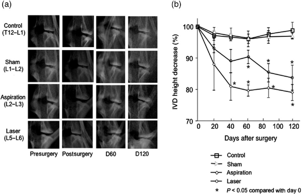
Change of IVD T2-weighted signal intensity
The T2wsi in MRI showed an intense decrease postsurgery time in the aspiration group compared with the laser group. After this postsurgery time, the T2wsi in MRI showed a progressive decrease in aspiration and laser-treated discs compared with control discs and the sham-operated group (Figure 3a). The time-course analysis also showed that T2wsi decreased significantly in the aspiration group as early as 20 days postsurgery, while in laser-treated discs, a significant decrease in T2wsi was observed 40 days later (Figure 3b).
MRI images (T2-weighted midsagittal) and analysis of rabbit lumbar spines. Intervertebral discs (L2-L3; L5-L6; L1-L2) of one-year-old rabbits were either treated according to the aspiration technique (aspiration), the laser procedure (laser) and the sham procedure as described in the Materials and methods section. Untreated IVDs (T12-L1; control) were used as an internal control. (a) After the indicated times, a magnetic resonance imaging (MRI) scan of the spine was performed. Representatives MRI scans are shown. (b) T2-weighted signal intensity was measured after the indicated times as described in the Materials and methods section. *P< 0.05 as compared with day 0. D60, day 60; D120, day 120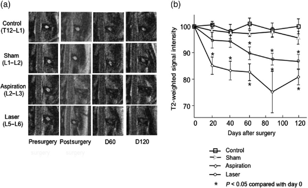
Histological analysis
To further address whether the experimentally-induced degeneration observed by radiological changes may be correlated with some tissue changes, we then performed histological stainings.
Histological analysis of non-injured discs at day 0 and sham-operated discs at day 60 (Figures 4a and f) revealed considerable cell density. Mucous degeneration and granular changes were rarely present, and tears and clefts were absent in non-injured discs at day 0 (Figure 5a) and in sham-operated discs at day 60 (Figure 5f). The injured IVD analysis revealed a decrease in cell density in both conditions (aspiration and laser) (Figures 4e and j). This decrease in cell density occured early in the aspiration condition, in contrast to the laser condition in which the density gradually decreased (Figures 4b–e and g–j). The process of aspiration-induced degeneration was characterized by the formation of tears and clefts associated with mucoid and granular changes (Figures 5b–e). Interestingly, the process of IVD degeneration following laser treatment (Figures 5g–j) showed major mucoid and granular changes but associated with a low formation of tears and clefts.
Histological analysis of rabbit intervertebral discs (IVDs) (hematoxylin phloxine saffron staining). IVDs of one-year-old rabbits were either treated according to the aspiration technique (aspiration) (b–e) or by the laser procedure (laser) (g–j) described in the Materials and methods section. IVDs were processed for histological analysis 20, 60, 90 and 120 days after treatment (aspiration and laser) as described in the Materials and methods section. Untreated IVDs (a) and sham-treated IVDs at day 60 (f) were used as internal controls. Representative hematoxylin phloxine saffron stainings are shown. Bar: 250 μm. (A color version of this figure is available in the online journal). D20, day 20; D60, day 60; D90, day 90; D120, day 120 Histological analysis of rabbit intervertebral discs (IVDs) (Alcian blue staining). IVDs of one-year-old rabbits were either treated according to the aspiration technique (aspiration) (b–e) or by the laser procedure (laser) (g–j) described in the Materials and methods section. IVDs were processed for histological analysis 20, 60, 90 and 120 days after treatment (aspiration and laser) as described in the Materials and methods section. Untreated IVDs (a) and sham-treated IVDs (f) were used as internal controls. Representative hematoxylin phloxine saffron stainings are shown. Bar: 250 μm. (A color version of this figure is available in the online journal). D20, day 20; D60, day 60; D90, day 90; D120, day 120

As a comparative control, we also observed spontaneous age-dependent IVD degeneration in 30-month-old rabbits (Figure 6). Our observations confirmed recent data generated in our lab
9
and revealed a decrease in cell density (Figure 6a) comparable to that observed in the laser condition as well as a high level of mucoid and granular changes (Figure 6b).
Histological analysis of rabbit intervertebral discs (IVD) (30-month-old). IVDs of 30-month-old rabbits were harvested and processed for histological analysis as described in the Materials and methods section. Representative hematoxylin phloxine saffron (a) and Alcian blue (b) stainings are shown. Bar: 250 μm. (A color version of this figure is available in the online journal). D20, day 20; D60, day 60; D90, day 90; D120, day 120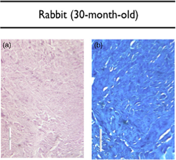
To quantitatively assess tissue changes, modified Boos’ scoring was used
15
(Figure 7). In sham-operated samples, no significant increase was observed as compared with control. Conversely, a significant increase (P < 0.05) in both laser and aspiration-induced IVD degeneration was observed. Interestingly, Boos’ scoring after laser treatment increased more progressively compared with aspiration treatment, with values of about 12 and 14 at day 20, and 15 and 16 at day 90, respectively. Tears and clefts were the main tissue changes observed in the aspiration-treated group while mucous degeneration and granular changes were essentially observed in the laser-treated group (data not shown). Boos’ scoring in the 30-month-old rabbits was estimated at 15.5.
Evaluation of Boos’ scoring for rabbit intervertebral discs (IVDs). IVDs of one-year-old rabbits were either treated according to the aspiration technique (aspiration), by the laser procedure (laser) as well as the sham procedure described in the Materials and methods section. Untreated IVDs and IVDs from 30-month-old rabbits were used as an internal control. Boos’ scoring was determined for the different conditions 0, 20, 60, 90 and 120 days after the procedure as described in the Materials and methods section (max/18). *P < 0.05 compared with day 0. D0, day 0; D20, day 20; D60, day 60; D90, day 90; D120, day 120
Immunohistochemical analysis revealed the presence of type I and II collagens in the extracellular matrix of the AF and NP (Figure 8). In intact IVDs, type I collagen was essentially found in the outer AF, and type II collagen was mainly found in the inner AF and NP. Laser treatment did not induce significant change in the distribution and intensity of the staining.
Immunohistological characterization of lumbar intervertebral disc (IVD) sections of one-year-old rabbits. Rabbit lumbar IVDs were processed for immu-nohistological detection of type I (a–n) and II (c–p) collagens as described in the Materials and methods section. (a–d) Untreated IVD (Control). (e–h) IVD after aspiration at day 120 (aspi D120). (i–l) IVD after laser treatment at day 90 (laser D90). (m–p) IVD after laser treatment at day 120 (laser D120). Bar: 100 μm. OAF, outer Annulus fibrosus; IAF, inner Annulus fibrosus. (A color version of this figure is available in the online journal)
Discussion
The current therapeutic strategies for patients with lombal-gia focus primarily on pain relief. A better understanding of the physiopathology of IVD degeneration has underlined regenerative medicine as a promising strategy.4,7 Accordingly, increasing attention has been paid to the regeneration of functional tissue by cell and biomaterial-based tissue engineering. Nevertheless, despite recent advances in biomaterial sciences and cell biology, many issues still need to be addressed before this concept can be fully evaluated. Particularly, animal models that exhibit appropriate mechanical, anatomical and physiopathological characteristics are needed to enable preclinical testing. Several spontaneous or experimentally induced models have thus been developed in animals, including the mouse, rat, sand rat, rabbit, dog, sheep, pig, goat and primate.10,11 Whereas some of them are useful for studying specific aspects of disc biology or pathology, most of these models fail to meet all the criteria required for the preclinical testing of IVD tissue engineering.
Currently, among the various experimentally induced models of IVD degeneration, the needle aspiration technique is the most widely used, especially in studies that aim to demonstrate the relevance of tissue engineering approaches.16–25 This technique, which consists of three successive 10-s NP aspirations using a 10-ml syringe and a 21-Gauge needle, 13 was used as a reference technique in our study. Our results using this technique confirmed the establishment of a degenerative process characterized by a decrease in IVD height and T2wsi by X-ray and MRI, respectively. The histological data and the elevated Boos’ scoring also confirmed the alteration of the NP extracellular matrix. Unfortunately, this degeneration process occurred rapidly with a dramatic decrease of the T2wsi and a high value of Boos’ scoring at early time. The non-progressive tissue damage triggered by aspiration contrasts to what can be observed during the natural process of degeneration that takes place over several years with a progressive alteration of the extracellular matrix and not the NP destruction.1,4
Contrary to the almost instantaneous effect of aspiration, the effect of laser treatment on the NP appears to be more progressive and less deleterious for the NP extracellular matrix. In fact, imaging analyses showed a later decrease in IVD height and T2wsi compared with the results obtained with aspiration. Analysis of histological sections confirmed the interest of the laser treatment compared with needle aspiration. The absence of a significant decrease in T2wsi and disc height in the control group and the sham-operated group confirms the absence of induced-degenerative processes associated to the AF puncture. This is further confirmed by analysis of the Boos's scoring.
Unlike aspiration, the comparison of the histological data for the laser-treated disc with that of 30-month-old rabbits strongly suggests that laser treatment may lead to tissue degeneration similar to that observed during natural aging, notably in terms of cell density decrease and ECM disorganization.1,4 One can assume that the less prominent destructive effect of laser treatment compared with aspiration is likely to closely mimic the natural degeneration process. The Boos’ scoring confirmed these data with a value at day 90 following laser treatment similar to that of IVD from 30-month-old rabbits. The exact mechanisms by which laser irradiation induces IVD degeneration remain poorly understood, but are likely to involve the high temperature resulting from the irradiation, as reported previously to explain the changes in ultrastructure and composition observed in laser-irradiated dentin. 26
The rapid occurrence of IVD degeneration constitutes a disadvantage for future tissue engineering strategies. This study demonstrates that our innovative experimentally-induced animal model of IVD degeneration meets several criteria that should characterize a relevant animal model of IVD degeneration, notably for tissue engineering experiments, namely: (1) rabbits are easily obtainable and not expensive; (2) rabbit IVDs exhibit an adequate size and anatomical characteristics compatible with the development of biomaterial-assisted surgical procedures; and (3) the onset of disc degeneration occurs reliably and quite quickly following laser treatment.
Conclusion
This study shows that IVD degeneration can be induced in the rabbit by a new laser process. Radiological, MRI and histological data confirmed the relevance of this new experimentally induced animal model of IVD degeneration. This model could be a useful tool to help validate a cell-based strategy for the prevention and treatment of IVD degeneration.
Footnotes
Acknowledgements
This study was supported by grants from Société Française de Rhumatologie, Fondation de l'Avenir pour la Recherche Médicale Appliquée (ET8-491 and ET9-541), Société Française de Neurochirurgie, Agence Nationale de la Recherche AAP TeCSAN (Chondrograft project), Agence de la Biomédecine, Institut National de la Santé Et de la Recherche Médicale (INSERM), Fondation pour la Recherche Médicale-MESCLE (FRM-MESCLE), Région des Pays de la Loire (BIOREGOS project) and the University Hospital of Nantes. The authors also gratefully acknowledge the technical assistance of Pierre Weiss, Pierre Monmousseau, Patrice Roy, Stéphane Madec, Dominique Rouleau and Christian Raphael, as well as Joanna Ashton-Chess and Christopher Bartlett for correcting the manuscript. We also thank KaVo Dental GmbH (Biberach, Germany) for providing us with a laser KaVo GENTLEray 980® diode.