




Abstract
Despite the large use of the Plantago major and Siparuna guianensis in traditional medicine, there are no studies demonstrating the effectiveness from extracts of these plants in the healing process by the present methodology. This study reported the effects and toxicity of the P. major and S. guianensis extracts in the wound healing compared with a commercial product used in Brazil by macroscopic and microscopic analysis. Following injury in cervical dorsal area of the mice, the extract from P. major and S. guianensis and ointment was applied after an injury in cervical dorsal area of the mice. Wound healing rates were calculated at 4, 9, 15 and 21 d after the wounding, and tissues were obtained on the ninth day for histological analysis. Moreover, mutagenic assay of extracts was performed. Mutagenicity studies carried out with plant extracts showed not mutagenic with or without metabolic activations. Reduction of the wound area occurred earlier in mice treated with P. major and control treatment. On the 15th day, the complete wound closure occurred in P. major-treated wounds. Throughout ointment and S. guianensis treatment it was not observed the wound closured. Microscopic analyses of the wound, on the ninth day, showed the more efficient formation of the neoepithelium and skin appendages in animals treated with S. guianensis and P. major, while ointment treatment presented no re-epithelialization and absent skin appendages in wound. Thus, P. major extract showed good effects on wound healing processes rendering it a promising candidate for the treatment of wounds what also justified its traditional usage in wound treatment.
Introduction
The wound healing is a dynamic and continuous process, performed and regulated by complex signaling cascades, involving three well-defined phases with each phase overlapping the next: inflammatory phase, proliferative phase and re-modeling phase. 1 Inflammatory cells migrate into the wound attracted by chemotactic factors and promote the inflammatory phase, which is characterized by the sequential infiltration of neutrophils, macrophages and lym-phocytes. 2 In the proliferative phase, re-epithelialization, formation of pilous follicle and angiogenesis are important activities determining the wound lesion. 3 In the re-modeling phase, the imbalance of collagen synthesis and degradation, resulting in excess accumulation of dermal collagen4,5 is frequently associated with scar complications. Chemical factors (drug substances), physical and biological (growth factors, cytokines, chemokines and nutritional status of the patient), can accelerate or delay the cicatrization.6,7 It is known that the goal for wound treatment is a fast and scar-less healing and among the substances used to accelerate the induction of this process is the use of medicinal plants. 8
A large number of plants are used by tribal and folklore in many countries for the treatment of wounds and it showed auspicious effects. 9 Over the years, various natural extracts were tested in the healing process such as the extract of Schinus terebinthifolius Raddi, 10 Calendula officinalis L. 11 Centaurea iberica 12 and the extract of Ixora coccinea. 13 In this sense, Brazil, for having one of the richest biodiversity on the planet, has many natural resources that could be used for curative purposes. 14 However, there are few controlled studies demonstrating the effectiveness of extracts in the healing process of lesions.
The Plantago major is a perennial plant that belongs to the Plantaginaceae family. It is a wild plant found worldwide and its leaves have been associated with various biological properties ranging from astringent, anesthetic, antihelmin-tic, analgesic, analeptic, antiviral, antihistaminic, anti-inflammatory, antirheumatic, antitumor, antiulcer, diuretic, hypotensive and expectorant.15,16 The Siparuna guianensis is an upright shrub or small tree 3–5 m tall, aromatic, almost all native Brazil, and that belongs to the Siparunaceae. The leaves and flowers of the S. guianensis are considered carminative, aromatic, stimulant, antidispépticas and diuretic. It is also used for back pain, rheumatism and arthritis. 17
Despite the large use of the P. major and S. guianensis in traditional medicine, there are no studies demonstrating the effectiveness from extracts of these plants in the healing process by the present methodology. The aim of the present study was to evaluate the toxicity and effects of the P. major and S. guianensis extracts in the wound healing compared with a commercial product used in Brazil.
Materials and methods
Chemicals
Dimethylsulfoxide (DMSO, CAS no. 67-68-5), methanol (CAS no. 67-56-1), dichloromethane (CAS no. 75-09-2), nico-tinamide adenine dinucleotide phosphate sodium salt (CAS no. 11-84-16-3), D-glucose-6-phosphate disodium salt (CAS no. 3671-99-6), magnesium chloride (CAS no. 7786-30-3), L-histidine monohydrate (CAS no. 7048-02-4), D-biotin (CAS no. 58-85-5), sodium azide (CAS no. 26628-22-8), 2-anthramine (CAS no. 613-13-8) and 4-nitro-o-phenylenediamine (CAS no. 99-56-9) were purchased from Sigma Chemical Co (St Louis, USA). Nutrient Broth No. 2 (Oxoid, UK), Difco Bacto Agar (Difco Laboratories, Detroit, MI, USA) and Mueller Hinton agar (Difco Laboratories) were used as bacterial media. D-Glucose (CAS no. 154-17-6), magnesium sulfate (CAS no. 7487-88-9), citric acid monohydrate (CAS no. 5949-29-1), ointment Collagenase ([DCB 0341.01-0] + Chloranphenicol [DCB 0296.01-5]), potassium phosphate dibasic anhydrous (CAS no. 7758-11-4), sodium ammonium phosphate (CAS no. 13011-54-6), sodium phosphate monobasic (CAS no. 7558-80-7), sodium phosphate dibasic (CAS no. 7558-79-4), sodium chloride (CAS no. 7647-14-5) were purchased from Merck (Whitehouse Station, NJ, USA).
Plant material and extraction
Samples of P. major and S. guianensis were collected in Minas Gerais State (Brazil) and vouchers were deposited at Universidade Federal de Minas Gerais (UFMG) Herbarium and Universidade Federal de Uberlândia (UFU) Herbarium (No. 143405, HUFU 49.454, respectively). Leaves (100 g) were washed with clean control at room temperature, dried, triturated and added to a hydro-alcoholic solution (70%). After maceration (48 h) the extract obtained was filtered and lyophilized (Liobras equi-pament, model K 105). The treatment was prepared as 10% (w/w) concentration, e.g. 5 g of extract was incorporated in 45 g of ointment base (lanolin and liquid paraffin).
Phytochemical assay
Phytochemical tests to detect the presence of secondary metabolites, such as tannins, flavonoids, esteroids, triter-pens, coumarins, saponins and alkaloids were performed according to Matos. 18 These tests are based on the visual observation of color modification or precipitate formation after the addition of specific reagents.
Ames mutagenicity assay
The Ames/Salmonella mutagenicity assay was performed using the plate incorporation protocol with the Salmonella typhimurium strains TA98 and TA10019,20 and five different concentrations of the hydroalcoholic extracts, obtained from leaves of S. guianensis (0.68, 1.36, 2.72, 5.45 and 10.90 mg plate-1) and P. major (1.78, 3.56, 7.11, 10.67 and 14.22 mg plate-1) were evaluated in this assay, with or without metabolic activation (S9 fraction). The various extract concentrations were added to 500 μL of phosphate-buffered saline (pH 7.4), plus 100 μL of bacterial culture and 2 mL of top agar. After agitation, the mixtures were poured on to plates containing minimum agar. The plates were incubated at 37°C for 48 h and the his
Animals
A total of 56 male mice of Swiss line (26-30 g) with six weeks were housed individually in plastic cages for one week prior to the experiment. The animals were fed with a standard commercial diet (Purina® Labina, Campinas -SP, Brazil) and control ad libitum. All of the experimental procedures were in accordance with the Brazilian College of Animal Experimentation (COBEA) ethical guidelines for the welfare of experimental animals. The experimental protocols were approved by the UFSJ-CEPEA protocol no 08/2009. The mice were anesthetized with Ketamina 50 mg kg-1 and Xilazina 20 mg kg-1. After trichotomy, a cutaneous incision with 2 cm of diameter was made in the cervical dorsal area. Following the injury, the animals were divided in four groups with 14 individuals each. They received local treatment with 100 μL of: group 1 control (water); group 2 P. major extract; group 3 S. guianensis extract; and group 4 ointment. Except for the drugs under study, no local/systemic therapy was provided to animals bearing any of the wounds.
Macroscopic analysis
The wound closure was measured every day using a pachy-meter from first after injury until 21 days. The data were converted in percentage. The period of cicatrisation was calculated as the number of days required for the wounds to heal completely without any raw wound left behind.
Microscopic analysis
Skin samples were collected on the ninth day, 23 fixed in 10% formalin, embedded in paraffin, sectioned with 5 μm thickness and stained with hematoxylin–eosin (HE). For analysis of wound healing, it was considered the presence or absence of epithelium and skin appendages throughout the wound, amount of edema and inflammatory infiltrate. To quantify the edema, it was used a score: 0 (physiological), 1 (tenuous), 2 (moderate) and 3 (severe). The score for inflammatory infiltrate intensity ranged from 0 (physiological), 1 (tenuous), 2 (moderate), 3 (severe) and 4 (very severe). For mast cells quantification, 10 consecutive fields of histo-logical sections stained with toluidine blue-sodium borate were acquired using an optical microscope and the AxioVision Rel 4.8 software (Carl Zeiss).
Statistical analysis
Statistical analysis was performed using GraphPad InStat software (Graphpad Software Inc., San Diego, CA). The data was statistically analyzed by ANOVA followed by Tukey–Kramer for multiple comparison and t Student's t-test was used followed by Kolmogorov and Smirnov correction for two groups comparison. The difference was considered significant when P < 0.05. All the values were expressed as mean ± standard deviation (SD).
Results
Phytochemical assay
The extracts of P. major and S. guianensis were subjected to phytochemical analysis for the identification of its major components. The presence of steroids and triterpens, and condensed tannins were different in extracts (Table 1).
Phytochemical tests with the hydroalcohol extract of plant P. major and S. guianensis

- absent, + low presence, + + medium presence, + + + high presence
Ames mutagenicity assay
The mutagenicity studies carried out with plant extracts obtained from P. major (Table 2) and S. guianensis (Table 3) showed that these compounds were not mutagenic with or without metabolic activations and there is no statistical difference between assays. The negative effects observed in all concentrations evaluated (MI < 2.0) indicated that these extracts did not present in its constitution substances able to induce substitutions, additions or deletions of bases in DNA structure.
Revertant/plate, standard deviation and mutagenicity index (MI) in strains TA98 and TA100 of S. typhimurium after treatment with various doses of P. major extract with (+ S9) and without (- S9) metabolic activation
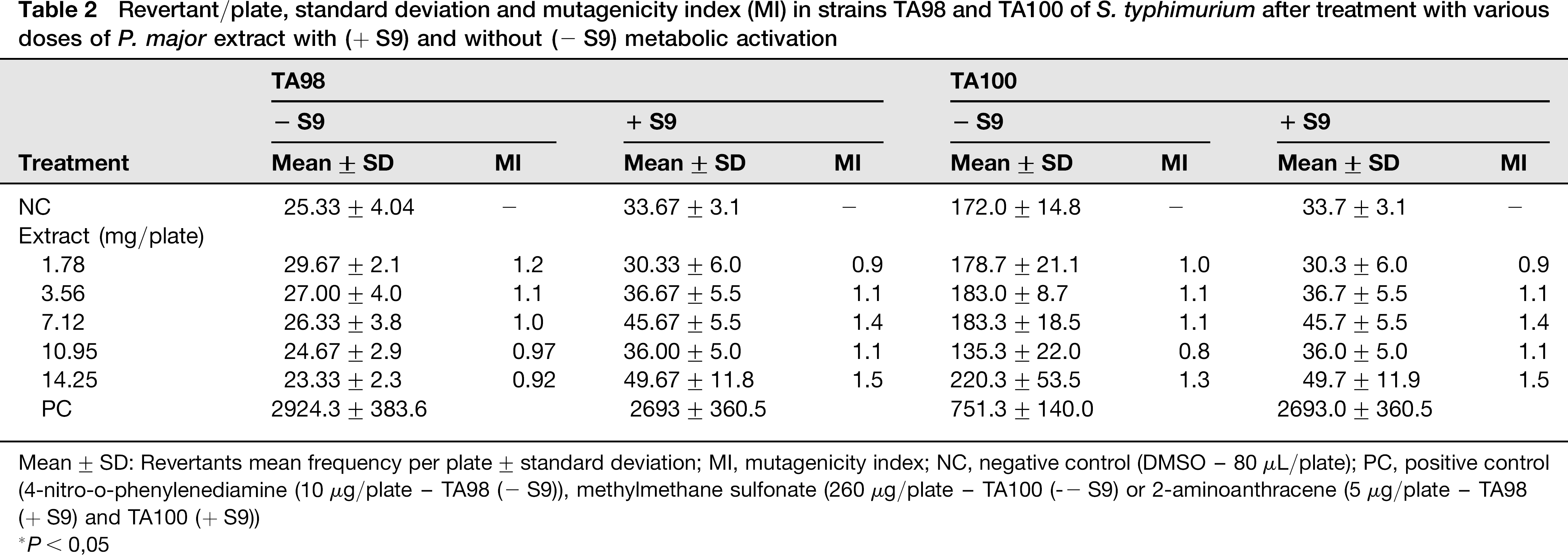
Mean ± SD: Revertants mean frequency per plate ± standard deviation; MI, mutagenicity index; NC, negative control (DMSO - 80 μL/plate); PC, positive control (4-nitro-o-phenylenediamine (10μg/plate - TA98 (
*P< 0,05
Revertant/plate, standard deviation and mutagenicity index (MI) in strains TA98 and TA100 of S. typhimurium after treatment with various doses of S. guianensis extract with (+ S9) and without (- S9) metabolic activation

Mean ± SD, Revertants mean frequency per plate ± standard deviation; MI, mutagenicity index; NC, negative control (DMSO - 80 μL/plate); PC, positive control (4-nitro-o-phenylenediamine (10 μg/plate - TA98 (- S9)), methylmethane sulfonate (260 μg/plate - TA100 (– S9) or 2-aminoanthracene (5 μg/plate - TA98 (+ S9) and TA100 (+ S9))
*P< 0.05
Macroscopic analysis
Wound healing of the skin incision was determined by the percentage of wound surface covered by regenerating epidermis at different treatments and the appearance of the wound was observed during the period studied (Figure 1a). The reduction of the wound area occurred earlier in mice treated with P. major and control treatment (no statistically significant differences). In the mice submitted to ointment treatment it was observed an increase in the wound healing area on the fourth day. On the 15th day, the complete wound closure occurred in P. major and in the control group. Throughout ointment and S. guianensis treatment it was not observed the wound closured (Figure 1b).

Wound healing effect after different treatments. (a) photographic representation of wound contraction and appearance observed throughout of the period studied G1 (control); G2 (P. major); G3 (S. guianensis); G4 (ointment); (b) curve of wound closure percentage, with P. major contributed to the wound healing compared with control, S. guianensis and ointment treatment. (A color version of this figure is available in the online journal)
Microscopic analysis
On the ninth day, regarding edema, P. major and control were classified as moderate, the S. guianensis was between tenuous and moderate, ointment was between moderate and severe with accumulation of fluid in the extravascular compartment and in the connective tissue (Figure 2a). The inflammatory infiltrate intensity of the S. guianensis was tenuous, P. major and control presented moderate, and ointment were between moderate and severe with remarkable presence of neutrophils and lymphocytes which were identified by nuclear morphology (Figure 2b). The mast cells with normal and degranulated morphology were recorded surrounding the wounds in all treatments. The quantitative analyses of the mast cells population showed the following values: control (range 0.0–46.0; mean 17.7 ± 1.3 cells/field), P. major (range 0.0–45.0; mean 18.5 ± 1.3 cells/field), S. guianensis (range 3.0–42.0; mean 22.0 ± 1.5 cells/ field) and ointment (range 0.0–43.0; mean 15.8 ± 1.4 cells/ field). There was a statistically significant difference only between the group treated with S. guianensis extract (Figure 2c) and the ointment treatment (Figure 2d).
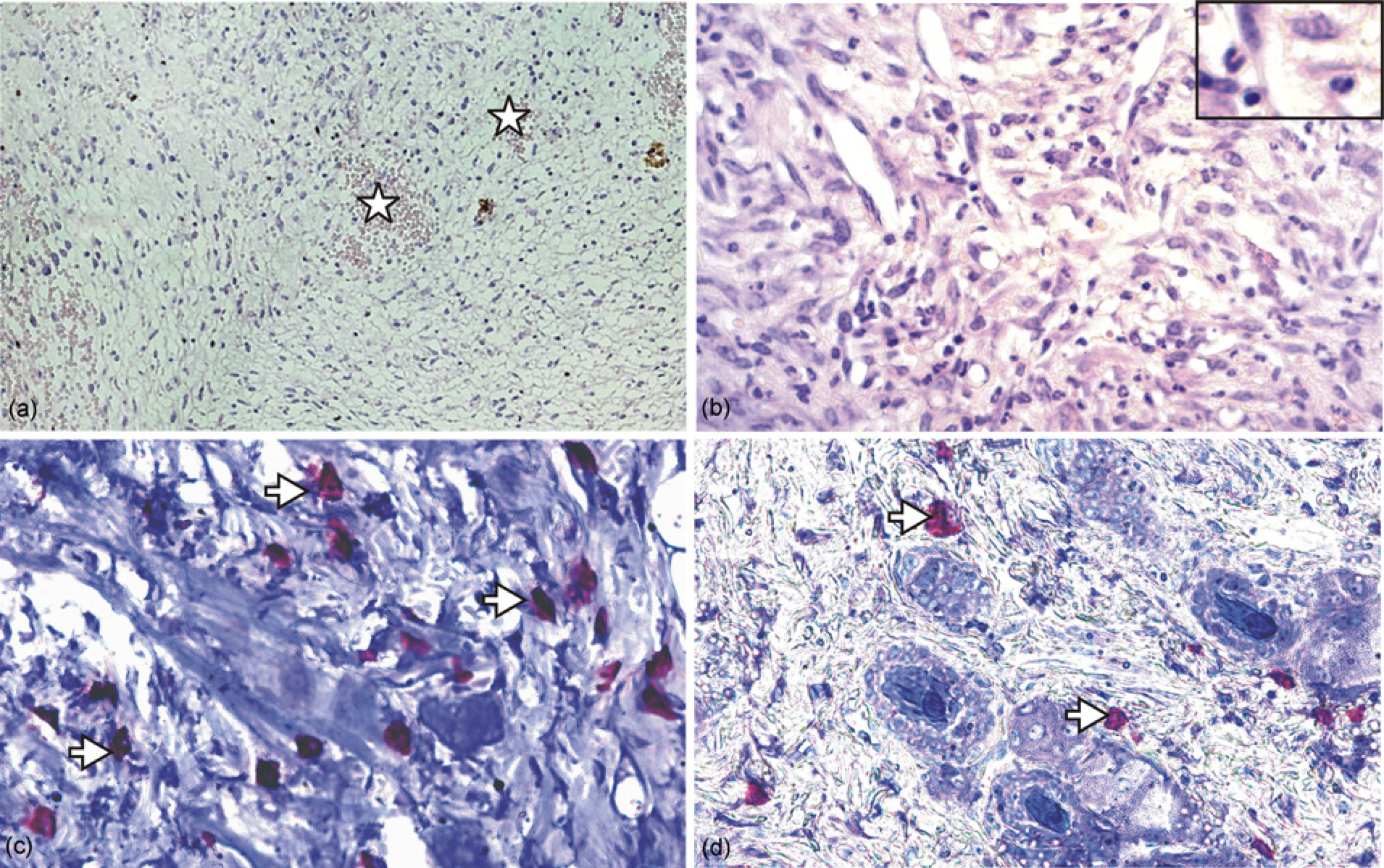
Histological sections of wound area at ninth day evidencing the remarkable findings, stained with hematoxilin–eosin (a, b) and toluidine blue (c, d). (a) edema and hyperemia (star) in individuals of the G4; (b) presence of inflammatory infiltrate in G4, detail with neutrophils and lymphocytes; (c) mast cells (white arrow) in G3; (d) mast cells (white arrow) in G4. (A color version of this figure is available in the online journal)
About the neoepithelium in skin repair, 42.9% of S. guianensis, 14.3% in control, 33.3% for P. major animals presented closed wound (Figure 3a) while ointment treatment showed no re-epithelialization (Figure 3d). Considering the skin appendages in wound, 42.9% in control, 28.6% for S. guianensis, 16.7% of P. major of the mice showed cutaneous structures in formation (Figure 3b) and completely established (Figure 3c) while in ointment this appendages were not observed.
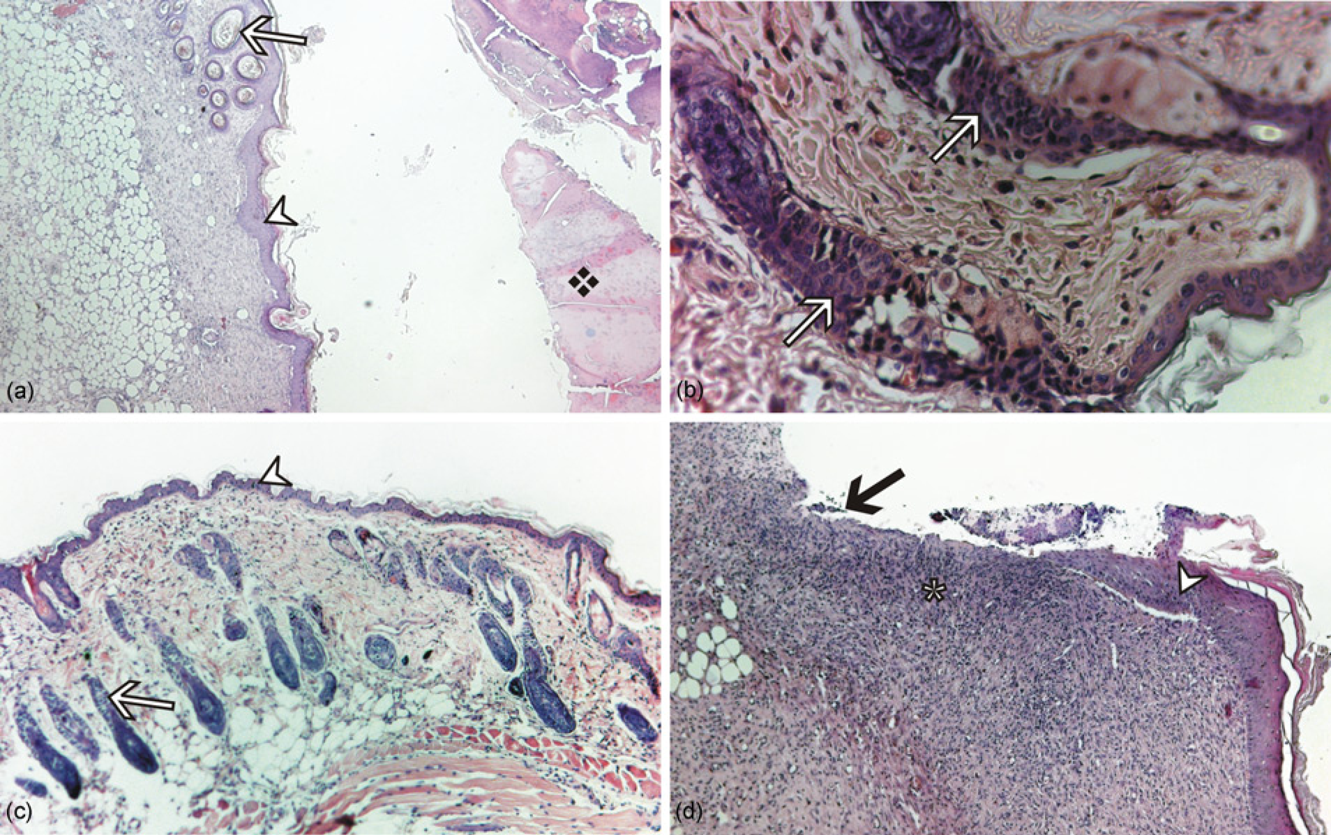
Histological sections of wound area at ninth day, stained with hematoxilin-eosin (a-d)and picrosirius red(e,f). (a) re-epithelialization: epithelium (white arrowhead) stratified with many layers and presence of crust (❖) in G1, G2 and G3 treatment; (b) pilous follicles and sebaceous glands (white arrow), formation early in G2; (c) neoepithelium with skin appendages; (d) ointment treatment showed incomplete epithelialization (black arrow) and inflammatory infiltrate (✲). (A color version of this figure is available in the online journal)
Discussion
This study reported the effect of P. major and S. guianensis extracts on the wound healing by macroscopic, microscopic analysis and mutagenic activity. The reduction of the wound area occurred earlier in mice treated with P. major. Histological analyses of the wound, on the ninth day, showed neoepithelium and skin appendages formation a more efficient in animals treated with P. major and S. guianensis, while ointment treatment presented no re-epithelialization and absent skin appendages in wound. Mutagenicity studies carried out with plant extracts showed not mutagenic with or without metabolic activations.
The P. major extracts showed a high presence of steroids and tripertenes, while the S. guianensis extracts presented condensed tannins. The steroids and tripertenes were related with anti-inflammatory activity. 24 The triterpenoids inhibits cyclooxygenase-1 and -2 catalyzed of prostaglandin biosynthesis in vitro while the structural isomer oleanolic acid is less active. 25 Tannins promote wound healing through several cellular mechanisms: scavenging of free radicals and reactive oxygen species, promoting contraction of the wound and increasing the formation of capillary vessels and fibroblasts. 26 However, the wound healing potential of these extracts is a result of the mixture of the phyto-constituents and the real contribution of each one needs to be better elucidated. Coumarins have multiple biological activities including disease prevention, growth modulation and antioxidant properties. These compounds are known to exert antitumor effects and can cause significant changes in the regulation of immune responses, cell growth and differentiation. 27 The alkaloids have been described as being responsible for the improvement in the healing of incisions by increasing the tensile strength of the scar tissue. 28 Moreover, it enhanced the wound healing by hastening the period of epithelization and wound contraction on fourth and 16th days. 28 Furthermore, it accelerates soft tissue repair in a dose-dependent manner at initial phases of the process, probably by chemotactic properties toward fibroblasts. 29
The mutagenicity studies carried out with plant extracts obtained from S. guianensis and P. major showed that these compounds were not mutagenic with or without metabolic activations. Mutagenic activity is an important parameter to evaluate the viability of topical administration, 30 as the epithelial tissue has a high-proliferation rate during the regeneration process. 31 The absence of a mutagenic response by plant extract is a positive step forward in determining the safe use of plants in traditional medicine. 32 The Ames test is a widely accepted method to identify various chemicals and drugs that can cause gene mutations and has a high-predictive value for in vivo carcinogenicity. 20
In the present study, the animals treated with S. guianensis presented the inflammation stage tenuous, while P. major presented moderate inflammation, and with the ointment it showed inflammatory response between moderate and severe. The time of inflammation can range from 24–48 h after injury, but in chronic inflammation the inflammatory response can be prolonged for weeks, months, or even indefinitely. This extended time course is caused by persistence of the causative stimulus to inflammation in the tissue, repetitive use of anti-inflammatory drugs, a weakened immune system, or an improper nerve supply. Thus, wound healing delayed by healing of chronic wounds often results from an imbalance in the wound preventing progression from one phase to another. 33 The stage of new tissue formation (proliferative phase), that happens from two days up to three weeks after the inflammatory phase, is characterized by a re-epithelialization process, activation and migration of fibroblast, production of collagen, glycosa-minoglycan and proteoglycans by fibroblasts, angiogenesis and later, some of the fibroblasts differentiate into myofibro-blasts. 34 In this work, on the ninth day, the neoepithelium and skin appendages formation was more efficient in animals treated with S. guianensis and P. major while ointment treatment showed no re-epithelialization and absent skin appendages in the wound. This step becomes the center point of wound healing, since the closure of the wound prevents the entry of pathogenic organisms that can cause various disorders including contamination and delay in the healing process. 35 The findings for S. guianensis and P. major, indicate that the compounds presents in the extracts of this species probably promote a proliferative phase more efficient, preventing a delay in phase and consequently the healing.
In the present work it was analyzed mast cells population during a wound healing where the animals treated with ointment had a lower density (15.8 ± 1.4 cells/field) since that the treatment with S. guianensis showed higher density for mast cell (22.0 ± 1.5 cells/field). There was a statistically significant difference only between the group treated with S. guianensis extract and the ointment treatment. These findings were expected since the ointment is a pharmaceutics industrial product, which decreases the allergen effects. The mast cell is involved in the various phases of skin repair (mainly proliferative and re-modeling phases), secreting pro-inflammatory molecules and also regulating the process of the healing.36,37 In fact, in mast cell deficient-mice, the healing process was partially impaired after skin scald injury. 37 The significance of this cellular type in the process of wound healing is in fact that an increased or a decreased of degranulated biological mediators causes impaired skin repair, with the formation of exaggerate granulation tissue, delayed closure and chronicity of the inflammatory stage. 36 The re-modeling phase is featured by the regression of most of the newly formed capillaries, thus the vascular density of the wound returns to normal. Another critical point of the re-modeling step is extracellular matrix re-modeling to an architecture that is similar to that of the normal tissue. Moreover, the wound undergoes physical contraction throughout the entire wound healing process, which probably can be performed by contractile fibroblasts (myofibroblasts) that appear in the wound. 35
The wound area was measured up to 21 days to determine whether the treatment with P. major and S. guianensis extracts affects the progress of wound healing. The control and group treated with P. major showed the wound closure on the 15th day. However, from the fourth day until the 15th day it was observed that the wound closure evolution in P. major treatment was better than others fact evidenced on ninth day. In the ointment and S. guianensis treatment the wound closure was not observed. If the goal for wound treatment is the fast healing, the P. major extract was more effective than ointment treatment. In fact, the P. major was suggested related with wound healing and some of these effects may attribute to the use of this plant in folk medicine. 15
Conclusions
In summary, the study has showed that the P. major extract effectively stimulated wound-healing processes as compared with the others treatments. These finding could justify its traditional usage in wound treatment.
Footnotes
Acknowledgements
Our thanks to Gregory Kitten who has kindly gave his technical support. This work was supported by grants from FAPEMIG (TECH APQ 03742/09).