
Abstract
To understand the effect of mechanical stimulation on posterior scleral reinforcement (PSR), rabbit scleral fibroblasts after PSR were subjected to stretch in vitro, and the viscoelastic behavior of scleral fibroblasts was evaluated. Three-week-old rabbits were monocularly treated by eyelid suturation randomly to prepare the experimental myopia eyes. After 60 days, the experimental myopia eyes were treated by PSR. After six months, the posterior pole scleral fibroblasts (normal sclera – group A, sclera after operation – group B and fusion region of sclera and reinforcing band – group C) were isolated and cultured in vitro. The cells were subjected to cyclic stretch regimens (sine wave, 3% and 6% elongation amplitude, 0.1 Hz, 48-h duration) by an FX-4000 Tension System. The micropipette aspiration technique was used to investigate the viscoelasticity of scleral fibroblasts. The cellular viscoelasticity (E 0, E ∞ and μ) of group C was significantly lower than groups A and B (P < 0.05), and there was no significant difference between groups A and B (P > 0.05). The results show that the viscoelasticity in different regions of sclera after PSR is different. Following a 48-h stretch, the cellular viscoelastic parameters were significantly decreased when compared with the respective static groups (P < 0.05) in groups A and B. For group C, the viscoelasticity of the stretch group was significantly higher than the static control group (P < 0.05). There was no difference between the 3% and 6% stretch groups in each group (P > 0.05). The changes of viscoelasticity suggest that different regions of sclera have different responses to mechanical stimulation in the process of treating high myopia by PSR and that mechanical stimulation plays an important role in the treatment of axial myopia by regulating the viscoelasticity of scleral fibroblasts.
Introduction
The sclera is a dense, fibrous, viscoelastic connective tissue that forms the outer coat of the eye and it is a dynamic tissue, capable of altering extracellular matrix (ECM) (e.g. collagen and proteoglycan) composition and its biomechanical properties in response to changes in the visual environment to regulate ocular size and refraction. The composition, structure and mechanical behaviors of sclera rest on, to some extent, the biological function and mechanical properties of scleral fibroblasts, which are the main cell composition of sclera. 1 High myopia development is accompanied by scleral thinning, the loss of scleral tissue and the weakening of the scleral mechanical properties. 2 In vivo, the sclera and scleral fibroblasts are in a mechanical environment because of the internal pressure and eye movements; the mechanical stimulation plays an important role in sclera remodeling during myopia development and recovery by means of regulating the biochemical and biomechanical properties of the sclera and scleral fibroblasts. Previous studies have shown that the mechanical stimulation had effects on the proliferation, ECM synthesis, gene expression and growth factor expression of scleral fibroblasts. 3–7 However, little is known about the effects of mechanical stimulation on the biomechanical properties of scleral fibroblasts.
Posterior scleral reinforcement (PSR) surgery is an effective way of treating pathological myopia 8 with improvement in elastic modulus and collagen content of sclera. 9 Therefore, we hypothesize that the biomechanical behavior of scleral fibroblasts is changed in the treatment of myopia and regulated by biomechanical stimulations. The aim of this study was to investigate the effects of mechanical stimulation on viscoelasticity of scleral fibroblasts after PSR surgery.
In this experiment, we obtained the scleral fibroblasts from different scleral regions of rabbit models after PSR surgery and cultured them in a mechanical environment using an FX-4000 Tension System (Flexcell Int. Corp., Hillsborough, NC, USA), and then examined the viscoelasticity of scleral fibroblasts by the micropipette aspiration technique (MAT). The results indicate the changes in scleral fibroblast viscoelasticity and the role of mechanical stimulation in the process of treating high myopia with PSR surgery.
Methods
Animal model of high myopia and PSR
Three-week-old New Zealand white rabbits (we selected the rabbits for this study because they are mammals and their eyeballs are big enough for PSR surgery) were monocularly treated by eyelid suturation randomly to establish an experimental high myopia eye model; eyes that were not operated on were the normal control. After 60 days, the experimental myopia eyes were treated by PSR with polyester fiber pericardium patches. Before and after the operation to establish an experimental high myopia eye model, eye axis measurement was performed with an A-ultrasonic (MEDA Corp. Ltd, Tianjin, China) to show the success of the experimental high myopia eye model. 10
Primary culture of scleral fibroblasts
After six months, the therapeutic effect of PSR was demonstrated by measuring the eye axis and posterior pole scleral fibroblasts were obtained by the tissue piece culture method. Between the sclera and reinforcing band, there was an obvious fusion region. The scleral fibroblasts were divided into three groups: group A (normal sclera), group B (sclera after operation) and group C (fusion region of sclera and reinforcing band after operation) (Figure 1). The scleral fibroblasts were plated in a culture bottle containing Dulbecco's modified Eagle's medium (DMEM) with 15% fetal bovine serum (FBS) and then incubated at 37°C (5% CO2). The cells were trypsinized after reaching confluency and centrifugated (1000 rpm×10 min) in an equal volume of DMEM with 15% FBS. The supernatant was removed, the cell pellet was reconstituted with DMEM with 15% FBS, transferred into 35 mm six-well BioFlex collagen type I-coated stretch plates (Flexcell Int. Corp.) and incubated at 37°C (5% CO2). After 24 h, the cells had adhered to the rubber elastic membrane and the medium was changed for mechanical stretch. The cells of the three groups were identified using an immunocytochemical method (detected the expressions of vimentin, desmin, keratin and S-100).
10
Schema of scleral tissue structure after performing posterior scleral reinforcement surgery. After six months, there was an obvious fusion region between the sclera and reinforcing band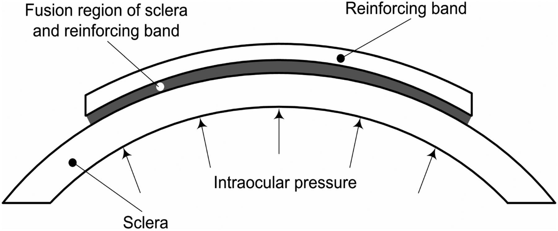
Mechanical stretch with FX-4000 Tension System
The scleral fibroblasts in six-well BioFlex stretch plates (Flexcell Int. Corp.) were subjected to cyclic stretch regimens (sine wave, 3% and 6% elongation amplitude, 0.1 Hz, 48-h duration) using the FX-4000 vacuum-driven Tension System together with the Bioflex Loading Stations baseplate (Flexcell Int. Corp.) at 37°C (5% CO2) (Figure 2). Cells plated on Bioflex plates but not subjected to stretch served as controls for each group.
Mechanical stretch scheme. (a) The FX-4000 Tension System can control the generation of interested mechanical stretch driven by the vacuum pump. (b) Sine wave. The mechanical loading mode inflicted on the cells using smooth stretch waveform – sine wave, smaller elongation amplitude – 3% and 6% (only the 6% is shown), minor frequency – 0.1 Hz. (c) Six-well culture plate. The cells were inoculated into the six-well BioFlex collagen type I-coated stretch plates. (d) Schematic diagram of FX-4000 Tension System. The negative pressure acts on the flexible bottom wells using the vacuum pump. The cells plated on the silicone membrane are affected by mechanical stimulation due to the deformation of the silicone membrane. (A color version of this figure is available in the online journal)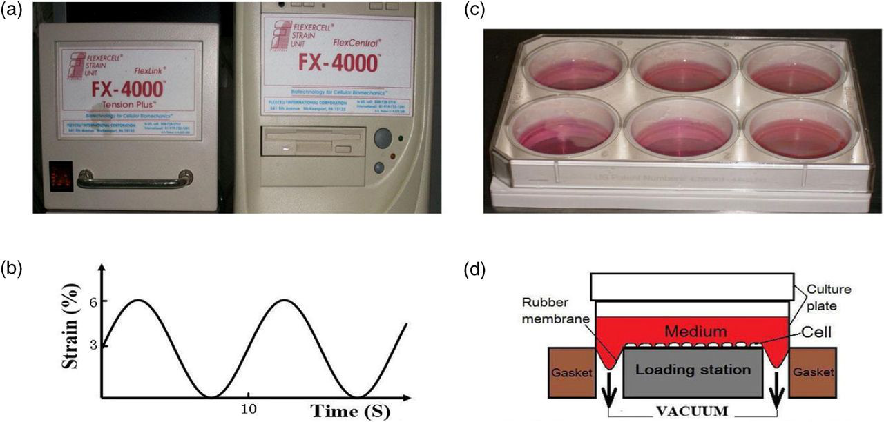
Micropipette aspiration technique
The viscoelasticity of scleral fibroblasts was tested with the MAT. After mechanical stretch, the scleral fibroblasts were harvested and made into single cell suspensions for examination of viscoelasticity. The scleral fibroblast suspension was loaded into a small chamber. The tip of the micropipette was controlled to approach the surface of a spherical cell by a micromanipulator, and the process was viewed with an inverted microscope. The negative pressure was fixed to a certain value (range from 0.4 to 0.5 kPa), which caused a time-dependent deformation of the scleral fibroblasts. The course of cell aspiration was maintained for 150 s and recorded by an image capture system. The pictures were analyzed with Image-Pro Plus 5.1 software (Olympus Corporation, Tokyo, Japan). All experiments were performed at 37°C within two hours.
Viscoelasticity of scleral fibroblasts
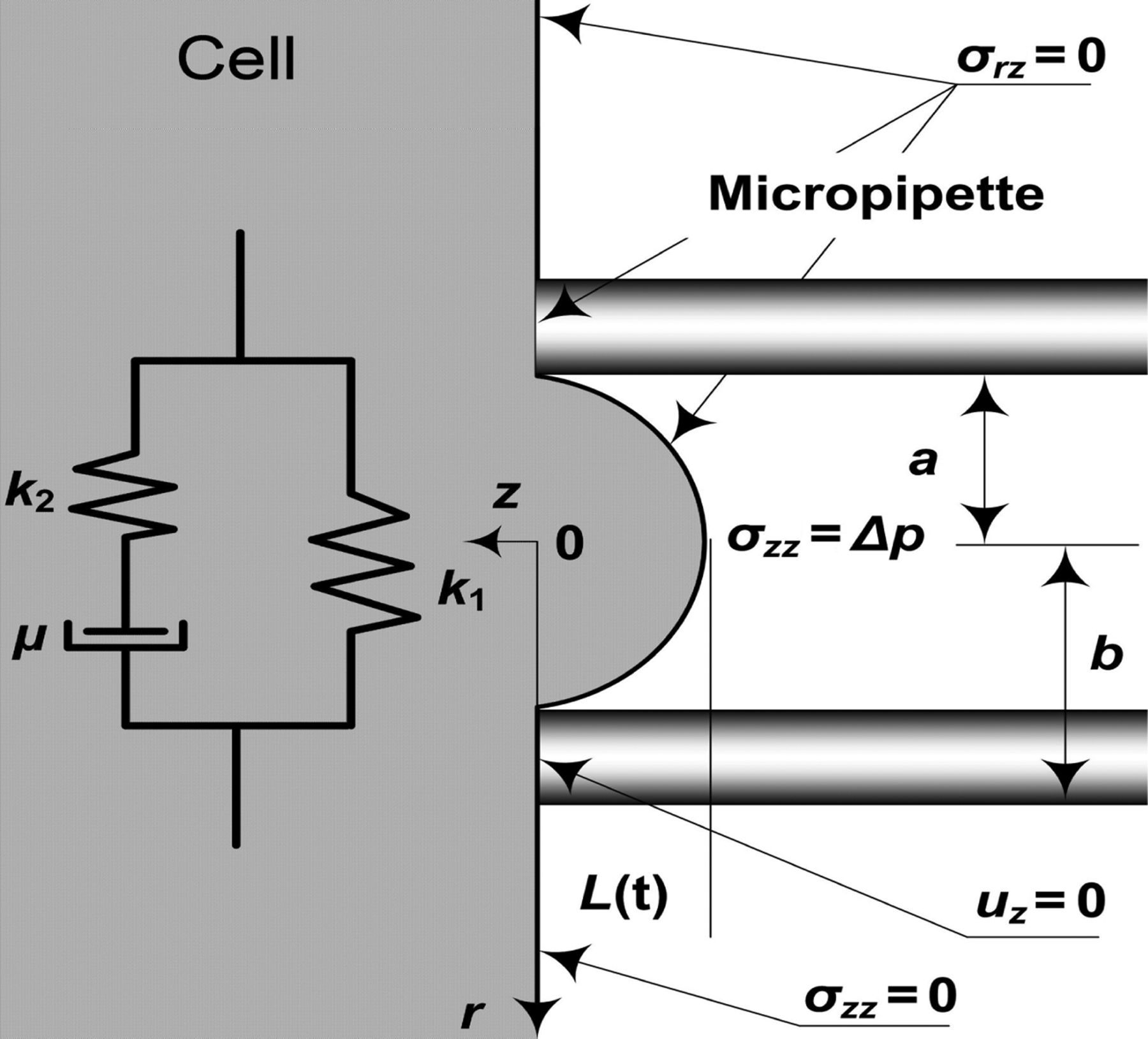
A standard linear viscoelastic solid model was used to analyze the viscoelastic behaviors of scleral fibroblasts (Figure 3)
11–13
. The relation between aspirated lengths (L) and time was given by the following equation: Schematic representation of a standard linear viscoelastic model: k
1
, k
2 and μ are the viscoelastic parameters of the model, a and b are the inner and the outer radii of micropipette, respectively, L is the aspirated lengths and Δp is the negative pressure (see ref.
11
)
Statistical analysis
Data in figures are shown as mean ± standard deviation. Statistical significance between the groups was estimated using t-test. Differences were considered significant when P < 0.05.
Results
Axes changes of rabbit eyes
Eye axes changes were used to show the success of the experimental high myopia eye model and the efficacy of PSR, using an A-ultrasonic measurement. The axes of the eyes which were treated by eyelid suturation extended by 2.85 ± 0.37 mm (15% longer than normal axes) and there was a significant difference when compared with the respective normal eyes (n = 10, P < 0.05). After six months of PSR, there was no significant difference in axial length between the operated eyes and respective normal eyes (n = 10, P > 0.05). As expected, the experimental high myopia eye model was successful.
Cell identification
The cells from three groups were identified with anti-vimentin, desmin, keratin and S-100 protein antibody by immunocytochemical methods. Experimental results: vimentin (+), desmin (−), keratin (−) and S-100 (−). The results indicated that the obtained cells were scleral fibroblasts.
The viscoelasticity of scleral fibroblasts after PSR surgery
The viscoelastic parameters (E 0, E ∞ and μ) of scleral fibroblasts from each group
*P < 0.05 compared with static group of groups A and B. † P < 0.05 compared with respective static groups
Effects of mechanical stretch on viscoelasticity of scleral fibroblasts
The viscoelasticity of scleral fibroblasts was evaluated following 48 h of stretch (Table 1). For groups A and B, the viscoelastic parameters (E 0, E ∞ and μ) of scleral fibroblasts were significantly decreased after 48 h of 3% and 6% elongation amplitude stretch when compared with the respective static groups (P < 0.05); there was no difference between the 3% stretch and 6% stretch groups in each group (P > 0.05). The viscoelastic parameters of 3% stretch and 6% stretch groups were significantly higher than the static control group in group C (P < 0.05); there was also no difference between the 3% and 6% stretch groups (P > 0.05). After a 48-h stretch, the viscoelastic parameters of the three groups of cells had no differences (P > 0.05).
Discussion
High myopia is characterized by axial elongation of the eye and scleral thinning of the posterior sclera, with scleral fibroblasts being involved in scleral remodeling during axial elongation. The changes in the biomechanical properties of the sclera are important in facilitating the increase in axial length that results in myopia. 14 In vivo, the mechanical stimulation (i.e. stresses and strains) caused by intraocular pressure and muscular tension have an effect on the ocular wall, especially on the posterior pole. 7 In the development of myopia, scleral collagen accumulation is reduced because of both decreased collagen synthesis and accelerated collagen degradation, 15,16 the biomechanical properties of sclera become lower, 17 and the scleral fibroblasts become more stiffer (the viscoelasticity of the scleral fibroblasts were higher). 13 In the process of myopia recovery and therapy, the scleral collagen content and sclera elastic modulus are both increased. 9 Therefore, the mechanical factor plays an important role in the development and therapy of myopia by regulating the biological properties and biomechanical behavior of sclera, and even the biomechanical behavior of scleral fibroblasts.
Our findings showed the changes in the viscoelasticity behavior of scleral fibroblasts in the process of high myopia treatment. The scleral fibroblasts of the fusion region of the sclera and reinforcing band after the operation exhibited lower E 0, E ∞ and μ than the scleral fibroblasts of the normal sclera and sclera after operation. After mechanical stretch, the viscoelasticity of scleral fibroblasts in the normal sclera and sclera after the operation was decreased, which means that the cells become soft and were easily deformed in order to adapt to the mechanical environment; the viscoelasticity of scleral fibroblasts in the fusion region was increased and the cells had great biomechanical properties to bear mechanical stimulation. The effect of 3% stretch on the viscoelasticity of scleral fibroblasts of the three groups was not significant when compared with 6% stretch, which shows that the scleral fibroblasts have the same response in terms of viscoelasticity to stretch in the range of 3–6%.
After PSR surgery, the viscoelasticity of the new scleral fibroblasts was lower than normal. When culturing in the mechanical environment, the normal scleral fibroblasts become softer and the scleral fibroblasts of the fusion region become stiffer. The viscoelastic behaviors of all the scleral fibroblasts become more identical. This could promote the fusion of the sclera and reinforcing band, the sclera remodeling and hyperplasia, enhance the biomechanical property of sclera and thus control the development of high myopia. Therefore, the mechanical stimulation can regulate the viscoelasticity of the scleral fibroblasts after PSR, accelerate fusion and thicken the posterior pole sclera, and achieve the efficacy of the treatment for high myopia.
In vivo, the force that the scleral fibroblasts suffer, is weak and changes with the fluctuation of intraocular pressure, eye movement and variations of vision surroundings, etc.; therefore, we selected a dynamic stretch regimen (smooth stretch waveform–sine wave, smaller elongation amplitude − 3% and 6% and minor frequency − 0.1 Hz) for scleral fibroblasts in vitro using the FX-4000 vacuum-driven Tension System. Our in vitro mechanical culture project does not model intraocular forces as the scleral fibroblasts occur in vivo, and the results of this study may be different when compared with the condition in vivo; however, our results do demonstrate that scleral fibroblasts respond to mechanical stretch by regulating the viscoelasticity. Mechanical stimulation changes the viscoelasticity of scleral fibroblasts, especially cells in the process of treating high myopia by PSR, which would be expected to change the biochemical and biomechanical properties of sclera. The results suggest that the biomechanical factor is important for the biochemical and biomechanical properties of scleral fibroblasts and sclera in the normal physiological condition, myopia development and treatment.
Footnotes
ACKNOWLEDGEMENTS
This study was supported by grants from the National Nature Science Foundation of China (10872140, 11032008) and the Natural Science Foundation of Shandong Province (ZR2012CQ005). The authors have no proprietary interest in any of the materials discussed in this article.