
Abstract
The aim of this work is to explore the feasibility and therapeutic effect of repairing rabbit articular cartilage defects using thermo-sensitive chitosan/poly (vinyl alcohol) composite hydrogel engineered Ad-hTGF-β1-transfected bone marrow mesenchymal stem cells. Rabbit's bone marrow stromal cells (BMSCs) were obtained and cultured in vitro and transfected with a well-constructed Ad-hTGF-β1 vector, the cartilage phenotype of the transfected cells was tested by reverse transcription polymerase chain reaction (RT-PCR) and Western blot. Twenty-four New Zealand white rabbits with articular cartilage defects were randomly divided into four groups: group A was treated with CS/PVA gel and transfected BMSCs; group B received CS/PVA gel and un-transfected BMSCs; group C was treated with CS/PVA gel alone and group D was the untreated control group. Experimental animals of each group were killed at 16 weeks after operation. General observation, Masson's trichrome staining and collagen II immunohistological staining of the specimens were performed to evaluate the repair effect. The Wakitani scoring method was used to evaluate the repair effect. RT-PCR and Western blot confirmed that the hTGF-β1 gene was expressed in BMSCs and triggered the expression of specific markers of cartilage differentiation such as aggrecan mRNA and Collagen II in BMSCs after transfection with Ad-hTGF-β1. Sixteen weeks after operation, the defects in group A had smooth and flat surfaces, and the defects appeared to have completely healed, exhibiting almost the same color and texture as the surrounding cartilage. Masson's trichrome staining showed that the cell arrangement and density of regenerated cartilage tissue in group A was not significantly different from that of normal cartilage tissue. The immunohistochemical staining of Col II showed a strong expression in group A and weak expression in group B, but no expression in groups C and D. According to the Wakitani score, the difference between experimental group A and all of the other groups was statistically significant (P, 0.01). To conclude, as a thermosensitive and injectable scaffold material, CS/PVA gel engineered with BMSCs transfected with hTGF-β1 can effectively repair rabbit articular cartilage defects.
Keywords
Introduction
Damaged articular cartilage has limited potential for self-healing. 1 When a cartilage defect covers a large area, no reliable approach is currently available for complete restoration of damaged articular cartilage with regard to morphological, biochemical and biomechanical characteristics. Therefore, the repair of articular cartilage injury is a hot topic in clinical and experimental research. Currently, using tissue engineering methods, seed cells have been combined with scaffold materials and transplanted into articular cartilage defects and this method has achieved success in animal experiments.2–4 Meanwhile, gene therapy5,6 has opened up a new method for treating articular cartilage defects, by providing the cartilage defect area with sustained and limited bioactive protein or gene products, promoting cell-directional migration or differentiation, improving the self-repair capability of cartilage and performing biological functions conducive to cartilage repair during the course of treatment.7–9 Injectable gel is a new scaffold material for cartilage repair which has been developed in recent years. Some researchers have combined chitosan gel, 10 collagen 11 or fibrin gel12,13 with cartilage cells or mesenchymal stem cells to repair cartilage defects and have achieved success. However, these gels share common shortcomings of weak mechanical strength and fast degradation rate. We prepared an injectable chitosan/poly-vinyl alcohol (CS/PVA) gel in a pilot study and explored its structure and physical and chemical properties; we found that the gel has low cytotoxicity and good biocompatibility.14,15 On this basis, CS/PVA gel was combined with rabbit bone marrow stromal cells (BMSCs) trans-fected with hTGFβ-1 and used to repair rabbit articular cartilage defects and the repair effect was evaluated.
Materials and methods
Experimental animals and materials
CS (deacetylating degree DA = 91%, Mw = 2.7 × 10⁵): Zhejiang Yuhuan Marine Biochemistry Co., Ltd; polyvinyl alcohol (n = 2400-2500): Shanghai Chemical Reagents Co., Ltd; β-sodium glycerophosphate (Sigma Chemical Company, St Louis, MO, USA), fetal bovine serum and Dulbecco's modified Eagle's medium (DMEM) culture medium (Gibco, Invitrogen, Carlsbad, CA, USA); rabbit anti-human TGFβ-1 (Beijing Biosynthesis Company, Beijing, China), mouse anti-rabbit Col II (Oncogene, San Diego, CA, USA); SP Kit (Wuhan Boster Company, Wuhan, China); Masson trichrome staining kit (Leagene, Beijing, China); Ad-hTGF-β1 was constructed and stored in our laboratory; other reagents were of analytical grade.
Experimental animals: Four one-month-old New Zealand white rabbits, 24 healthy adult New Zealand white rabbits (Wuhan University Center for Animal Experiment), weight: 2.5 - 3 kg, male and female are equal, both hind legs were used for experiments. This process was approved by the Medical Animal Care & Welfare Committee of Zhongnan Hospital of Wuhan University.
Culture of rabbit BMSCs and Ad-hTGFβ-1 transfection
Rabbit BMSCs were obtained by femoral aspiration from one-month-old New Zealand white rabbits. The bone marrow aspirate was combined with 40 mL of Dulbecco's phosphate- buffered saline (DPBS; Gibco) and centrifuged at 900 × g for 10 min at 20°C. The cells were re-suspended and gently layered onto Percoll (density 1.073 g/mL; Gibco), at a density of 1 × 10 6 nucleated cells/25 mL. The low-density BMSC-enriched fraction was collected, combined with 25 mL DPBS and centrifuged. The cells were re-suspended in BMSC culture medium (10% fetal bovine serum in DMEM with antibiotic/antimycotic supplements (Gibco) and plated at 3 × 10 7 cells/25 cm2 . Cultures were maintained at 37°C in a humidified atmosphere containing 95% air and 5% CO2. When the cultures were near 80-90% confluence, the cells were detached by treatment with trypsin and ethylenediaminetetraacetic acid (EDTA). Third-passage BMSCs were collected for experiments. BMSCs were seeded into six-well cell culture plates, into which cover glasses had been placed beforehand at 2 × 10⁵/mL; when the cells reached 80% confluence, the culture medium was changed to serum-free medium and the cultures were transfected with adenovirus at a multiplicity of infection (virus/cell, MOI) of 100. After incubating for four hours, the culture medium was changed to DMEM complete medium, and the cells were returned to culture. No-load adenovirus transfection was used as a control group.
Reverse transcription polymerase chain reaction assay
Reverse transcription polymerase chain reaction (RT-PCR) analysis was used to determine the mRNA expression of hTGF-β1 and aggrecan in the transfected cells. Total RNA was isolated from cells using an RNeasy mini kit (Qiagen, Valencia, CA, USA). Cells were harvested into 1 mL TRIzol reagent (Invitrogen). Homogenates were left at room temperature for five minutes to facilitate mRNA extraction and then centrifuged at 12,000 × g for 15 min. cDNA was reverse transcribed using a cDNA synthesis kit (Qiagen) with 1 μg mRNA/20 μL reaction. RT-PCR analysis was carried out with 1 μg of total RNA using a Qiagen One Step RT-PCR kit (Qiagen). The following primers were synthesized on the basis of the sequences reported in the Genbank database. Aggrecan: sense, 5
Western blot
To further validate the endogenous gene and chondrocyte phenotype expression in the transfected cells, Western blotting methods were used to detect the hTGF-β1, aggrecan and Collagen II proteins. Cells were collected at one week after transfection and then homogenized in cell lysis buffer containing 50 mmol/L Tris, 10 mmol/L NaCl, 1% NP40, 0.02% sodium azide and protease inhibitor cocktail (Roche Inc, Basel, Switzerland) at pH 7.4. Aliquots containing 20 μg of protein were dissolved in Laemmli buffer and boiled at 95°C for five minutes, and the proteins seperated on a 4-12% SDS-PAGE (sodium dodecyl sulfate poly-acrylamide gel electrophoresis) gradient gel, transferred onto nitrocellulose membranes, blocked and then incubated with primary antibodies at 4°C. Primary antibodies included antibodies against transforming growth factor beta 1 (TGF-β1) (1:200, Santa Cruz, CA, USA), aggrecan and Collagen II (1:200, Santa Cruz Biotechnology). Samples were then incubated with horseradish peroxidase (HRP)-conjugated secondary antibody, followed by enhanced chemiluminescence detection (Image J). Chemiluminescence detection values were used to quantitate the Western blot results. A ratio of each band of interest to the internal control was obtained and the statistical significance of differences between control and experimental groups determined.
Animals
Twenty-four New Zealand white rabbits, with both their hind legs, were used for the experiment. The animals were anesthetized intravenously with 3% pentobarbital sodium (30 mg/kg). After sterilization with 1% povidone iodine, a medial incision was made into the joint cavity, the patella was pushed outward to expose the articular surface of the femoral condyle and a drill was used to create a full-thickness cartilage defect model with a diameter of 4 mm and a depth of 2.5 mm at the center of the articular surface of the femoral condyle. The CS/PVA gel was prepared as we have reported before14,15 and the cells were prepared. The gel and cells were mixed at 4°C, and cell density was adjusted to 5 × 10⁷/mL. The animals were randomly divided into four groups, six rabbits per group. The defects of group A were injected with a mixture of CS/PVA gel and transfected cells, group B were injected with a mixture of CS/PVA gel and untransfected cells, group C were injected gel alone and group D were the control groups, without any treatment of the defects. 15-20 min after gel injection, the defects could be seen to be filled with an elastic solid, at this point, the surgical incision was rinsed with normal saline, bleeding was stopped and the incision was sutured layer by layer. After the animals regained consciousness, they were placed in a cage allowing free movement and fed with general feedstuff.
General specimen observation
Two rabbits of each group were killed at four weeks after surgery to observe the defects, others were killed at 16 weeks after surgery and the tissue of the defect area and surrounding normal cartilage was removed for general observation. General specimen observation involved: checking the smoothness, gloss and density of the articular cartilage surface and the condition of the joint with the surrounding normal cartilage.
Masson's trichrome staining
In order to observe the celluar components and expression of cartilage matrix of the specimens, Masson's trichrome staining was used. The specimens were fixed with 10% formaldehyde, decalcification with EDTA, embedding in paraffin and the sections were stained with Masson's trichrome staining (carried out following the manufacturer's protocol provided), so as to observe the cell types and the morphology of repaired tissue, matrix staining and cartilage density.
Collagen II immunohistochemical staining
For immunohistochemistry, first the sections were deparaffinized and washed in phosphate-buffered saline, then digested with 1% trypsin for 30 min and pretreated with 3% bovine serum albumin for 15 min at 37°C. The sections were incubated with a mouse anti-rabbit antibody to Col II (1:100) for 60 min at 37 and 4°C overnight, then incubated with secondary antibody(goat anti-mouse, 1:50). The sections were treated with AEC chromogenic and hematoxylin counterstain. Sections stained without primary antibodies were used as negative controls. Under a microscope, the image appears in clear brown granules metachromatic is considered positive.
Repair assessment
A histological score was given to the repaired cartilage tissue using the Wakitani method, 16 which involves assessment of the cell types in the repaired tissue, matrix staining, surface smoothness, cartilage density and integration into the surrounding normal cartilage tissue.
Statistical analyses
The experimental data are shown in the form of mean ± standard deviation (
Results
Culture of BMSCs and Ad-hTGF-β1 transfection
When initially plated, BMSCs appeared as heterogeneous groups of large flat cells, smaller spindle-shaped cells and small round cells. Five days after plating, most of the adherent cells showed typical BMSC fibroblastic morphology with large flat and spindle-shaped cells (Figure 1). After 10∼12 days, the cells were generally near 80-90% confluence. BMSCs tend to be identical morphologically, and fibroblast-like cells with regular morphology and uniform size could be seen under the microscope. After Ad-TGF-β1 transfection, in the early stage, cells changed shape from a long spindle shape to polygonal and round shapes, but cell morphology recovered after 24 h.
Bone marrow stromal cells at generation P3, with uniform cell morphology and fibroblast-like growth (×200)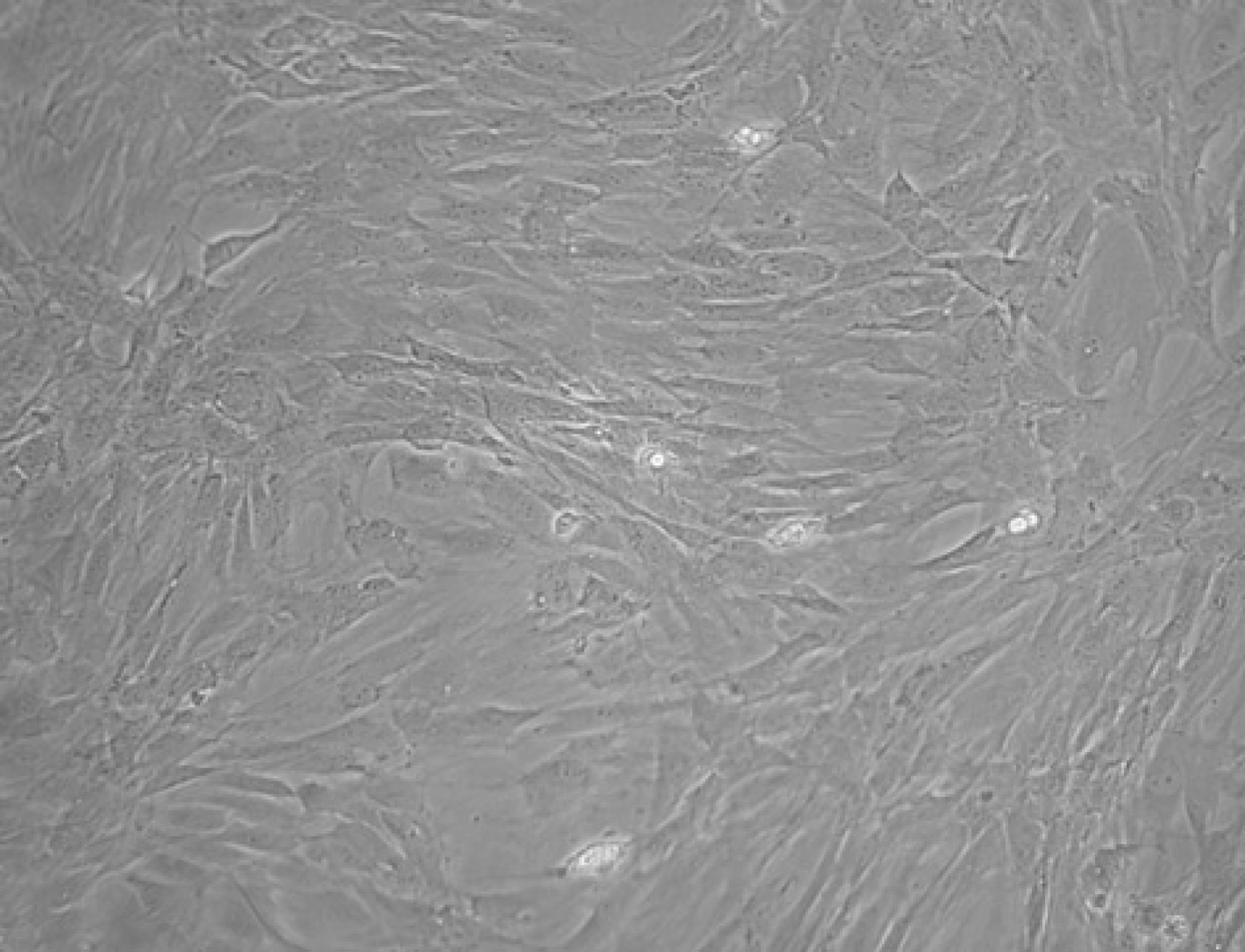
Reverse transcription polymerase chain reaction
RT-PCR analysis showed that a hTGF-β1 target gene band of 476 bp could be detected in Ad-hTGF-β1 transfected cells with strong expression than the non-transfected BMSCs group and no-load adenovirus transfected BMSCs group (P < 0.05, n = 6). The aggrecan mRNA was highly expressive in the Ad-hTGF-β1 transfected cells in 258 bp. However, no expression of aggrecan mRNA was detected in the non-transfected BMSCs and no-load adenovirus transfected cells (P < 0.05, n = 6) (Figures 2a and b).
mRNA expression of hTGF-β1, aggrecan and glyceraldehyde-3-phosphate dehydrogenase (
Western blot
One week after Ad-hTGF-β1 transfection of BMSCs, protein was extracted from the BMSCs, and the expression of hTGF-β1 and collagen type II protein was detected by Western blot method. It was found that the hTGF-β1 protein band was located at approximately 175 kDa and the aggrecan protein band was located at 25 kDa; the Collagen II protein band was located at 180 kDa. The expression of hTGF-β1 was higher than those in non-transfected BMSCs and the no-load adenovirus transfection group (P < 0.05, n = 6), the Collagen II protein and aggrecan protein could not be seen in the no-load adenovirus transfection group and the untransfected control BMSCs. The GAPDH protein band, located at approximately 36 kDa, was used as an internal loading control (Figures 3a and b).
hTGF-β1, aggrecan and Collagen II protein expressed in the cells: (a) lane 1, Ad-hTGF-β1 transfected; lane 2, non-transfected 
General specimen observations
Among the 24 experimental animals, one rabbit died after surgery, while the others exhibited normal feeding and activity. Among the 46 knee joints of living rabbits, three cases showed joint adhesion, and their knees could not move normally after repair, while the rest did not show any obvious abnormal flexion or extension of the knee joint. After surgery, a diameter of 4 mm could be seen in the surface of the femoral condyle, and after one month, the groups of injection were found to be filled with a porcelain-white gel material. The filler was tightly bonded with the surrounding femoral articular surface, and the articular surface was smooth and flat. A clear boundary line was evident between the filling tissue and the normal articular cartilage, and its surface was smooth due to movement and automatic shaping of the joint. In contrast, the cartilage defects in group D were filled with blood clots, showing an obvious defect. Sixteen weeks after operation, the defects in group A had smooth and flat surfaces, and the defects appeared to have completely healed, exhibiting almost the same color and texture as the surrounding cartilage (Figure 4a). The defects in group B had a smooth surface, similar to that seen in group A (Figure 4b); the defects in group C also had a smooth surface, but the color and texture were significantly different from the surrounding cartilage tissue (Figure 4c); in group D, though the defection existed, some proliferative chondroid tissues emerged on the edge. (Figure 4d).
Repair effects in each group 16 weeks after operation. (a) The defects had smooth and flat surfaces, and the defects had completely healed, having almost the same color and texture as the surrounding cartilage. (b) The defects had smooth surfaces and the surface was covered by white chondroid tissue, there was no clear boundary between the edge of the defects and normal tissue, although the texture of the defects was slightly different from that of normal cartilage tissue. (c) The defects also had a smooth surface, but their color and texture were significantly different from the surrounding cartilage tissue. (d) The defects were obvious, filled with chondroid tissue and a clear boundary line between the defects and the surrounding normal cartilage tissue could be seen. (A color version of this figure is available in the online journal)
Masson's trichrome staining
Sixteen weeks after operation, Masson's trichrome staining showed that the cell arrangement and density of regenerated cartilage tissue in group A was not significantly different from that of normal cartilage tissue. The boundary between regenerated and normal cartilage tissue was unclear. Matrix staining showed the equivalent levels of coloring in regenerated and normal cartilage tissue (Figure 5). The cartilage cell density in regenerated tissue of group B was lower than that of group A. The regenerated cartilage tissue in group B contained a mixed cell population of cartilage cells, fibroblasts and bone cells. The cartilage defects in group C were mainly filled with fibrous tissue and contained a small number of immature cartilage cells in the surrounding area, and a smaller quantity and lower density of cells than normal cartilage tissue. Most of the cells were found at the surface and in the bottom layer.
Results of Masson's trichrome staining in each group 16 weeks after operation. (a) The cellular organization and density of regenerated cartilage tissue was not significantly different from that of normal cartilage tissue. The cartilage cell density in the regenerated tissue of group B was lower than that of group A. (b) The regenerated cartilage tissue contained a mixed population of cartilage cells, fibroblasts and bone cells. (c) Immature cartilage cells could be seen in the surrounding area, but they were present in lower numbers and at a lower density than normal cartilage tissue. Most cells were located at the surface and in the bottom layer, and the main area of the defect was largely filled with fibrous tissue. (d) The defects were filled with fibrous tissue, with many fibroblasts and newly generated vessels, but no cartilage cells (×200). (A color version of this figure is available in the online journal)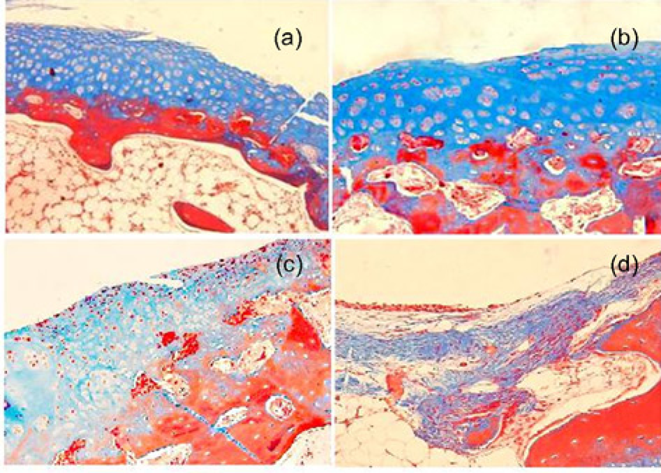
Collagen II immunohistochemical staining
A positive immunohistochemical staining of Col II was observed in group A and a small amount of expression in group B (Figures 6a and b), but not in groups C and D (Figure 6c and d). It was much stronger in group A. Positive staining was mainly localized in the matrix around the lacunae in the regenerated cartilage.
Regenerated cartilage tissue immunohistochemistry of collagen II: group a was strongly positive cells and the matrix is brown metachromatic. Group b was weakly positive than group, and group c and group d were negative (×200). (A color version of this figure is available in the online journal)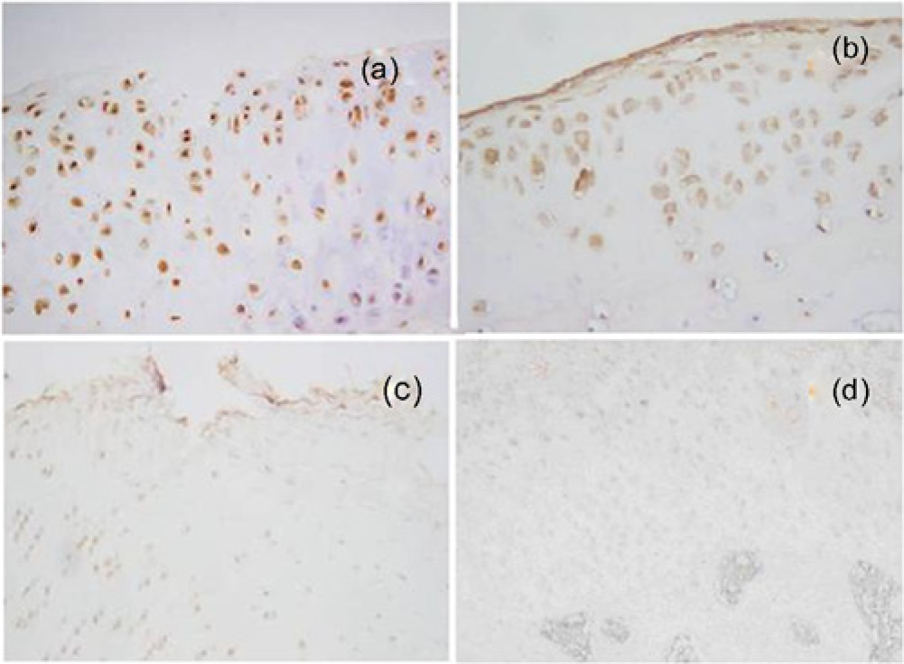
Repair assessment
The regenerated cartilage tissue in each group was given a histological score by the Wakitani method. According to statistical analysis (Figure 7), the difference between experimental group A and all of the other groups was statistically significant (P < 0.01), while the difference between group B and the control group was also statistically significant (P < 0.05); however the group treated with gel alone (group C) was not significantly different from the control group.
Wakitani score 16 weeks after operation, *P<0.01, **P<0.05 (n = 6)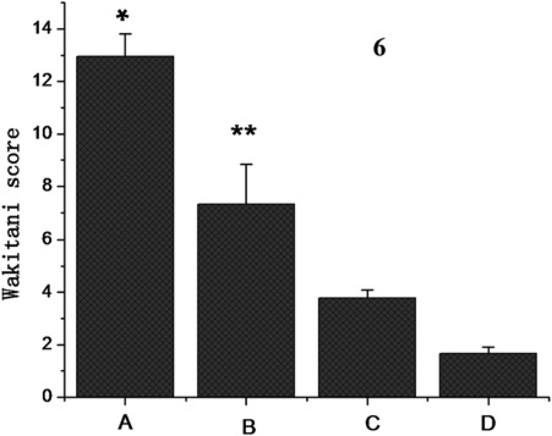
Discussion
Chitosan hydrogel is a hydrophilic natural hydrogel with good degradability and biocompatibility. Both Chenite et al. and Hoemann et al. used chitosan as the main material in the preparation of thermosensitive hydrogel, combined it with animal cartilage cells and successfully constructed tissue-engineered cartilage. However, pure chitosan gel has poor mechanical characteristics and degrades quickly, hence its mechanical performance is greatly inferior to that of normal cartilage tissue. PVA has a microporous structure similar to natural cartilage, containing large amounts of water and consists of a permeable rubber-like material, characterized by high elasticity, good mechanical performance, stable chemical properties and ease of molding, good biocompatibility and physical properties similar to articular cartilage.19,20 Various composite materials using PVA as the main component have been widely used in cartilage repair, including HA-PVA, 21 PVA-Collagen 22 and PVA-PLGA 23 and all of these materials have achieved a certain level of repair effect. In our study, PVA was introduced into CS gel to form a CS/PVA physically-mixed gel. While retaining the temperature sensitivity of CS gel, CS/ PVA gel has improved structural and mechanical properties, 15 slowing down the degradation rate of CS in the body, hence it is more applicable to cartilage repair as an in situ molding and implantation material.
Currently, the main cell types used in cartilage tissue engineering are cartilage cells and BMSCs. Cartilage cells are difficult to obtain and are highly likely to cause allograft rejection. They are also likely to lose cell phenotype during in vitro culture, and have certain limitations when used as seed cells. In contrast, BMSCs are considered adult stem cells and are mesoderm-derived mesenchymal cells like bone and cartilage cells. BMSCs can not only proliferate but also achieve cross-phenotype differentiation in certain external environments and under the influence of stimulating factors.24–26 BMSCs always maintain the capacity to differentiate into cartilage cells, and serve as lifelong cartilage precursor cells and seed cells frequently used in cartilage tissue engineering.27,28 In this study, BMSCs were isolated by density gradient centrifugation in vitro, and their number was increased by subculture to achieve a higher initial density of seed cells. The microenvironment in which BMSCs are located can determine their direction of differentiation. After modification by TGF-β1, BMSCs will differentiate into hyaline cartilage cells under the combined effects of a large number of inducing factors.
It has been proven that TGF-β has multiple biological effects. It can not only regulate the proliferation but also direct the differentiation of BMSCs,29–32 it is also a strong immunosuppressant, making transplantation of allogeneic chondrocytes more secure.33,34 After transfection into receptor cells with an adenoviral vector, the TGF-β1 target gene can achieve long term and efficient expression in cells. In our study, we used RT-PCR to confirm that the trans-fected BMSCs expressed exogenous TGF-β1 mRNA. The expression of TGF-β1 mRNA proves that the exogenous gene has been successfully transferred into cells, and is a prerequisite for the expression of TGF-β1 protein and the performance of biological functions in cells. The expression of aggrecan mRNA was also detected after BMSC transfection. Aggrecan is a key promoter gene for the differentiation of BMSCs into cartilage cells.35–37 The expression of aggre-can is regulated by TGF-β1, and this is a pivotal sign of BMSC differentiation into cartilage cells and one of its key differentiation mechanisms. It is also one of the most distinctive surface markers of cartilage cells, hence was used for the identification of cartilage cells in this study. According to our experimental results, BMSCs transfected with Ad-hTGF-β1 and combined with composite gel in group A after in vivo culture formed regenerated cartilage tissue in the gel which had a uniform cell morphology, contained evenly distributed cells and was able to secrete cartilage matrix, the expression of collagen II was detected in the matrix and the positive expression rate was higher than other groups, confirming that TGF-β1 can strongly induce the formation of cartilage cells. Moreover, analysis of the specimens collected 16 weeks after surgery showed that the cartilage defect filled with gel alone was generally covered by cartilage tissue, and histological observation proved that the main component of this regenerated cartilage was fibrocartilage, with little normal hyaline cartilage. The gel is able to induce the repair of cartilage tissue through space conduction and occupation of part of the articular cartilage defect area. In the degradation process, the gel also provided three-dimensional support for cartilage growth toward the defect center. Cartilage cells can exhibit very slow migration toward the defect as the gel gradually degrades, and ultimately forms fibrocartilage tissue on the surface of the defect, repairing the cartilage defect to some extent. Generally, pure CS gel degrades completely in vivo after 12 weeks. However, when combined with PVA, the non-degradable PVA can postpone the degradation of CS gel, and thus prolong the self-repair term of this tissue. The cartilage defect is repaired by a combination of cartilage cells migrating from the surrounding tissue and BMSCs from the marrow cavity.
This research provides preliminary evidence that CS/PVA gel can be applied to the repair of articular cartilage defects as an injectable material in tissue engineering, and the regenerated cartilage can secrete cartilage matrix and perform the functions of hyaline cartilage. Use of this gel for cartilage repair has advantages such as minor surgical procedure required, tight bonding with the damaged tissue and lack of rejection. However, knee adhesion also occurred in a few experimental animals, suggesting that the mode of fixation between the gel and the defect still needs further improvement. In addition, further studies need to be carried out on the long-term biomechanical stability of regenerated cartilage tissue and whether the newly generated cartilage cells are likely to undergo dedifferentiation.
Footnotes
Acknowledgements
This study was supported by the National Natural Sciences Foundation of China (No. 30770574) and Research Fund Project of Health Department of Hubei Province (No. JX3B21).