
Abstract
Introduction
Forearm-based splints have been traditionally used for extensor pollicis longus (EPL) tendon repairs for zones T II–T V (T is used to represent thumb extensor tendon zones). Limited literature exists on hand-based splinting in the rehabilitation of zone T II EPL tendon repairs. This retrospective review of five case studies highlights the anatomical justification and the outcome of rehabilitation of zone T II EPL surgical repairs using a static hand-based thumb extension splint.
Methods
In this study, five patients were retrospectively reviewed. All patients attended hand therapy for initial treatment within three days postoperatively. The postoperative interphalangeal joint mobilization regimen utilized in this study was early active motion (EAM)
Results
The results demonstrated that a hand-based splint did not create undue stress on the EPL tendon repair, as there was no incidence of rupture. Hyperextension of EPL was within 8° compared with the non-injured thumb. ‘Excellent’ and ‘good’ categories were achieved when applying TAM criteria, White's assessment and Dargan's criteria.
Conclusion
A hand-based splint with an EAM regimen is a viable treatment option for zone T II EPL surgical repairs instead of a long forearm-based splint. Further research is warranted with a larger sample and using a control group.
Introduction
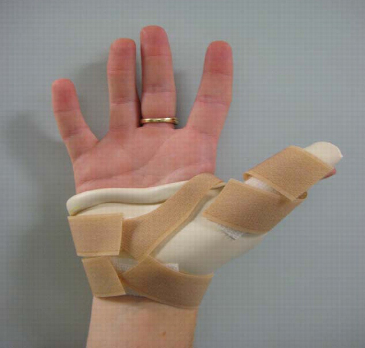
The preferred postoperative treatment within The Northern Hospital for patients following zone T II extensor pollicis longus (EPL) repair is a hand-based thumb extension splint, with the interphalangeal (IP) joint positioned in hyperextension. This regimen is widely accepted and requested by the Plastic Surgery Department within The Northern Hospital (Figures 1 and 2). Minimal evidence exists for the use of a hand-based thumb extension splint for zone T II EPL repairs. Please refer to Figure 3 for the thumb extensor tendon zones. Mobilization of the wrist in a hand-based splint after EPL lacerations in zone T II is controversial. 1 Historically, a long forearm-based splint has been fabricated to treat EPL zone T II repairs. 1,2 In this paper we discuss the functional anatomy of the dorsum of the thumb to justify using a hand-based splint for zone T II EPL repairs and report the outcomes from a case series to show that EPL repairs at zone T II were not compromised by utilizing a hand-based splint in place of the forearm-based splint.

Dorsal view of hand-based thumb extension splint
Volar view of hand-based thumb extension splint

Thumb extensor tendon T zones
Our approach is based on evidence for early active motion (EAM) literature, which illustrates that tendons actively mobilized have improved intrinsic healing, resulting in a stronger tendon repair and reduced adhesions maximizing active range of motion (AROM). 3–9 Various factors for suitability of an EAM regimen are considered in the clinical reasoning process, including repairs compromised by infection, fragile repair as stated by the surgeon, compliance of the patient and experience of the therapist.
On examination of the literature, there was limited recent research to support the use of either forearm or hand-based splinting approaches for treatment of zone T II EPL tendon repairs. The majority of the existing literature regarding EPL tendon rehabilitation consists predominantly of clinical outcomes for dynamic splinting combined with controlled passive motion protocols, 5,9,10–12 and two papers on EAM. 3,4 Both versions implemented a forearm-based splint, with little discussion on variation of splint design.
The evidence available for hand-based splinting is contradictory. Hunter and colleagues 7 recommend a hand-based splint for zone T I and T II EPL tendon repairs. However, their justification for a forearm-based splint is opinion based with no evidence or outcome measures documented. Rosenthal and Stoddard 1 discussed their concerns with risk of rupture regarding the use of a hand-based splint during rehabilitation of zone T II EPL injuries. They suggested that when employing a hand-based splint, there is a risk of stress to the repair from wrist tenodesis, for overactive or non-compliant patients, preferring to include and immobilize the wrist as a precautionary measure.
Crosby et al. 2 also recommended that all thumb extensor tendon lacerations required forearm-based splinting. In distal lacerations of EPL, they recommended dynamic passive mobilization of the IP joint with a metacarpophalangeal (MP) joint immobilized in extension. They stated that hand-based splinting was not used for any thumb EPL lacerations because the thumb sagittal band structure is not sufficient support compared with those of the fingers to protect a distal repair of EPL without including the forearm in the splint. This statement is not supported with any clinical study or cadaver kinematic dissection as to why the sagittal bands are not sufficient support. Crosby et al. 2 continued to state that the sagittal bands of the fingers will prevent proximal retraction if laceration is distal to them, which they acknowledge is also true of the thumb's sagittal bands at the MP joint level.
Anatomically, it is known that as the EPL travels distally over the metacarpal joint of the thumb, it is supported laterally by the extensor expansion, including tendon fibres from abductor pollicis brevis, flexor pollicis brevis and adductor pollicis brevis. 4 Following a laceration, this expansion prevents proximal migration of the EPL, with the sagittal bands having a similar effect to extensor digitorum communis of the finger. 1 Jaibaji et al. 13 investigated the functional anatomy of the thumb sagittal band using 24 cadaveric amputated specimens. Jaibaji et al. 13 also report that proximal migration during isolated and composite MP and IP joint flexion is restricted at zone II due to the thenar muscles inserting onto the EPL expansion. Given these are intrinsic muscles, tension on the repair will not change with alteration of the wrist position. Hence, wrist motion in all directions with a hand-based splint is not considered to create undue stress on the EPL repair. It was further discussed that although the anatomy of the thumb sagittal band is different from that of the fingers, its basic kinematic and stabilizing function for the extensor apparatus is similar. 13
A hand-based splint for zone T II EPL repairs is therefore in accordance with the anatomical arrangement of EPL. The prime function of EPL is hyperextension of the IP joint and retropulsion. Evans and Burkhalter 6 found that with the wrist in neutral and MP of the thumb extended, that 60° of IP joint motion affected 5 mm of tendon excursion. Brand 14 considers 5 mm of tendon glide to be safe stress to prevent adhesions while minimizing the risk of tendon rupture. Brand 14 recommends that if the IP joint is flexed beyond 60°, the tendon may be at risk of rupture. As the hand-based splint does not allow IP joint motion there is no clinical risk of rupture.
The five retrospectively reviewed cases aim to demonstrate a static hand-based splint as a safe splinting alternative to a long forearm-based splint in the rehabilitation of surgically repaired zone T II EPL tendons.
Method
We retrospectively reviewed five patients with an isolated laceration of the EPL tendon at zone II greater than 80%. To allow direct comparison, a consistent population group was selected, resulting in the exclusion of those treated with additional injuries such as significant skin or soft tissue loss, bone injuries, EPL repairs less than 80%, postoperative infection and re-plants/re-vascularizations or premorbid injuries that affected AROM of the thumb. Review of the medical histories and privacy protection were applied in accordance with Northern Health Policy. As this rehabilitation regimen is standard practice at our facility, ethics approval and informed consent was not necessary.
The EPL was repaired by various rostered plastic surgeons adopting a standard repair technique for this injury of a two-strand mattress EPL tendon repair. The five patients were treated within three days following surgery as per routine hand therapy department practice guidelines. The patients’ postoperative dressings were changed and minimized to allow for ease of mobility of the thumb during the EAM guideline. One hand therapist assessed and treated all five patients. The hand therapist routinely educated the patients regarding their injury including anatomical biomechanics, surgical repair, splinting regimen, home exercise programme including massage, hand hygiene, safe hand function, precautions and contraindications.
A hand-based thumb extension splint was fabricated with the IP joint in hyperextension equal to the non-injured thumb for active hyperextension. The fingers and wrist were free to mobilize. The splint was firmly secured in place with Velcro® strapping (Figures 1 and 2). Patients were permitted light functional activities of up to 0.5 kg and to mobilize the wrist freely. The splint was implemented full time for the first four weeks postoperatively, with exception to when conducting the exercise regimen.
The therapist advised the patient of exercises according to The Northern Hospital EAM guideline, recommending two-hourly five repetitions of removing the thumb straps to perform passive extension, place and hold into extension and active retropulsion from the splint. The patient was then advised to remove the splint with the wrist in neutral to perform isolated IP joint full hyperextension and flexion as far as tolerable but not flexing to full end IP joint range with the MP joint held in extension. The final exercise was isolated MP joint flexion and extension with the IP joint positioned in extension. For this exercise, patients were advised to flex to only half the full AROM of MP joint range.
Dosage of all exercises was potentially increased from 5 to 10 repetitions between days 15 and 28 postoperatively to reflect the wound healing process and stronger collagen formation. That is, the increase in exercises was clinically determined by the treating therapist dependent on the patient's individual scar formation process. For example, if the patient was mobilizing comfortably to IP joint 60° with minimal scar formation, then through clinical reasoning, the dosage of five repetitions remained the same.
After the initial four weeks of treatment, the splint was applied for precautionary wear during high-risk activity and when sleeping for an additional two weeks.
As per The Northern Hospital guideline, all patients were reviewed weekly by the treating hand therapist for the initial four weeks postoperatively then either weekly or fortnightly (depending on clinical reasoning based on assessment of thumb mobility and scar adhesions present) until the routine eight-week postoperative session. Continued sessions beyond week 8 postoperatively were again based on clinical reasoning depending on thumb mobility and strength required for the patient's activities of daily living and patient satisfaction of thumb function.
All measures of progress in this retrospective review were completed by the same therapist. Recordings of measures taken from the hand therapy sessions have been tabulated at four, six and eight weeks postoperatively to illustrate improvement in AROM. Measurements were performed using a goniometer. The goniometer was placed with the axis over the dorsum of the MP and IP joints for thumb flexion, respectively. Extension of the MP joint and hyperextension of the IP joint of the thumb were measured radio-laterally.
TAM, 15 Dargan's criteria 16 and White's assessment of interphalangeal range of motion 17 were applied for the final measures at eight weeks postoperatively to analyse the outcome measures, as depicted in Tables 1–3. Baseline measures were also taken of the non-injured thumb.
Outcome measures for total active motion (TAM) of injured thumb compared with contralateral thumb 15
Outcome measures for White's Assessment of intraphalangeal joint range of movement compared with the contralateral thumb 17
Outcome measures by goniometry for Dargan's criteria measuring intraphalangeal thumb extension, compared with the contralateral thumb 16
Results
All patients were treated and measured throughout the rehabilitation process by the same treating therapist. No patients suffered any postoperative complications, such as rupture or infection. They received weekly treatment sessions for the first four weeks following repair. Only case studies 1 and 2 had their exercises increased from five to 10 repetitions between 15 and 28 days postoperatively based on the clinical reasoning of the treating therapist.
All patients included in the study had the same mechanism of injury to the non-dominant hand and were greater than 80% EPL tendon repair. Case 2 did not attend at week 6; however, attended for the week 8 measurement. Case 5 failed to attend at week 8, although AROM measurement taken at week 6 demonstrated 3° from full IP joint hyperextension and an IP joint flexion deficit of 30°.
For the four cases that did attend at week 8, AROM measurements demonstrate that full flexion range was not achieved except for only one patient who obtained full IP joint hyperextension and flexion range compared with the non-injured hand, achieving ‘excellent’ in the TAM classification. The remaining three cases achieved ‘good’ in the TAM classification obtaining AROM greater than 75% of the non-injured thumb.
The four cases at week 8 achieved greater than 70% AROM compared with the non-injured thumb and therefore were considered ‘excellent’ according to White's assessment.
In comparing IP joint hyperextension of the injured thumb to the non-injured thumb at week 8, only two cases achieved full hyperextension, an ‘excellent’ result according to Dargan's criteria. The remaining two cases were less than 15% of extensor lag compared with the non-injured thumb hyperextension, and hence according to Dargan's criteria are considered a ‘good’ outcome (Table 4).
Case demographic details for all patients included in the study
The goniometry results are outlined in Tables 5–7.
Goniometry measures of intraphalangeal (IP) active range of movement of the non-injured and the injured thumb at weeks 4, 6 and 8
MP AROM was not included as there was no deficit in comparison with the non-injured goniometry measures
Final measurements at week 8 of the TAM of the injured and non-injured thumbs
TAM, total active motion
Final measurements of the injured thumb at week 8 assessed by White's and Dargan's criteria
Discussion
The five cases, all of which had a knife laceration to the non-dominant thumb EPL tendon zone T II, were rehabilitated using a hand-based thumb extension splint with no rupture.
A lack of full hyperextension compared with the non-injured hand was considered to be an EPL lag. By assessing hyperextension the true function of the EPL tendon is demonstrated by ruling out intrinsic thumb muscle fibres that insert onto the thumb extensor hood and are capable of IP extension to 0°.
At week 8 postoperatively, two patients had achieved full hyperextension with no lag when compared with the contra-lateral hand. Case 2 was 5° and case 3 was 8° from full hyperextension. Case 5, who failed to attend from week 8 onwards, was only 3° from full hyperextension at week 6 in comparison to the contra-lateral hand. It has been shown that hyperextension can often be lost after EPL repair in all injury zones. 3,5,9
According to White's criteria, 17 four of the five patients achieved ‘excellent’ IP joint extension by eight weeks postoperatively. In reference to Dargan's criteria, 16 two cases achieved ‘excellent’ and the remaining two ‘good’ results. For TAM classification, 15 one case achieved ‘excellent’ and the remainder three cases scored in the ‘good’ category. It is noted that although case 5 failed to attend for the remainder of the treatment after week 6, the outcomes for this patient at week 6 are ‘excellent’ under White's criteria 17 and in the ‘good’ category according to Dargan's 16 and TAM classification. 15
It is noted from our goniometry measures at week 8, a flexion deficit ranging from 5° to 25° was apparent in three of the five cases. The clinical reasoning for this is that scar adherence was responsible for reducing end range proximal glide for hyperextension and distal glide for end range flexion. Extensor tendon adherence by scar tissue largely explains the loss of thumb movement after EPL tendon repair. 3,5,9
A hand-based splint allows the wrist to mobilize freely without creating stress on the EPL repair to produce a rupture. The primary function of EPL is IP joint hyperextension, and by positioning the IP joint in hyperextension within the splint, tension is relieved from the distal aspect on the repair itself in zone T II. The tendon repair is also protected proximally through insertions of the abductor pollicis brevis, flexor pollicis brevis and adductor pollicis brevis onto the EPL itself forming the extensor expansion. 4,6,7 Given these structures are all intrinsic muscles and are retaining EPL preventing its retraction, it is unnecessary to include the wrist in the splint for postoperative protection.
Essentially, the case series’ focus was on reviewing EPL function (namely IP joint hyperextension), following use of a hand-based splint rather than on regaining full range into flexion. It is also argued that these flexion measures may not be improved or altered by having a forearm-based splint in place of the hand-based thumb extension splint. It is possible that full flexion range would eventually be achieved with ongoing functional use, scar management and further therapeutic interventions.
By practical investigation with the assistance of a trained sonographer at The Northern Hospital, our hand therapy staff investigated the thumb extensor anatomy on a healthy individual with an image ultrasound machine while wearing a hand-based thumb extension splint with the IP immobilized in maximal hyperextension and MP at neutral. EPL glide was viewed and palpated at zone T II with full wrist mobilization. Maximum EPL glide of less than 5 mm was viewed at zone T II with flexion and ulnar deviation of the wrist. Tendon glide of 5 mm or less is considered to be a safe amount of glide without risk of tendon rupture. 6,14 Hand-based thumb splinting by positioning the thumb MP in extension, the IP in hyperextension and enabling wrist motion, does not produce greater than 5 mm of glide of the EPL tendon at zone T II for risk of rupture with any wrist position.
In support of our study, Hunter et al. 7 recommended zone T II injuries be immobilized with a hand-based static splint that immobilizes the MP and IP in 0° and radially extends the thumb. The hand-based splint design for zone T II EPL repairs is further supported by Jaibaji et al. 13 who found that the orientation of the extensor hood was altered directly from the position of the MP joint and more specifically with the IP joint. Hence, splinting with the MP in extension, and IP joint in hyperextension, there would be minimal alteration of the extensor hood at zone T II, even with wrist mobilization.
Although there is a high prevalence of EPL injury, the literature on clinical rehabilitation studies of EPL repairs following trauma is small 3–5 and rarely discussed in isolation from the finger extensors. 3,5 There is also limited recent literature specifically investigating splinting options for zone T II EPL tendon repairs. There has been a collection of studies in the last six years that have addressed the benefit of adopting an EAM regimen postoperatively with the aim of achieving strong tendon healing and optimal AROM. 3–5 However, these papers did not investigate zone T II thumb injuries with the utilization of a hand-based splint as preference to a forearm-based splint.
Elliot and Southgate 5 summarized major studies on the primary repair of EPL from 1980 to 2004 and found that regardless of rehabilitation technique and method of assessment, all of the previous studies reported that 70–90% of EPL repairs achieved ‘excellent’ or ‘good’ results. The retrospective results in this paper are consistent with that summary. Elliot and Southgate 5 highlight with the exception of zone T I injuries that the majority of zone T II, T III and T IV EPL injuries were mobilized early in a forearm-based dynamic extension splint, which controlled MP flexion while allowing the IP joint to mobilize freely.
Chinchalkar et al. 4 proposed postoperative management of EPL near the vicinity of the extensor retinaculum zones T IV and T V using a dynamic splint with a wrist hinge that enabled EPL glide by controlled wrist mobilization. The paper was a practical forum reporting a cadaveric experiment and describing how to fabricate the splint. No patient measurements were documented in the paper, although future recommendations include a randomized clinical trial.
Burr and Pratt
3
described an EAM regimen for EPL repairs and compared static immobilization versus EAM protocol using a palmar blocking splint. The two case studies were actually zone T III EPL injuries that were fitted with a forearm-based static splint. The splint position of the IP joint was adjusted according to the individual patient's normal degrees of hyperextension
Both cases in the Burr and Pratt 3 article made a full recovery including recovery of hyperextension. The EAM subject regained motion and light function sooner and required four fewer hand therapy sessions than the subject who was immobilized postoperatively. The static immobilization subject had to work harder to regain thumb motion and also reported anxiety regarding reduced thumb movement after immobilization at four weeks postoperatively. A larger study was being compiled to further support the two cases and the effectiveness of the EAM regimen. 3
In terms of standardized measurement of EPL function, hyperextension is difficult to measure. 5,9,16,17 This study utilized White's assessment of IP tendon function, the most appropriate standardized measure of IP joint range available. 5,9,17 In addition to AROM measurement of the unaffected side as a comparative measure, Dargan's criteria 16 of finger extensor lag and TAM 15 of the injured and non-injured IP joints were utilized.
The average IP joint AROM reportedly ranges from +15/90°.
5,9
Khandwala et al.
9
recommend excluding hyperextension when measuring EPL repair, as it is too difficult to determine if hyperextension is due to the EPL glide or the bony shape and mechanics of the IP joint. To rectify this, we have included a direct comparison to the unaffected thumb as a baseline measure. Furthermore, Khandwala et al.
9
consider measuring hyperextension as insignificant. Their rationale was that a loss of hyperextension results in minimal functional disturbance when compared with the loss of flexion. White's assessment and TAM have been recommended by Khandwala et al.
9
as valid and sensitive for IP joint AROM, reporting it as the most affected joint in all zones of EPL repair
Retropulsion and MP joint extension were not included in Table 2. These particular motions were not compromised with all the five cases, who demonstrated full MP extension and flexion and retropulsion. The clinical reasoning for retropulsion and MP joint AROM not being affected in the five cases with a zone T II injury, is that the extensor hood fibres at the MP joint of the thumb prevent proximal retraction of a repaired EPL distal to the MP joint. 13,17 Therefore, including a measurement of the MP joint would have only assessed integrity of the extensor hood fibres involving abductor pollicis, flexor pollicis brevis and adductor pollicis and extensor pollicis brevis, not the EPL function as an isolated unit, which is the only tendon that hyperextends the IP joint.
There are limitations to this case series. Firstly, it was conducted retrospectively on a small sample size of five cases. Results could have possibly been influenced by uncontrolled factors such as the gender, age and occupation of the cases reviewed.
Secondly, given this is a retrospective review, it was difficult to obtain the one surgeon for all cases given the patients are on a trauma list with a different Plastic Surgeon rostered on each day. This possibility allows a difference in technique of the repair. On the other hand, it also shows that the hand-based splint can be implemented on all repairs, regardless of the surgeon.
In addition, the number of hand therapy sessions and the number of exercise repetitions were not equally set among the five cases between 15 and 28 days postoperatively, where only case studies 1 and 2 increased the number of repetitions from five to 10, in comparison to the remaining three patients included in the study. Hand therapy treatment was based on the clinical reasoning of the one treating hand therapist, adjusting treatment to the patient's needs. The same hand therapist treated and measured all five patients throughout the intervention.
As mentioned earlier, not employing a palmar block template to guide the amount of flexion performed is a limitation. The instructed EAM IP joint and MP joint exercises are subjective allowing variability in both therapist and patient interpretation. Despite this, both authors have not ever experienced a patient who has ruptured an EPL over the last nine years when implementing the EAM regimen. In a prospective study design, it would be essential that the IP joint and MP joint EAM exercises be performed in a controlled manner using a palmar block template.
Another recognized limitation was that follow-up was only until eight weeks postoperatively and that both the flexion and hyperextension range may have demonstrated further improvement if measurements at 12 weeks postoperatively were included. Recording results at eight weeks underestimates the final outcome of treatment of these injuries. 9 The difficulty faced as a clinician is the loss of patients to follow-up once functional range of motion is achieved and perceived by the patient. This is supported by Burr and Pratt 3 who reported that their patient on an EAM regimen regained movement and function sooner than the patient who had their thumb immobilized and was discharged at eight weeks postoperatively. A similar experience of patients unwilling to attend beyond eight weeks postoperatively was documented by Elliot and Southgate. 5 This was the rationale for choosing eight weeks postoperatively for final measurement.
There were no tendon ruptures and the five cases obtained ‘good’ to’ excellent’ results on the three classifications at only week 8 postoperatively. This reflects that hand-based splinting regimen for zone T II EPL surgical repairs with an EAM regimen is a viable rehabilitation option. With the wrist mobile, wrist stiffness may be avoided and light function may be easier than if the wrist was kept immobile.
The results of this study can only be generalized to zone T II EPL uncomplicated traumatic lacerations greater than 80% of injury. The five cases presented in this study were selected retrospectively based on only EPL zone T II laceration >80%. There was no compromise in integrity of the EPL zone T II tendon repair using a hand-based splint, as no rupture was identified in any of the five cases. We intend to further investigate this postoperative treatment regimen as a viable safe treatment regimen for all patients recovering from zone T II EPL tendon repair. With these early promising outcomes, we plan to conduct a controlled study with a larger sample size to enable statistical analysis and comparison of a hand-based splinting group with an EAM regimen to a control group, who have their rehabilitation completed in a forearm-based splint with an EAM regimen. A longer follow up time frame of 12 weeks postoperatively would be attempted to illustrate final outcome measures. A standardized functional assessment would also be included to quantify the functional impact of allowing wrist mobilization during the splinting phase of treatment.
Conclusion
Overall, the five cases in this retrospective review demonstrated no adverse outcome in employing a hand-based splint combined with an EAM regimen to treat uncomplicated EPL zone T II tendon repairs greater than 80%. No rupture was identified throughout the eight-week rehabilitation process. The results suggest that traditional forearm-based splinting for treatment of zone T II EPL repairs may not be necessary and a hand-based splint combined with an EAM regimen can be a viable safe treatment option for this injury.