




Abstract
We report two cases who attended the genitourinary medicine clinic for the treatment of epididymo-orchitis. They did not respond to treatment and subsequent ultrasound sonography (USS) of testis confirmed microlithiasis and excluded any inflammatory process or carcinoma in situ. Because of its association with testicular cancer, these patients should be followed up by USS at least once a year on a long-term basis after diagnosis. Patients should be encouraged for testicular self-examination.
Report of Two Cases: Case Number 1
A 23-year-old male patient (KB) attended the genitourinary (GU) medicine clinic in August 2005 complaining of testicular pain on the right side for two months’ duration. He had only one regular girlfriend over two years who was asymptomatic. He never had any sort of sexually transmitted infections in the past. He did not take any antibiotics prior to his presentation at the GU medicine clinic. He had no family history of any testicular cancer. On examination, there was no visible urethral discharge. The right testis was tender, but not swollen. The spermatic cords on both sides were normal. There was no inguinal lymph adenopathy. The endo-urethral swabs were negative for both gonorrhoea and chlamydia. The mid-stream urine was sterile. Two glass urine test was clear. Routine full blood counts, urea and electrolytes and liver function tests were all within the normal limits. A clinical diagnosis of epididymo-orchitis was made on the first visit and he was treated with two-weeks’ course of 100 mg doxycycline twice a day and pain killers. He returned after two weeks with no improvement when an ultrasound sonography of the right testis was carried out. This showed microcalcification of the right testis (Figure 1). The ultrasound sonography (USS) showed normal epididymides on both sides. Then, the patient was referred to the urology department. There is a Trust guideline on the management of microcalcification of the testis. According to this guideline, no treatment was given to him, but followed up for two years. After two years, the patient was discharged from the hospital with the advice of self-testicular examination. During this two-year period of ultrasound observation, the patient had time-to-time testicular pain, but was controlled with paracetamol.
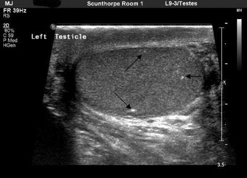
Ultrasonography of testis showing micro-calcification
Case Number 2
A 21-year-old male patient (PM) attended the GU medicine clinic in July 2005. He complained of left testicular pain. He had chlamydia a year ago. He and his only girlfriend at the time were treated adequately. There was no significant family history. The clinical examination was insignificant other than swollen lower pole of the left testis along with the testicular tenderness. A diagnosis of epididymo-orchitis was made and he was treated with two-weeks’ course of doxycycline. He improved about 50%, but still experienced pain in the left testis. Then, an ultrasound scan of the left testis was arranged that revealed microcalcification of the testis and a 3 mm cyst of left epididymal head. The patient was referred to urology department, who was managed according to the Trust guideline and was discharged in July 2007 with the advice of self-testicular examination. It was decided not to excise the cyst because of its small size.
Discussion
Testicular microcalcification also known as testicular microlithiasis is a harmless benign condition. In a study of 1504 healthy men, sonographic studies of their testes showed that only 84 (5.6%) had microlithiasis and their ages were between 18–35. 1
On the other hand, in a urology department, 500 ultrasound scans, taken over three years, only two cases (0.4%) of testicular microlithiasis were found. 2 When patients are selected from a specialized clinic i.e. infertile male patients undergoing routine USS of testes, the incidence of microlithiasis could be higher, i.e. in a study it was 6.2%. 3 All these cases were unilateral. However, in our two cases, we did not investigate for infertility as our two patients were under 25 and did not attend the GU medicine clinic complaining in sub-fertility. In the opinion of the other authors, the testicular microlithiasis can be found in normal testes and the patients are often asymptomatic. 4 In contrast, a high percentage of testicular tumours is known to be associated with microcalcification. In their series, 44% of cases of microcalcification were associated with testicular tumours. 3 They suggested sonographic follow-up for all these patients to exclude malignancy. In another study, 46% of cases with the testicular microlithiasis were associated with testicular cancer. 5 The authors suggested a regular follow-up with USS. This condition should be regarded as pre-malignant. 5
Although microlithiasis is seen commonly in urology clinics and investigated for it in sub-fertility or when testicular cancer is suspected, it is not reported from GU medicine clinics. It was coincidental to find microlithiasis in cases of epididymo-orchitis. Once found, because of its association with testicular cancer, it should be carefully followed up and patients should be taught about self-testicular examination for early detection of cancer in future. Testicular cancer if detected early the prognosis is very good. It is reported that testicular cancer has a age-dependant decline. Its prevalence is practically zero at the age of 60 and minimal after age of 50. 6 Other authors have also advocated a long-term follow-up.7,8 Many experts, therefore, adhere to the practice of annual follow-up with clinical examination backed up with USS for a long term. Our view is that these patients should be followed up annually until 50.