




Abstract
Imiquimod has immune-stimulant properties that can precipitate autoimmune conditions like eczema, psoriasis and lichenoid conditions. We report two cases here where imiquimod induced florid lichen sclerosus in one patient and lichen planus in another. In both patients the condition was so aggressive and unresponsive to steroid treatment that circumcision was necessary.
Keywords
INTRODUCTION
Imiquimod is a unique immune response modifier that is licensed for the treatment of viral warts, actinic keratosis and superficial basal cell carcinomata. Imiquimod creates a chemical gradient that induces immune cells into the area and increases production of interferon α and other cytokines. The use of imiquimod has transformed our management of genital warts, allowing the vast majority of patients to do home treatment. 1 Also, as it is a self-induced immune clearance, there is a low risk of recurrence.
Its immune-stimulant properties can however precipitate other autoimmune conditions like eczema, psoriasis and lichen sclerosus. We report two cases where imiquimod induced florid lichen sclerosus in one patient and lichen planus in another. In both patients, the condition was so aggressive and unresponsive to steroid treatment that circumcision was necessary.
PATIENT 1
A 37-year-old man presented in mid April 2006 with a markedly white warty area on the reflex prepuce (Figure 1). There was also a small area of adherence; hence there was concern that this might be lichen sclerosus or Bowen's disease. He was referred to the dermatology outpatient department (OPD) where a biopsy of the white area was performed. This was reported histologically as changes consistent with a viral wart. He was therefore commenced on imiquimod in mid-May 2006 and when seen three weeks later, the wart had cleared but there was a lot of adherence and inflammation. It looked like aggressive lichen sclerosus (Figure 2). A repeat biopsy showed changes suggestive of lichen sclerosus. He was treated with Dermovate®, but this only seemed to promote wart recurrence (Figure 3). The lichen sclerosus was responding poorly; hence he was referred to plastic surgery in September 2006 and had a circumcision in May 2007. When seen in January 2008 the penile skin was normal (Figure 4).
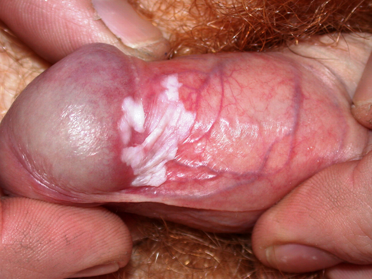
White warty area on prepuce. Just above this, there is a hint of adherence of prepuce to glans penis
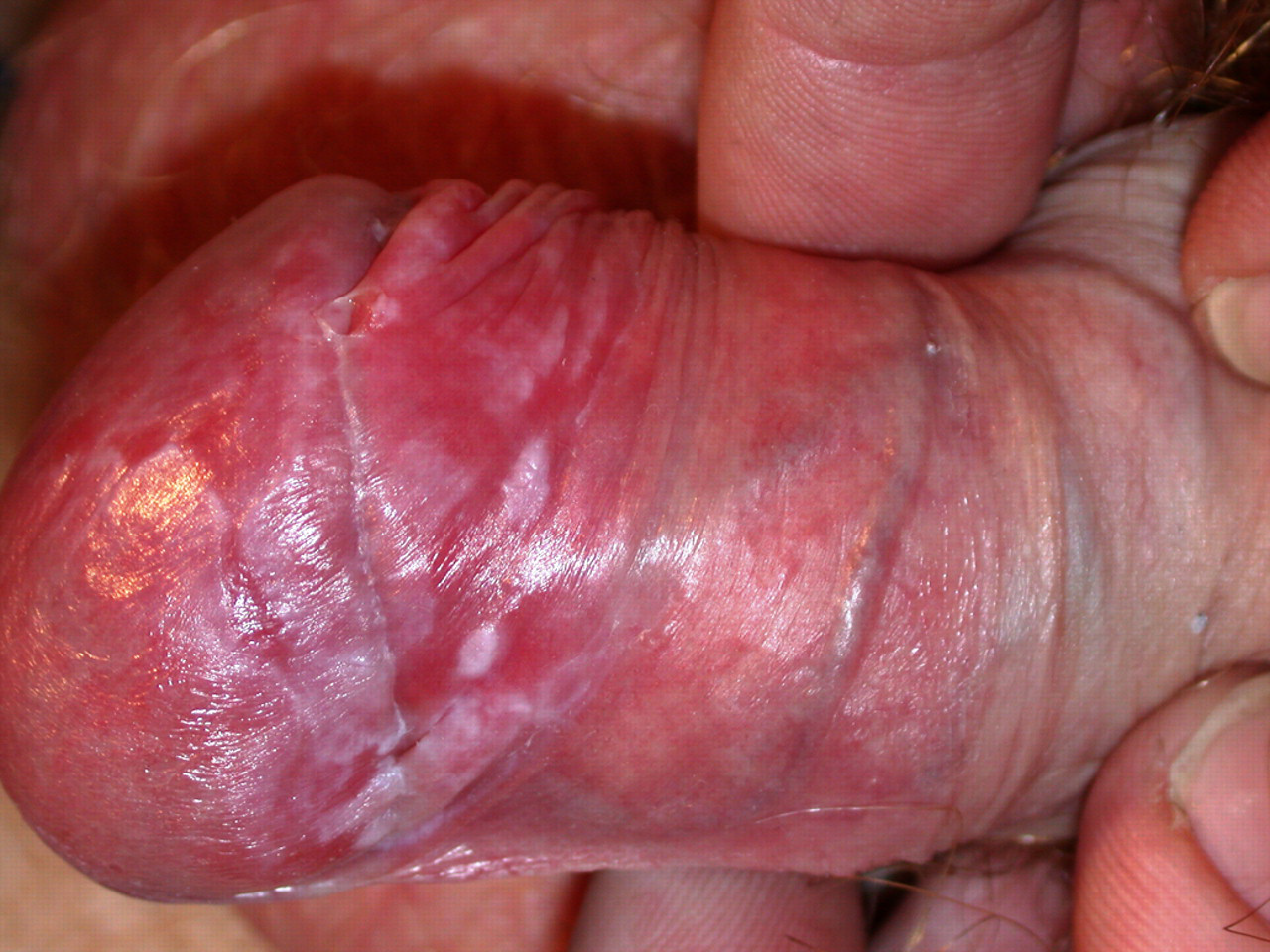
Inflammation after three weeks of imiquimod
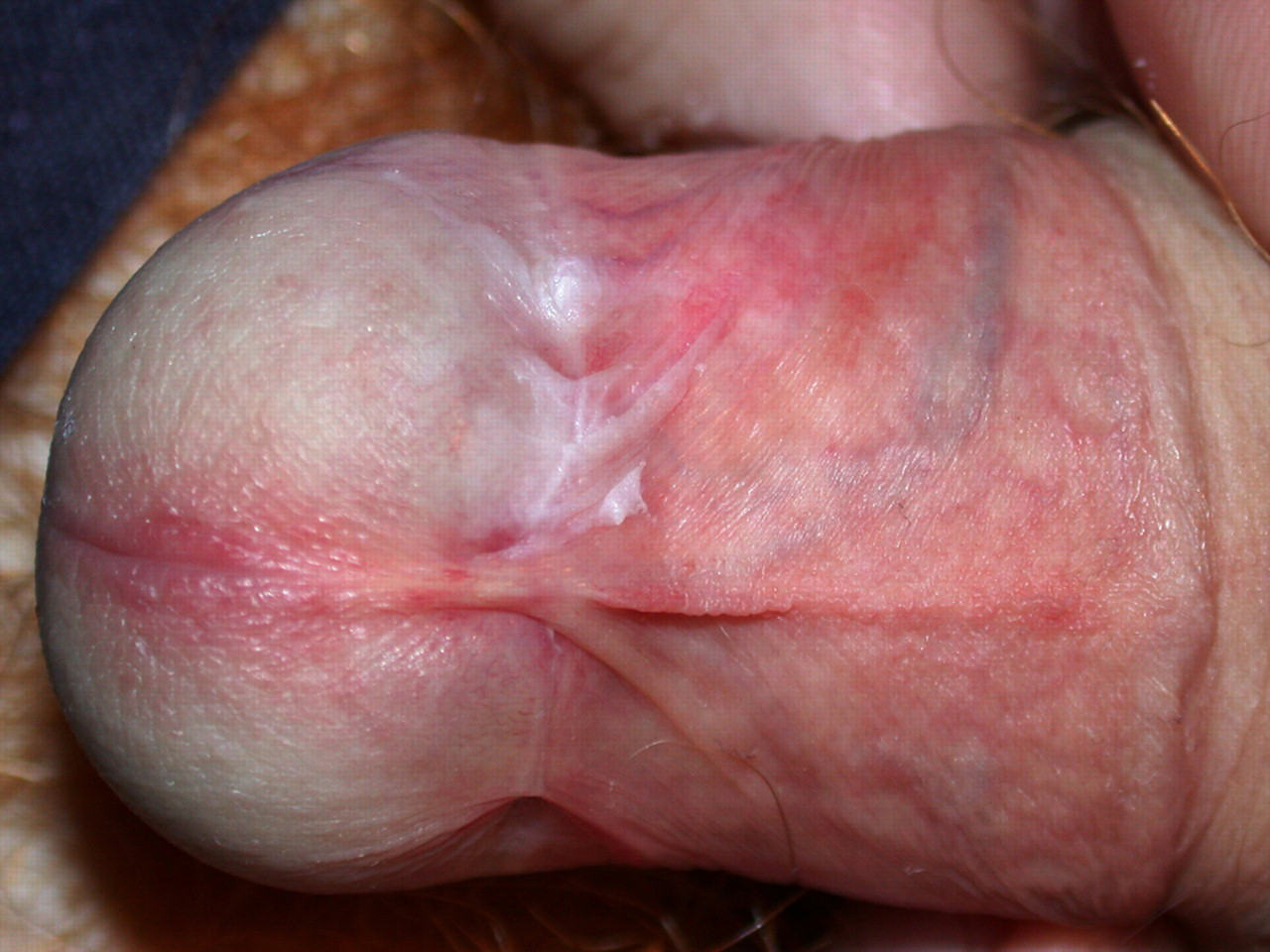
Possible wart recurrence
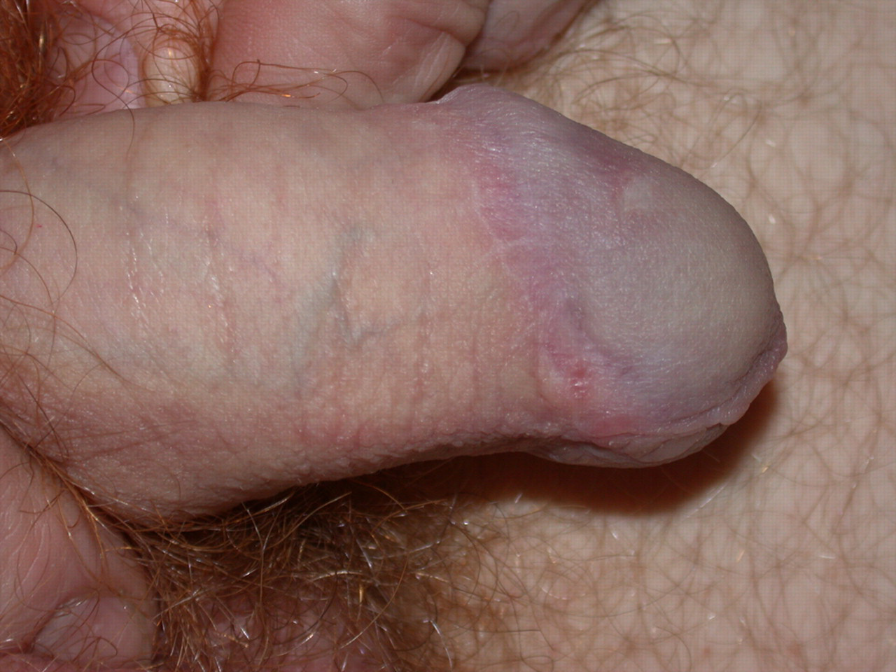
Postcircumcision, normal skin
PATIENT 2
A 21-year-old man presented in August 2007 with a few flat keratinized warts in the preputial area. He was given imiquimod. Two weeks later he reattended and the prepuce was red and inflamed. He was advised that this was a reasonable reaction and to continue to use Aldara® but reduce it to once or twice a week. He reattended five weeks later. The warts had resolved, but the penis was still inflamed. He was advised to stop the imiquimod. Six weeks later he had obvious extensive lichen planus with typical lesions on the shaft (Figure 5) and on the glans and prepuce (Figure 6). Some small ulcers were also noted. He was treated with clotrimazole/hydrocortisone cream and referred to the dermatology OPD. This was clinically agreed to be lichen planus and he was given Dermovate® ointment. However, a month later, the lichen planus was still extensive and the recurring warts were not responding to topical trichloroacetic acid. He was referred to plastic surgery and a circumcision was done in April 2008. In August 2008 he had two small persisting warts that responded to cryotherapy. He was last seen in September 2008 and his condition remains under excellent remission with no recurrence of the viral warts.
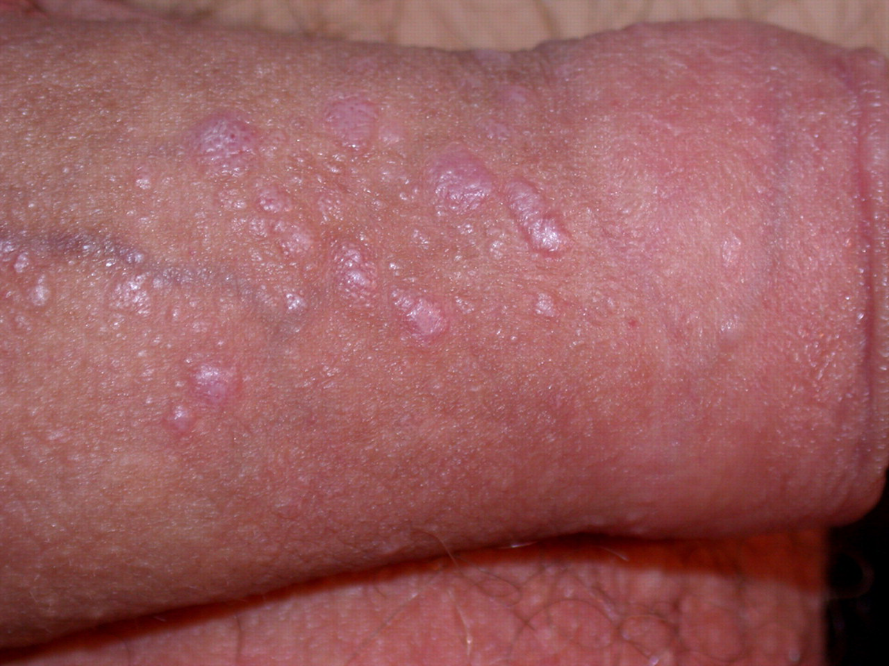
Typical lichen planus violacious plaques on shaft of penis
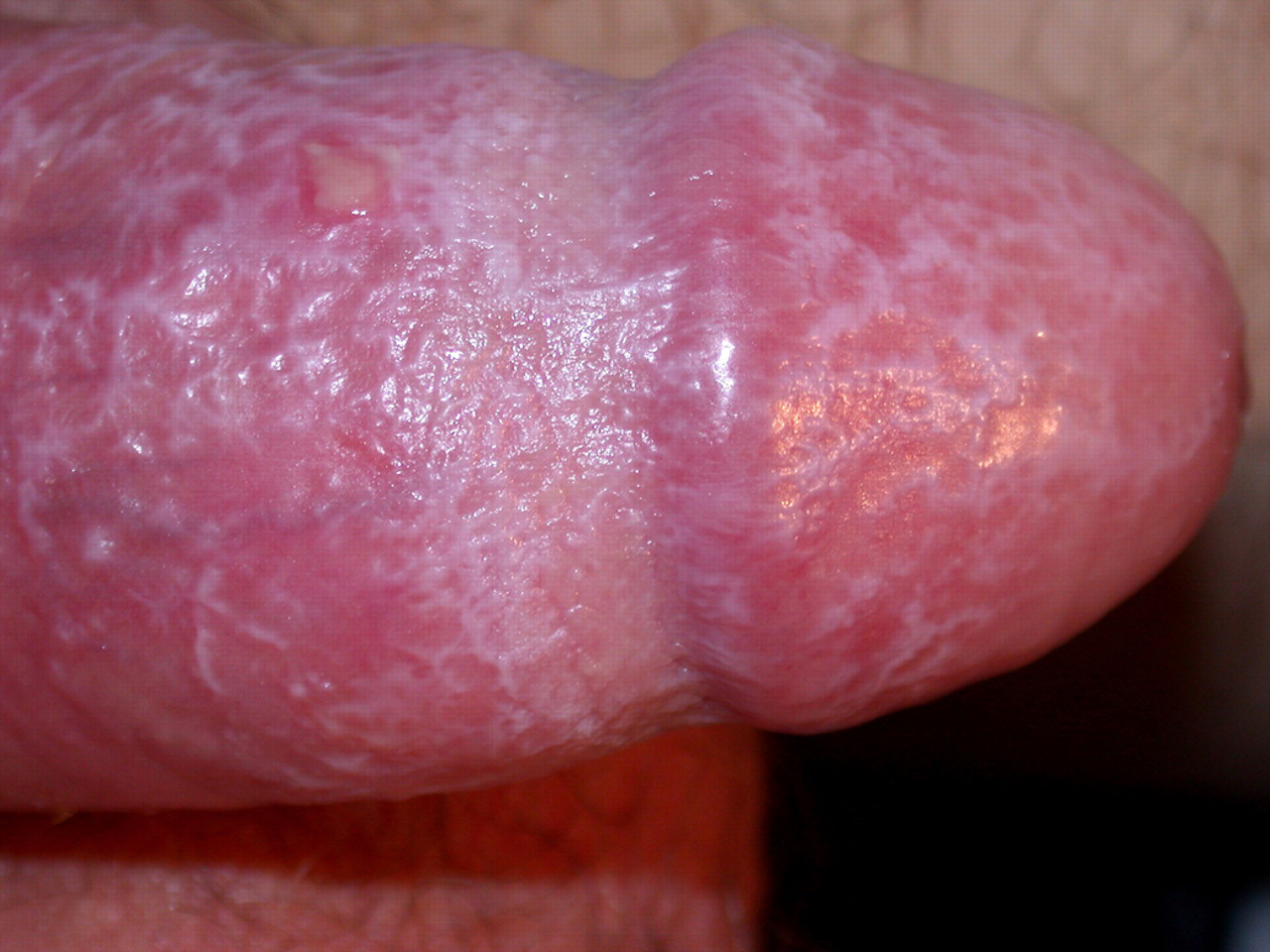
Typical reticulate lichen planus pattern on glans and reflex prepuce. Note herpetiform ulcer also
DISCUSSION
Topical imiquimod interacts with Toll-like receptors, activating immune cells and increasing production of interferon α that in turn activates cytotoxic T cells. This has beneficial effects in treating various skin conditions but could also potentially unmask other inflammatory dermatoses. 2 Vitiligo, eczema, psoriasis, localized pemphigus vulgaris and aphthous ulcers are some of the conditions precipitated or exacerbated by the use of topical imiquimod. 3,4
There is good evidence that a type I interferon (IFN) associated cytotoxic, cellular immune response is a feature of lichen planus. There is strong expression of the IFN-inducible proteins MxA and IF127 at both the mRNA and protein level and IFN-α mRNA in lichen planus lesions. 5,6 Furthermore the IFN-inducible chemokines CxCl9 and CxCl10 and the chemokine receptor CxCR3 are strongly expressed in lichen planus. 7
The evidence implicating type 1 IFNs in lichen sclerosus is less comprehensive but a recent study 8 showed significant expression of MxA closely associated with the expression of the interferon-inducible protein IP10, and the recruitment of CxCR3+ve cytotoxic T lymphocytes, many of which contained IP10 in their intracytoplasmic granules.
It appears that in certain conditions there is a common IFN-driven cytotoxic attack on skin cells and it is probably the genetic make up of the patient that determines whether this manifests as lichen planus, lichen sclerosus or some other autoimmune disease. This form of activation is important for dealing with viral infections or malignant cells but is inappropriately activated in autoimmune skin disorders. 5
It seems likely that imiquimod-induced cell-mediated cytotoxic attack was an aetiological factor in our patients.