
Abstract
Detecting acute HIV infection is important, but often the infection is difficult to recognize. We report the case of a 23-year-old man with persistent genital ulceration; all microbiological examinations from the ulcers were negative. HIV-1/2 antibodies were positive, but the HIV-1 Western blot (WB) was indeterminate and HIV-1 p24 antigen was persistently negative, with a low HIV-1 RNA level. One month later, the genital ulcerations disappeared and the WB test showed complete seroconversion; nonetheless HIV p24 antigen remained negative and HIV-RNA was persistently low. HIV-1 proviral DNA was detected by nested polymerase chain reaction (PCR) from the initial ulcers swabs. This case was notable due to genital ulceration being the only sign of primary HIV infection. Furthermore, the serological pattern was unusual, and HIV-RNA was unexpectedly low. This underlines the importance of screening for HIV being infection in the setting of sexually transmitted infections (STIs), and also in cases of atypical STI-like presentations.
INTRODUCTION
The diagnosis of HIV infection in the early stages following viral acquisition is very important for clinical and epidemiological purposes. However, primary HIV infection may be asymptomatic or present with non-specific or minor symptoms. Nevertheless, the available diagnostic tools have suboptimal sensitivity and early diagnosis is not easy to confirm before the seroconversion is complete.
CASE REPORT
A 23-year-old heterosexual man was seen in our Institute presenting with persistent genital ulcerations, lasting one month, unresponsive to oral aciclovir and doxycycline treatment. The patient had unprotected sexual intercourse with a casual female partner of unknown HIV serostatus three weeks before the appearance of the lesions. On medical examination he had a 2 cm ulcer and several smaller ulcers on the foreskin and on the corona of the glans penis (Figure 1). They were deeply painful, soft-based, whitish and with erythematous haloes. Bilateral inguinal nodes and spleen were enlarged. Behcet's disease was excluded. All bacterial and yeast cultures from the swabs were negative, using blood, Sabouraud's, eosin methylene blue, and Thayer-Martin agars. Direct examination of smears and cultures for Haemophilus ducreyi were negative. Specimens from genital ulcers analysed with molecular tests (polymerase chain reaction [PCR]) for herpes simplex virus (HSV), varicella-zoster virus and human papillomavirus were also negative. Treponema pallidum was negative by darkfield microscopic examination, PCR and serology. Chlamydia trachomatis cultures, PCR and serology also showed negative results. The patient refused biopsy of the ulcers.
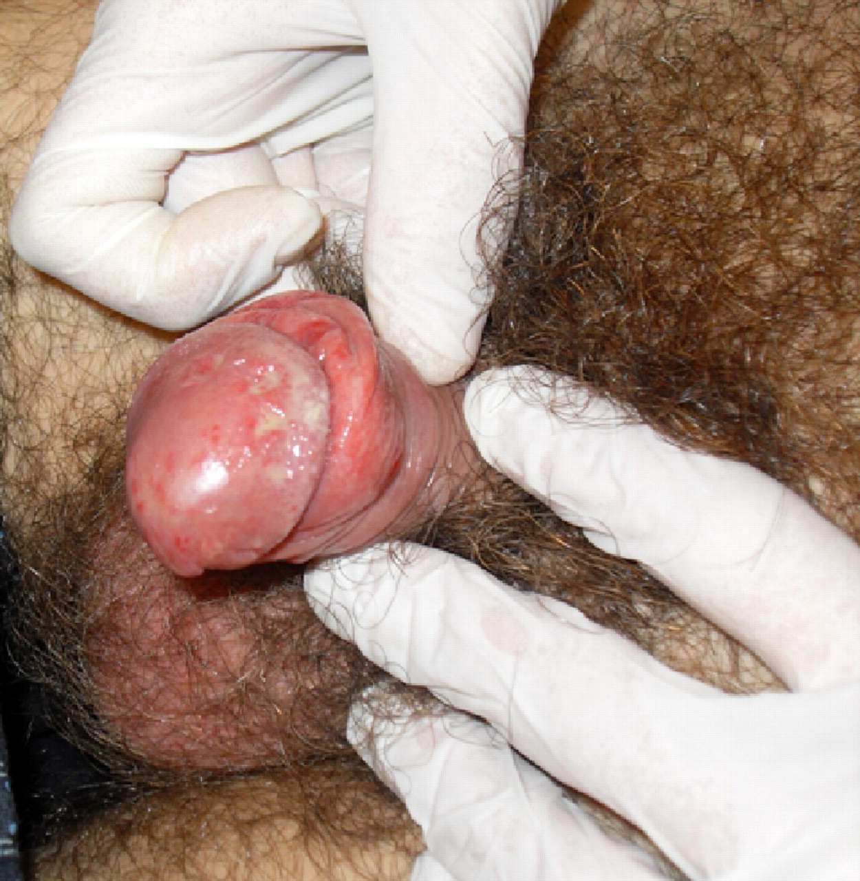
Multiple genital ulcers present at the initial medical examination
Blood cell count was normal and the erythrocyte sedimentation rate was 7 mm at the first hour. The lymphocyte count was normal, and CD4+ and CD8+ counts were 479 (38.2%) and 413 (32.9%) × 10/L, respectively. Serologic tests for Epstein–Barr virus, cytomegalovirus and HSV-1 indicated past infections; no antibodies were detected against HSV-2. No antibodies against either syphilis or other pathogens were seen in convalescent sera.
HIV-1/2 antibodies were detected (HIV-1/2 Ab/Ag enzyme-linked immunosorbent assay [ELISA] Combo Biorad, HIV-1/2 Ab Bio-Rad Genscreen [Marnes-la-Coquette, France], HIV-1/2 Ab/Ag AXSYM System-Abbott Combo [Sligo, Ireland]), but HIV-1 Western blot (WB) was indeterminate and HIV-1 p24 antigen, detected by ELISA (sensitivity: 15 pg/mL), was also negative. The initial serum sample was tested a second time for p24 antigen with a more sensitive test (ELFA [VIDAS, BioMérieux, Marcy l'Etoile, France], sensitivity: 3 pg/mL), not available at the time of diagnosis, but provided a negative result. A low level HIV-1 RNA was detected (212 copies/mL) (Real Time Abbott [Des Plaines, IL, USA], linear range: 40–10,000,000 copies/mL). A similar pattern of results was observed in a follow-up visit 10 days later, with indeterminate WB, negative p24 antigenaemia and low-level HIV viraemia (355 copies/mL). One month after the first test, the WB showed complete seroconversion with a broad HIV-1 antibody response, but p24 antigen remained negative. HIV-RNA was 879 copies/mL with a wild-type pattern on HIV-1 genotypic resistance testing.
Nucleic acid extraction from the ulcer swab was performed in MDX BIOROBOT (Qiagen Inc, Valencia, CA, USA). HIV-1 proviral DNA was detected by a nested PCR using primers for the gag region as previously described, 1 using Taq Gold polymerase (Applied Biosystems Inc, Forster City, CA, USA). Positive control DNA was from 8E5LAV cell lines, and the negative control contained no DNA template. PCR testing for the presence of the β-globin gene was used to assess the adequacy of DNA sample. By one month after the first examination the genital ulcerations had disappeared.
DISCUSSION
Genital ulcers may be one of the primary signs of HIV acute infection in about 15% of cases. These are frequently associated with fever, malaise, lymphadenopathy and a skin rash. 2–4
The case described here is notable due to the genital ulcerations that were the only sign of acute HIV infection; in fact, unlike the majority of the cases, where more than one symptom or sign are present, in our case the genital ulcers were not accompanied by fever, rash, generalized lymphadenopathy or other symptoms, and this unusual clinical presentation makes this case report of interest. We performed extensive tests on the ulcers to exclude other sexually transmitted infections (STIs). The subsequent HIV seroconversion established the diagnosis of acute HIV infection.
Some other aspects of the present case are peculiar. Repeated HIV tests enabled detection of HIV seroconversion and correlation of the serology with the clinical picture. However, the first HIV-positive test was difficult to explain and did not allow a conclusive diagnosis, as it was accompanied by a negative p24 antigen result, even when repeated with the most sensitive test available. The subsequent serological pattern was also unusual because of the persistent absence of p24 antigen upto the time when the WB had completely converted, underlining the limitations of this method. Furthermore, in this case, the HIV-RNA load was unexpectedly low. Normally, during acute infection the wide and uncontested spread of HIV leads to very high levels of HIV viraemia. 5 Therefore, if clinical suspicion of acute HIV infection is high, it is advisable to repeat the HIV test in the presence of an unresolved seroconversion pattern, to definitively confirm or rule out the diagnosis. The present case underlines the importance of screening for HIV infection in the setting of any STI, as the guidelines suggest, 6,7 but also when the clinical suspicion of HIV infection is low, as occurred in our case, due to the presence of an unusual presentation. This case supports the need to perform HIV testing routinely, regardless of whether the patient is known or suspected to have specific behavioural risks for HIV infection. 6,7