




Abstract
Lues maligna is a severe and rare manifestation of syphilis, also known as malignant syphilis or ulceronodular syphilis and mostly occurring in patients with HIV infection. In this report, we present a case of lues maligna in an AIDS patient. Our report shows the typical clinical features of lues maligna and the important association between lues maligna and HIV/AIDS. Although the clinical manifestations of lues maligna are complicated and severe, the response to the therapy of penicillin is excellent.
INTRODUCTION
Lues maligna is a severe and rare manifestation of syphilis, also known as malignant syphilis or ulceronodular syphilis and characterized by papulopustular skin lesions that enlarge rapidly and evolve into round or oval ulcers with sharp borders, centrally covered by a dark, sometimes rupioid crust. Mucous membranes of the mouth and nose may be involved, and prodromal symptoms of fever, headache and myalgia are common. 1 Lues maligna mostly occurs in patients with HIV infection. In this report, we described a case of lues maligna in an AIDS patient.
CASE REPORT
A 36-year-old man presented to our department in November 2010 with a 20-day history of general malaise, headache, myalgia, intermittent fever of up to 40°C, and multiple erythematous papules and pustules on the face, scalp, neck, limbs and trunk, which progressed to nodules and ulcers. He also complained of fatigue and significant weight loss in the last year. The patient reported sex with other men for the past seven years and did not know his HIV status.
On physical examination, the patient was pyrexial at 39.8°C, there was bilateral cervical and axillary lymphadenopathy. The neurological examination was normal. The skin examination revealed multiple disseminated erythematous papules, pustules, round or oval nodules (Figure 1a) and deep ulcers with sharp borders, centrally covered by a yellow-brown or dark, rupioid crust (Figure 1b, c), mostly on the face, scalp and neck and partially on limbs and trunk.
Multiple skin lesions of lues maligna in an AIDS patient: multiple disseminated erythematous papules, pustules, round or oval nodules (a) and deep ulcers with sharp borders, centrally covered by a yellow-brown or dark, rupioid crust, mostly on the face and scalp (b, c)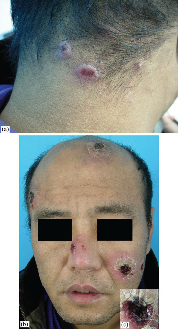
Laboratory investigations revealed the following: white blood cell count was normal, albumin 30 g/L (normal 35–55 g/L), C-reactive protein 64 mg/L (0–10 mg/L). Rapid plasma reagin (RPR) was positive at 1:32 and Treponema pallidum particle agglutination assay was positive. Enzyme-linked immunosorbent assay and Western blot for HIV were positive and CD4+ cell count was 136 cells/mm3 and CD4+/CD8+ was 0.18. Lumbar puncture was refused. Abdominal colour Doppler ultrasound indicated hepatomegaly with changes in liver parenchyma and splenomegaly. Head computed tomography was normal. Skin biopsy was performed and histopathology of the lesion from his left upper limb revealed mild epidermal hyperplasia, a diffuse dermal and perivascular inflammatory infiltration with lymphocytes, plasma cells and histiocytes, and proliferation of vascular endothelial cells (Figure 2).
Histopathology of biopsy specimen showing: mild epidermal hyperplasia, a diffuse dermal and perivascular inflammatory infiltration with lymphocytes, plasma cells and histiocytes, and proliferation of vascular endothelial cells (haematoxylin and eosin, ×200)
Based on the diagnostic criteria
1,2
the patient was diagnosed with lues maligna and AIDS. Treatment was started with intravenous aqueous crystalline penicillin G 24 million units (MU) per day, administered as 4 MU every four hours for 14 days, and preceded by prednisone 30 mg per day for three days. After completion of the regimens, the therapy of benzathine penicillin, 2.4 MU intramuscularly once per week for up to three weeks and combination antiretroviral therapy (zidovudine 300 mg/day + lamivudine 300 mg/day + nevirapine 400 mg/day) was used. The clinical response to penicillin was excellent; at one month after treatment, all skin lesions had healed, though some with residual scarring (Figure 3). Three months later, laboratory investigations showed that the RPR titre was 1:8, white blood cell 7.95×10⁹ cells/L and CD4+ cell count 140 cells/mm3. Follow-up of the patient continues.
At one month after treatment, all skin lesions had healed, though some with residual scarring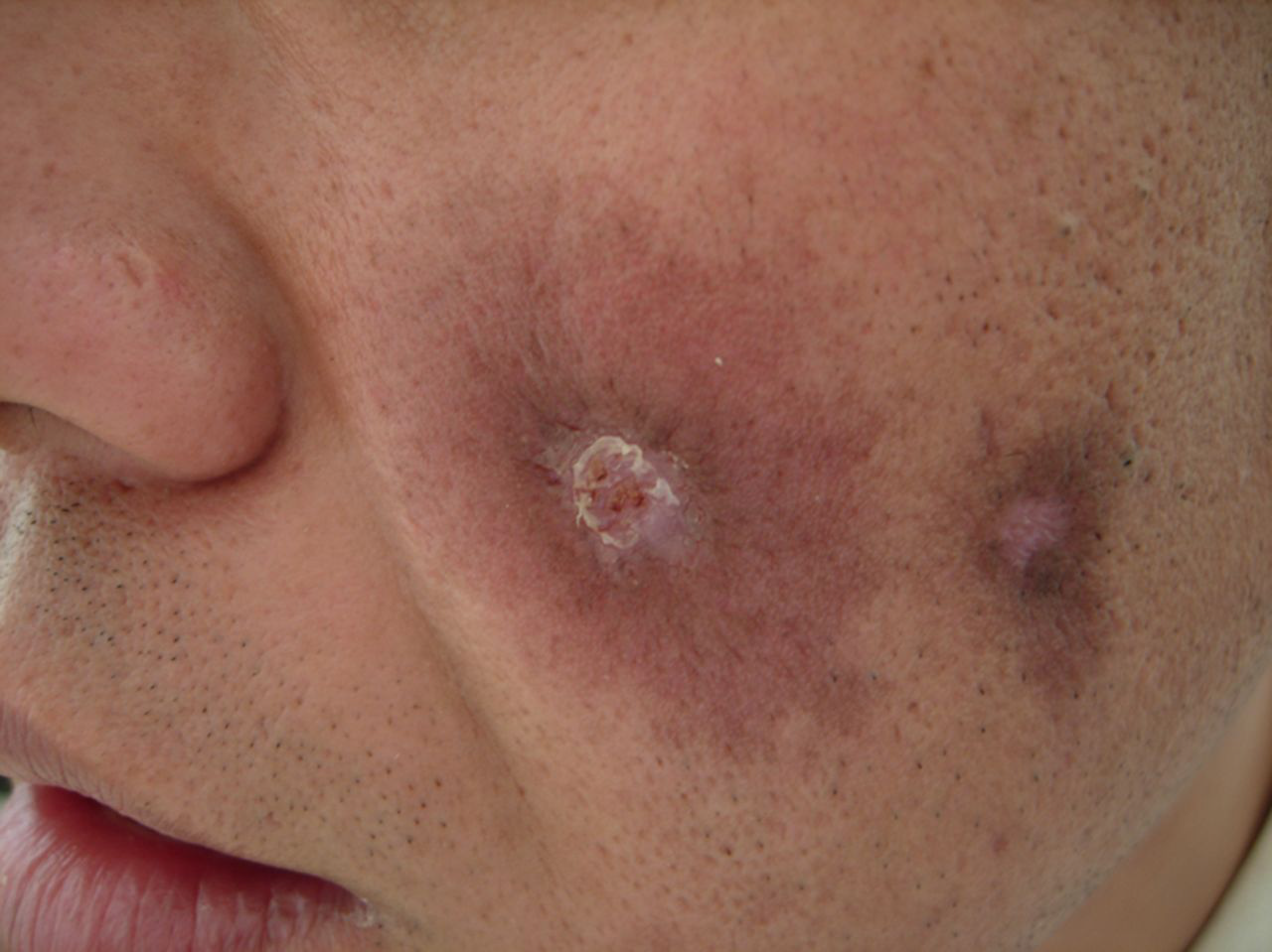
DISCUSSION
It is recognized that co-infection with HIV can modify the natural history of syphilis. 1 Diagnostic criteria for lues maligna include the following:
(1) strongly positive RPR titre; (2) a severe Jarisch–Herxheimer reaction; (3) characteristic manifestation and histopathology findings; and (4) an excellent response to antibiotic therapy. 1 Clinical differential diagnosis of lues maligna includes pyoderma gangrenosum, vasculitis, lymphoma, leishmaniasis, leprosy, yaws and mycobacterial or fungal infections. 3 The histological characteristics of lues maligna include a lymphocytic predominance with plasma cells in the upper and middle dermis; a significant perivascular infiltration and pronounced endothelial swelling, proliferation and fibrinoid necrosis of superficial and deep vessels. Granulomatous infiltration and giant cells have been seen in cases of lues maligna in HIV-infected patients. 4
The optimal antimicrobial regimen to treat syphilis in HIV-infected subjects remains controversial. Guideline recommendations in this population are based on few objective data. 5 Two studies found that the risk of abnormal cerebrospinal fluid laboratory findings was significantly higher when both risk factors (i.e. serum RPR titre≥1:32 and CD4+ cell count <350 cells/mm3) were present in the same patient. 6,7 After considering the clinical manifestations, serum RPR titre 1:32 and CD4+ cell count 136 cells/mm3, the patient was treated with a neurosyphilis regimen and antiretroviral therapy.
This case showed the typical clinical aspects of lues maligna and the important association between lues maligna and HIV/AIDS. Although the clinical manifestations of lues maligna in HIV are complicated and severe, the response to the penicillin therapy is still excellent.