




Abstract
We have investigated the changes in the workflow of the cytologist due to the introduction of telepathology. These changes occur in two stages. The first is the use of telepathology as a support methodology using external providers (i.e. outside the hospital) to digitize the slides. The second is the use of telepathology in routine laboratory operations using an internal scanner to digitize the slides. To improve courses in the Master of Cytology programme at the University of La Sapienza, new learning modules were designed, which were made available via the wide area computer network to familiarize students with the new technologies. The new methodologies had three benefits. The first was the high level of knowledge for the student. The second was the cost advantage to the student, who did not need to study in a medical laboratory to participate in the teaching. The third was the cost advantage to the hospital: as laboratories become freed from academic work, they become more available for clinical use.
Introduction
Over the last few years, the scenario of work has changed several times for the cytologist. Cytologists first had to learn to diagnose tissue placed on glass slides by spatula. They also had to learn to manage tissue placed on glass slides after centrifugation. Most recently they have had to learn to digitize and manage tissue on virtual slides, i.e. for use in telepathology.
We have investigated the changes in the workflow of cytologists due to the introduction of telepathology. This has allowed us to optimize various courses in the Master of Cytology programme at the University ‘La Sapienza'.
Telepathology
Telepathology uses virtual microscopy, which has three core aspects.
1–6
These are:
digitization, using a scanner, to obtain a virtual slide; storage of the virtual slide on a server and establishing an appropriate network connection to reach it; navigation of the virtual slide by means of appropriate software.
External scanner. In the first stage, an external service is used to obtain the virtual slides (Figure 1). Examples of such services are the remote web server at Leeds
7
or the Milestone centre at Bergamo.
8
These services digitize glass slides and store them on a server. They also store the images on a DVD that is sent back to the remote laboratory. The virtual slides can be consulted via the Internet using various low cost (or free) software tools. These tools (such as the Image scope tool
7,9
), installed in the telepathology clients, allow navigation of the virtual slide. This first stage represents a gentle introduction to the use of virtual slides and telepathology as a supporting methodology for traditional pathology using conventional glass slides;
In-house scanner. The second stage consists of the use of a scanner in the laboratory to obtain virtual slides directly in the laboratory (Figure 2). The virtual slides are then stored on a local server and on a DVD. Virtual slides can be consulted via the local network using low cost (or free) software.
The changes in the laboratory introduced by telepathology have occurred in two stages:

First stage of changes. The server and the scanner are external to the hospital, accessible via a wide area network

Second stage of changes. The server and scanner are located in the hospital itself, accessible via the local area network
Master in cytology
Students completing their Masters degree in diagnostic cytopathology and population screening at the University ‘La Sapienza’ need to make a certain number of successful diagnoses, in accordance with national laws. They also need to spend considerable time in the microscopy laboratories of the hospital. These laboratories usually operate under pressure because of the routine workload of the hospital, so complex scheduling problems often occur. Telepathology could therefore be a method of reducing the workload of the laboratory. The virtual slides, once prepared, could also provide a useful database of medical knowledge, as in the case of the experience in Leeds.
Training
In order to address the changes which have occurred in the practice of pathology, courses at the University ‘La Sapienza’ have been modified to include elements of telepathology. This starts from the first peer-to-peer two node WAN connections designed for dynamic telepathology applications 5,10–13 and covers a range of more advanced telepathology applications. By the end of their training in telepathology, cytology students are able to interact with each of the system elements shown in Figures 1 and 2.
The specific modules for telepathology can be accessed via the network and consist of:
training on virtual slides; training with the tools for navigation of virtual slides; familiarization with tools for navigating virtual slides using the intranet, extranet and Internet; setting up intranet, extranet and Internet telepathology connections; familiarization with the Picture Archiving and Communication System (PACS) connected to the hospital information system.
Familiarization with virtual slides is accomplished by specific training on the scanner to be used most, such as those made by Zeiss, Nikon, Olympus and Hamamatsu; this is arranged by contacting the manufacturers directly. This is a core attribute of virtual microscopy. In fact, the customization of the scanner is a function of the type of slides and thus a basic training matter.
We chose the tools proposed by Aperio.
5
Aperio, in fact, provides two useful tools for training. The first, Image Scope, allows the easy extraction of coordinates (X, Y, Zoom) from a virtual slide region; the second, Spectrum Web Viewer, provides the Internet path of a specific virtual slide region. For points (3) and (4) we have designed a specific tool for the training, an e-learning form. The teacher (Figure 3a) selects a region, by virtual navigation; pushes a button to generate the relevant Internet path (Figure 3b); and inserts the path in the e-learning form for the test (Figure 3c). Note that this interaction results in the exchange of an Internet link, rather than images or data. Once the student receives the e-learning form with the path, activates the intranet or Internet connection and reaches an area of a virtual slide (for example, the Area
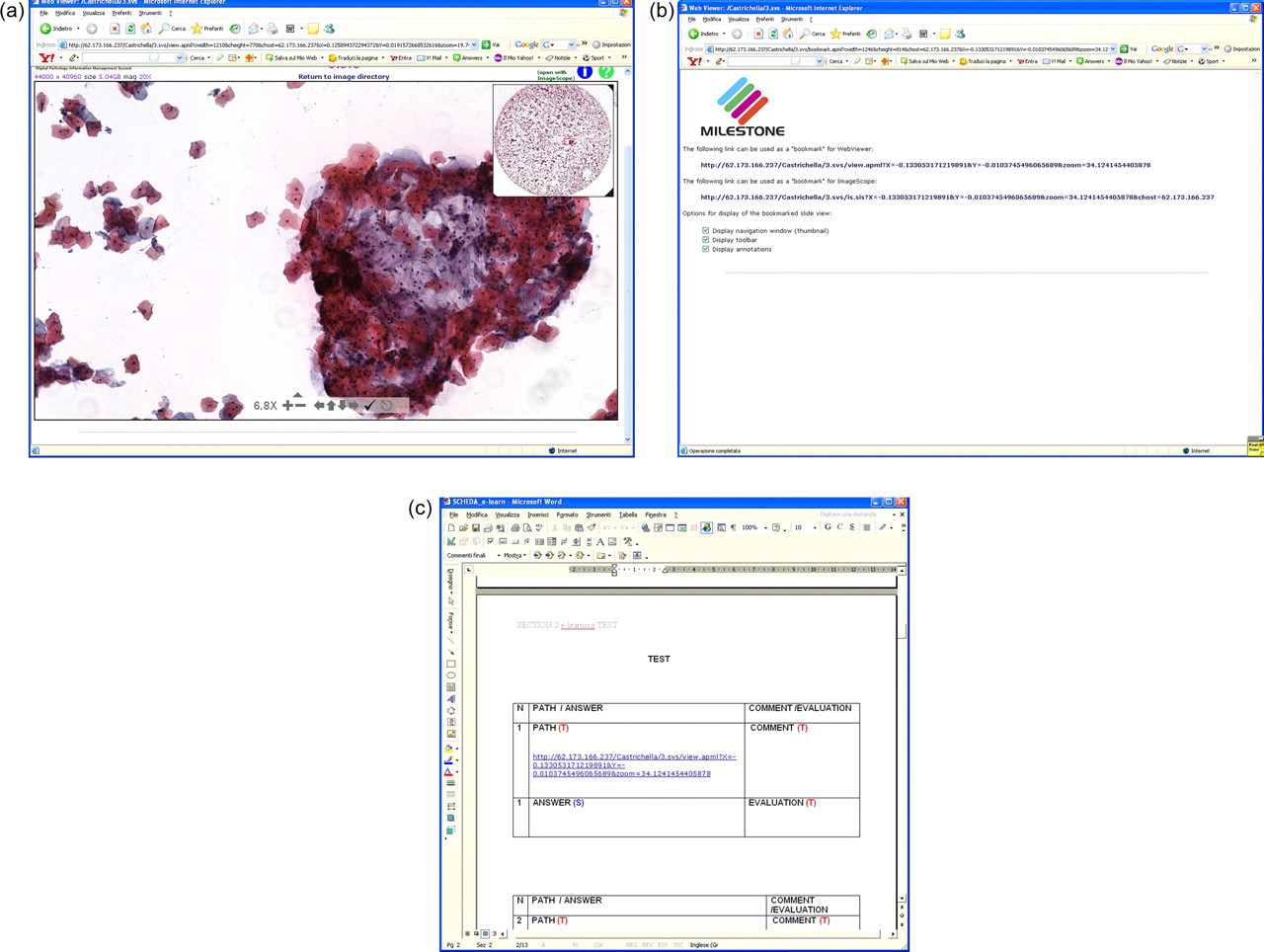
The teacher selects a region, by virtual navigation (Figure 3a); then pushes a button to generate the relevant Internet path (Figure 3b); and inserts the path in the e-learning form for the test (Figure 3c). See
We have tested this methodology on a sample of five subjects (teachers and students) involved in the master of cytology programme.
Regarding point (5) it should be considered that at present, no standard for PACS in telepathology exists. In telepathology, there are no standards for archiving images or standards about what to archive. A virtual slide requires a large amount of memory. A cytology virtual slide could require memory space similar to that of a medium-sized hard disk. The approaches followed by the different manufacturers to design a PACS vary. For these reasons, we have limited training to the standard PACS used in the hospital for radiology. This is satisfactory for training cytologists for future PACS designed for telepathology.
The connection failure rate was assessed in 5000 random trials of the connection and was equal to 0.1%. The acceptance of the e-learning form was very high, as shown in Table 1.
Acceptance of the methodology based on the e-learning form
*0 = minimum acceptance; 3 = maximum acceptance
Discussion
Universities need to respond to the technological changes caused by telepathology. Both in the case of an external scanner and server, and an internal scanner and server, the cytologist should be competent in using the software tools specific for telepathology navigation of a virtual slide. Cytologists should also be able to interact with picture archiving systems in a similar way to a radiologist who interacts daily with PACS to archive CT or NMR images. For these reasons we have designed and tested specific training for students at the University ‘La Sapienza’ that allows for familiarization with virtual slides, with the tools used for remote navigation of the virtual slides and with the PACS for integration with the hospital information system.
There are other benefits of this training:
advantages for the students, who can analyse virtual slides at home without needing to go to the hospital; advantages for the hospitals, as laboratories are used less for academic activities and thus remain more available for routine work of the hospital; the ability to perform cooperative diagnosis from the home or hospital desk without needing to go to the laboratory.