
Abstract
We examined the feasibility of distance diagnosis of oral diseases, using transmission of digital images by email. Twenty-five cases of oral lesions were documented during a 12-month study in a primary care public health clinic in Paraná in Southern Brazil. Clinical electronic charts and images were produced and sent by email to two oral medicine specialists with a median of 10 years experience in the field. The consultants provided a maximum of two clinical hypotheses for each case. In 15 of the 25 cases (60%) both consultants made a correct diagnosis; in seven cases (28%) only one consultant made a correct diagnosis; and in three cases (12%) neither consultant made a correct diagnosis. Thus in 88% of cases, at least one consultant was able to provide the correct diagnosis. The results suggest that distant diagnosis can be an effective alternative in the diagnosis of oral lesions and that the using two distant consultants improves diagnostic accuracy. Primary care public health clinics may benefit from the use of email and digital cameras for telehealth in remote areas where oral medicine specialists are not available.
Introduction
Little is known about the impact on public health or the economics of telehealth in dentistry. Some authors have reported that communication based on the Internet could be used in the public health system as a method of reducing costs and improving assistance in oral health. Tyndall et al. 1 reported that although there has been considerable research on the use of teleradiology in medicine, there has been little effort to evaluate its use in dentistry. Farman and Farag 2 reported that telemedicine should be an alternative to a second opinion in dental practice and that this system could provide economic benefit by allowing patients in remote areas access to specialized medical care without the difficulty and cost of travel to distant medical centres.
Telehealth practices in dentistry showed a 50% reduction in cost associated with preventing unnecessary transfers of patients for emergency maxillofacial surgery. 3 Similar studies using teledermatology verified a 51% reduction in referrals by general clinicians who would have otherwise referred the patients to a specialist. 4 It was also reported that a teleconsulting system for preoperative consultation reduced from 43 to 10 the number of patients requiring the physical presence of a specialist for consultation before dento-alveolar procedures. 5 In 1999, Leão and Porter 6 reported that there had been no investigation of the Internet potential for distance diagnosis of oral diseases, despite an impressive increase in computer applications in dentistry. Mistak et al. 7 and Baker et al. 8 compared the radiological interpretation of periapical lesions when analysed by a conventional method or teledentistry and found no significant differences between these two methods. Jacobs et al. 9 also studied the diagnosis of maxillofacial fractures using conventional radiology and digital radiology; they reported the usefulness of the electronic system and emphasized that clinical information was of crucial importance.
Stephens et al. 10 reported that email image transfer was a reliable form of orthodontic planning and advice for general dental clinicians in the UK. Other authors found that photographic documentation of oral lesions was considered acceptable by 75% of patients seen for diagnosis of the lesions. 6 The patients involved in this study also mentioned that the visualization of oral lesions helped them to understand and support clinical decisions. Almost 50% of the study sample said that the time required to document oral lesions was a problem of the teledentistry system.
We have investigated the feasibility of distant diagnosis in oral medicine. Telehealth has not yet been systematically investigated in that field and possible applications, as stated above, are mostly proposed from results obtained in similar specialties in dentistry or medicine, such as oral maxillofacial surgery and dermatology, respectively.
Methods
Data were collected from oral clinical examination and oral digital photography. Patients with oral lesions were selected over a 12-month period from a public primary care unit 100 km from Curitiba in Paraná, Brazil. Patients involved in the study sought oral consultation; individuals without oral lesions were excluded from the sample. The research was approved by the appropriate ethics committee.
Clinical data were recorded in an electronic form specially designed for the study and stored as a word-processing file. Oral lesions were documented using a digital camera (S7000, Fuji) with a 50 mm macro lens and circular illumination system. Clinical images were saved as JPEG files and then sent as an email attachment to two consultants. The images of oral lesions were 150–500 kByte in size, with a minimum resolution of 600 dpi. An Internet connection was obtained via a 56 kbit/s modem – it took about 15 s to transmit a 500 kByte image via the modem. No specific protocol concerning angles, number or views was designed for obtaining clinical images. A 43 cm colour monitor was used for lesion visualization.
Image and text files were renamed with sequential numbers to avoid patient identification. Two different referees, oral medicine specialists with a median of 10 years experience in the field, separately analysed the images and clinical information. They recorded a maximum of two clinical diagnoses for each case transmitted electronically. Clinical hypotheses were selected from a predefined list of terms designed to avoid misunderstanding and to establish a predictable data comparison. Biopsy specimens in cases requiring histological examination for definitive diagnosis were sent to the oral histopathology laboratory of the Universidade Positivo Dental Centre in Curitiba.
Final diagnoses were compared to the diagnoses of the remote clinicians to verify percentage of total, partial, or none correct hypothesis. Interexaminer agreement was measured by the kappa coefficient of agreement.
Results
Twenty-five cases of oral lesions were documented during the study period. The final diagnoses varied from benign and self-limiting diseases such as aphthous ulcer to malign and life-threatening conditions such as epidermoid carcinoma. The lesions were a very heterogenous group (Figures 1 and 2).

Epidermoid carcinoma. The size and classical characteristics of oral lesion produced complete agreement in distance diagnosis
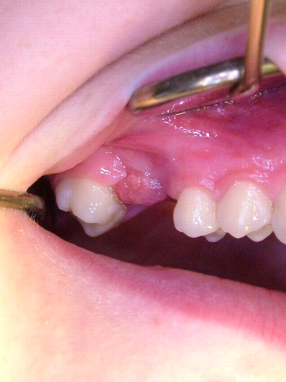
Pyogenic granuloma. This produced one correct diagnosis. One consultant hypothesized two other proliferative/reactive lesions: fibrous hyperplasia and giant cell granuloma
No problems were detected in the store-and-forward system during the study. In one case, however, one of the distant clinicians said he could not establish a diagnostic hypothesis because of poor image quality.
In 15 of the 25 cases (60%) there was complete agreement between the two examiners and the final diagnosis. In seven of the remaining 10 cases, one consultant made a correct diagnosis. Neither consultant made a correct diagnosis in 3 of 25 cases (12%). Thus in 88% of cases (22 of 25), at least one consultant was able to provide the correct diagnosis (Table 1). When only the first clinical hypothesis was considered, both clinicians performed similarly with 64% (n = 16) correct diagnoses.
Distant diagnosis of oral lesions
There was agreement between clinicians in 12 cases (48%) when only the first hypothesis was considered. The inter-observer agreement for the first hypothesis was fair (kappa = 0.30). When both hypotheses were considered and resulted in a correct diagnosis by both clinicians, little difference was observed: Clinician 1 had a 72% correct diagnosis rate (n = 18), and Clinician 2 had a 76% (n = 19) correct diagnosis rate. When both hypotheses were considered, there was 60% agreement between clinicians and the kappa values suggested fair agreement (kappa = 0.27). A final statistical analysis was conducted considering total, partial and no correct answers. The weighted kappa coefficient (0.28) suggested fair agreement between clinicians.
Discussion
The objectives of the present study were to examine the use of teledentistry by email and digital photography and to quantify the accuracy of diagnosis by two distant consultants. Vassallo et al. 11 described the use of a similar methodology with email and digital images in other medical specialties such as neurology, orthopaedics, nephrology, rheumatology and paediatrics. The use of teleradiology as an auxiliary method for distant diagnosis and treatment has also been studied in medicine and dentistry. Hayakawa et al. 12 conducted research in teleradiology in a fashion similar to that in the present study. Digital intra-oral radiographs were transmitted for distant analysis using email. Although there appear to be no similar studies in oral medicine for comparison, it was possible to identify studies in other fields that allow comparison with our results. The 60% total correct diagnosis rate observed in the present study was lower than that obtained by Corr et al. 13 who found a 94% correct diagnosis rate when digital radiographs were transmitted through email.
Prior studies in the field of teledermatology have reported similar results. Piccolo et al. 14 found a mean of 85% correct diagnoses using email. Lozzi et al. 15 compared teledermatology diagnosis with histopathological examinations and found a 79% correct distant diagnosis rate. In another dermatology study involving 46 patients, Massone et al. 16 found correct diagnoses in 73% and 74% of cases, compared to the presenting diagnosis and to the histopathological diagnosis, respectively. Complete agreement among teleconsultants was obtained in 20% of the cases. Lee et al. 17 found 100% agreement between presenting and distant diagnosis.
In the present study, clinicians missed the correct diagnosis in 3 of 25 cases (12%). Erroneous diagnoses were made in relatively common oral pathologies such as mucocele (Figure 3), hyperkeratosis and fibrous hyperplasia. In one case, the clinical image suggested a diagnosis of verruca vulgaris. Histologically, however, the case was found to be hyperkeratosis and acanthosis. In a previous study, incorrect diagnoses were seen in 6 of 96 images analysed and no attempt was made to discuss qualitatively why they happened. 13 The lower accuracy in the present study may be explained perhaps by the very heterogeneous group of oral diseases in our sample. Oral diseases are difficult to diagnose, even with the examiner present. Moreover, it is not unusual to have a histopathological inconsistency with the clinical diagnosis, even in conventional clinical practice.
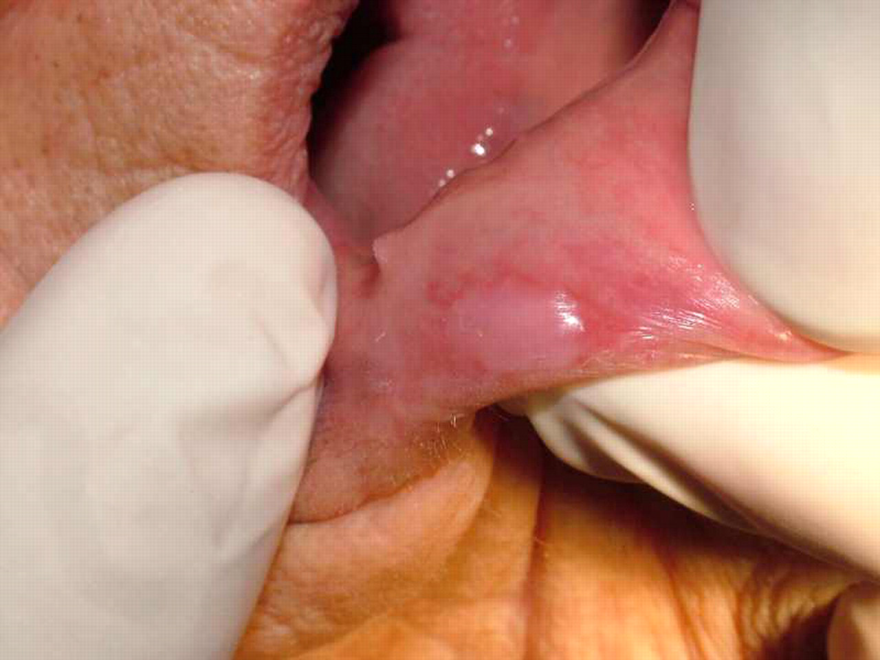
This misleading clinical image of a very common oral lesion produced an incorrect diagnosis from both consultants. The mucocele was misinterpreted as a fibrous hyperplasia or a leukoplakia
Although there are obvious limitations in comparing the results of telediagnosis in different specialties, the majority of papers favour transmission of clinical and radiographic images by email. Teledentistry has shown promising results in early studies, particularly in the fields of endodontics and maxillofacial surgery. There were no significant differences seen between clinicians who assessed periapical lesions using traditional radiographs or radiographs transmitted via email. 7,8 A study on the diagnosis of facial fractures showed similar results using telemedicine images and direct radiograph visualization. 9 Stephens et al. 10 suggested that many patients were being referred for specialized care in orthodontics without a real need for this type of treatment. They stressed that teledentistry could provide a more rational and precise approach to patient referral.
The present investigation suggests that distant diagnosis can be an effective approach for oral lesions and that the use of two distant clinicians improves the rate of correct diagnosis. Primary care services in dentistry could benefit from using email and a store-and-forward image system. Such a system could provide a screening approach to organizing a referral system for patients requiring specialty services in the field of oral medicine.
Footnotes
Acknowledgements
We thank the Brazilian National Research Council for financial support (grant 38-2004 Decit-Ministério da Saúde).