




Abstract
The diatom test is widely used by forensic pathologists as proof of drowning, notwithstanding some criticisms mainly concerning the occurrence of false-positive results (presence of diatoms in the tissues of subjects who died from causes other than drowning). The aim of the present study was to verify the claimed inaccuracy of the method caused by an excessive rate of false-positives related to inadvertent exposure to diatoms of the general population. The study was carried out to investigate the presence of diatoms in the tissues (lungs and sternum) of subjects who died from causes other than drowning. Two groups of cadavers that underwent an autopsy at the Institute of Forensic Medicine of the University of Verona were included in the study. Group A comprised 45 individuals who died from causes other than drowning, whereas Group B comprised 20 bodies which had been recovered from water. The extraction of the diatoms was performed by incubation of samples in nitric acid for 48 hours at 60°C. The analysis of the samples from Group A showed the absence of diatoms in both lung and sternum samples. In Group B all lung samples showed the presence of diatoms, whereas only six sternum samples were shown to contain diatoms. The difference between Groups A and B was statistically highly significant. The absence of diatoms in the samples collected from Group A falsified the hypothesis that false-positive results from the diatom test may occur due to diatoms entering living bodies through the respiratory and/or digestive tracts via air, water or food, supporting the validity of the diatom test as proof of drowning.
Introduction
Researchers first started investigating the presence of diatoms in the body tissues of those who died by drowning at the beginning of the 20th century. 1 After some decades of neglect, in the 1960s the first systematic studies on this subject were published, which opened a research field which still attracts the attention of forensic pathologists worldwide. 2 Among the different issues on which the debate is still open, certainly the most significant concerns the diagnostic specificity of the ‘diatom test’ (i.e. the occurrence of ‘false-positives’, whereby diatoms are found in the tissues of subjects who died from causes other than drowning).
Most authors ascribe the origin of the diatoms found in the tissues of non-drowned bodies to the environment to which the subjects had been exposed during life.3–5 Quite recently, Law and Jayaprakash 6 conducted a study on the diatom content of foods, namely shrimps, clams, fish, chicken and anchovies. After digestion in nitric acid, different types of diatoms were found in shrimps and clams, whereas no diatoms were found in fish, chicken and anchovies. On the basis of these findings, the authors concluded, but did not demonstrate, that it is theoretically possible that diatoms entering the digestive system with food could pass into the systemic circulation and thus arrive at the peripheral organs, thus interfering with the diatom test (i.e. producing false-positive results). Conversely, other studies support the reliability of the diatom test by demonstrating that in the bodies of ‘non-drowned subjects’ the presence of diatoms was irrelevant.7–9 It is worth mentioning a paper by Auer and Mottonen 10 reporting a study of 107 cases of drowned subjects and 15 ‘control cases’ in which the authors took scrupulous care to avoid contamination during autopsy, collection of samples and extraction of diatoms from tissues. The results clearly showed the absence of diatoms in the tissues from the control group. On the basis of these findings, the authors suggested that the main cause of the false-positive results is sample contamination by diatoms present on the surface of the body and/or in the clothing and/or in solvents, reactants or cellulose filters used for sample pretreatment.
In the effort to unravel the problem of false-positive diatom tests, a few authors suggested the use of quantification of the measurement of diatom elements in samples. In 1968, Timperman 11 reported a neat discrepancy between the number of diatoms per weight-unit found in the tissue-extracts of drowned subjects (ranging from 100 to 5000 per 100 g of digested lung tissue) and the number found in the tissue-extracts of non-drowned subjects (not detected in 13 out of 17 cases and ≤4 diatoms per 100 g in the remaining four cases). More recently, Ludes et al. 12 have suggested a cut-off concentration (i.e. the diatom concentration above which the test should be reported indicative of drowning) of 20 diatoms per 10 g in lung and five diatoms per 10 g in peripheral organs.
An interesting approach, reported again by Auer 13 and later by Ludes et al.14,15 and by Pollanen et al., 16 is based on the comparison of the diatoms found in the tissue extracts of bodies recovered from water with those present in the drowning water, or, as recently proposed by Horton et al., 17 with the diatoms recovered from the clothing of the submersed body.
On the other hand, the problem of false-negative results (no diatoms recovered from the tissues of drowned subjects) was less attractive for forensic pathologists, as evidenced by the paucity of the literature available on this topic. Different authors suggested as possible causes of false-negative results: (i) a too rapid death after submersion, due to the presence of concurrent diseases; (ii) reduced organ vascularization; (iii) low number of diatoms in the drowning medium; and (iv) inefficient/destructive extraction process.18–21 Last but not least, the limited sensitivity of light microscopy for diatom identification was pointed out in 1992 by Pachar and Cameron 22 and more recently by Bortolotti et al., 2 who showed that only scanning electron microscopy allows for the identification and characterization of the smallest elements or fragments of diatoms, which would not be identifiable by using standard light microscopy.
Because of the potential relevance of a validated diatom test for the practice of forensic pathology, the present study aimed to verify the claimed inaccuracy of the method caused by an excessive rate of false-positives, related to the inadvertent exposure to diatoms of the general population. The study was carried out with careful investigation of the presence of diatoms in the tissues (lungs and bone) of bodies of subjects who died of causes other than drowning.
Materials and methods
Cases
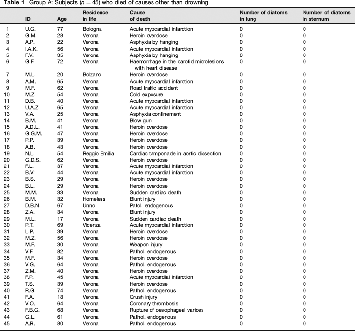
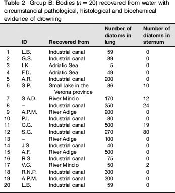
Two groups of cadavers that underwent autopsy at the Institute of Forensic Medicine of the University of Verona were included in the study. The first group (Group A) comprised 45 individuals, aged between 17 and 80, who died between October 2006 and January 2009 of causes other than drowning. All these subjects lived in the city of Verona or in surrounding areas (Table 1). The second group (Group B) comprised 20 bodies which had been recovered from water between August 2004 and January 2007: among them, three were found in the River Adige, 12 in industrial canals, two in the River Mincio, one in a small lake in the Verona province and two in the Adriatic Sea (Table 2). In Group B, the postmortem examination ruled out endogenous pathologies and injuries as the cause of death, while all the evidence (circumstantial, pathological, histological, biochemical) was consistent with the hypothesis of drowning. Furthermore, autopsy, anatomical-pathological and histological findings, as well as chemical-physical investigations, were all consistent with the diagnosis of death by drowning.
Group A: Subjects (n = 45) who died of causes other than drowning
Group B: Bodies (n = 20) recovered from water with circumstancial pathological, histological and biochemical evidence of drowning
Sample collection and sample treatment
Lung and sternum samples were collected from all the study subjects. The most careful precautions were adopted to avoid sample contamination during autopsy. In particular, the instruments used for sampling and the organs themselves were thoroughly washed with ultrafiltered water before sampling. Peripheral 2 × 2 × 3 cm samples were collected from the lungs of each body. The sternum samples were collected from the corpus sterni by cutting a 3 × 2 cm whole-thickness segment. The collected samples were stored in disposable polypropylene containers and kept at −20°C until analysis. Before digestion, both lung and sternum samples were cut into smaller fragments weighting 5 and 10 g, respectively, which were incubated in 40 mL of 65 % nitric acid (Carlo Erba) for 48 hours at 60°C in disposable polypropylene tubes. The digested material was then centrifuged at 1300g for 15 min in a bench centrifuge; the supernatant was removed and the precipitate was redissolved with 40 mL of ultrafiltered deionized water. The solution was again centrifuged and the cycle was repeated three times. At the end of the third cycle, the remaining pellet was resuspended with 300 μL of water, which was deposited on a microscope slide where it was dried by a hot plate. The dried extracts were examined directly by light microscopy using an Eclipse E400 microscope (Nikon, Tokyo, Japan) at × 200, × 400 and × 600 magnification.
Results
The analysis of the samples from Group A (non-drowned subjects) showed the absence of diatoms or diatom fragments in both lung tissue and sternal bone marrow.
In Group B (drowned subjects), all lung samples showed the presence of diatoms or diatom fragments. Quantitatively, between 5 and 500 diatoms/fragments (mean = 173.6; SD 150.5; median: 94.5) were counted in each 5 g lung specimen. On the other hand, between two and 80 diatoms (mean = 24.5; SD 28.2; median: 15.5) were found in six sternal bone marrow samples (10 g each), whereas 17 specimens were ‘negative’ for diatom presence.
A statistical evaluation of data using the χ2 test showed that the difference between the two groups was highly significant as far as the test was applied to the lung (P < 0.0009), whereas the data from sternum had a much weaker statistical significance (P < 0.027).
A possible correlation between diatom presence in the lungs and in the sternal bone marrow was evaluated using a non-parametric test (Spearman's rank test). This test was performed on the 20 cases of Group B and showed moderate significance with a P value <0.043.
Discussion
The total absence of diatoms in the tissue samples collected from Group A substantially falsifies the hypothesis, proposed by Otto, 3 Dayan et al., 4 Geissler et al. 5 and Law and Jayaprakash, 6 that false-positive results from the diatom test may occur due to diatoms entering living bodies through the respiratory and/or digestive tract via air, water or food. On the other hand, the data presented are in agreement with the alternative hypothesis ascribing the occurrence of false-positives to contamination during autopsy, sample collection and/or sample treatment.
On the basis of the data of the present study, little can be added to the controversy on the use of a quantitative cut-off to interpret the diatom test. Indeed the total absence of diatoms in the samples from Group A would suggest to report as positive any sample in which even a single diatom element is found. Otherwise, it looks fairly reasonable to take into account the possibility of passive penetration of diatoms in the first part of the airways of a submersed cadaver, even if not drowned, and/or the possibility of light contamination during the sample collection procedures. On the basis of these considerations, the use of a ‘reasonable’ cut-off would seem to be a wise decision.
Thus, adopting the cut-off values suggested by Ludes et al., 12 i.e. 20 diatoms per 10 g of lung and five diatoms per 10 g of tissue from other organs, the number of positive cases in Group B would be 19 out of 20 in the lung test and five out of 20 in the sternal bone marrow test. Using this approach, the statistical reevaluation (χ2 test) would lead to lower P, which anyway would be <0.0009 and <0.056 for the lung and the sternum, respectively.
Even if the study was not designed to assess diagnostic specificity and sensitivity, it is clear that the data highlight, in agreement with Pollanen et al., 16 the specificity of the method, particularly if performed on bone marrow. In this specific sample (bone) the assay sensitivity was fairly limited, being affected by the reduced perfusion of bone, reducing the chance of diatoms present in the bloodstream being trapped in this tissue.
Conclusions
The diagnostic validity of the diatom test is a long-debated issue in the international medicolegal literature. In fact, although grounded on a sound physio-pathological theory, the application of the test, typically in a forensic pathology environment, has raised considerable objections from the scientific community. The most radical criticisms are based on the presumption that the test is highly nonspecific, because of the presence of diatoms also in the general population. Other objections concern the sensitivity of the test, being based on the assumption that in many drowning cases death occurs without aspiration of a significant amount of water into the lungs.
Instead, the results of our study support the validity of the diatom test, excluding, in the studied cases, spurious presence of diatoms not only in the bone marrow but also in the lungs.
To assess the other possible cause of non-specificity, a further specific study should be carried out to verify the hypothesis of passive penetration of diatoms in the lungs of subjects who died from causes other than drowning and were submersed in water postmortem. However, physiological and anatomic considerations would suggest as highly unreliable the possibility of penetration of water up to the peripheral bronchial/ alveolar regions where sample collection is performed.
Interestingly, the present study showed for the first time, to the best of our knowledge, a significant correlation between the presence of diatoms in the lungs and the sternum, which supports the theory of diatoms passing from the water entering into the airways then to the peripheral tissues via blood circulation, which is also in agreement with a paper published by Lunetta et al. in 1998, 23 showing the penetration of diatoms into lung capillaries by scanning electron microscopy.