




Abstract

Varicose veins are dilated and tortuous veins, usually found in the legs, associated with valvular incompetence in adults. The congenitally defective valves may be responsible for chronic venous distension. 1,2
Evans et al. 3 classified ulceration associated with spontaneous haemorrhage into two types: acute perforative and chronic ulcerative. Haemorrhage may occur from the rupture of an intact varix, sometimes associated with minor trauma. The fragility of the skin in the elderly means that more significant injuries may result from relatively less severe trauma than in younger individuals. Coincidental ischaemic heart disease may predispose to death following significant exsanguination. 4
In such cases, failure to provide first aid may be linked to the lack of attention given to painless venous bleeding. Social isolation also plays a considerable role in terms of the availability of first aid. 4,5
Since the first study by Evans et al., 3 only a few further cases of sudden death due to fatal external haemorrhage caused by varicose veins have been described. 4 Varicose veins should be considered as a potential cause of sudden and unexpected death. 6
Bleeding is often profuse, and may be mistaken as arterial because of high venous pressure. 2 In these rare instances, bloodstain pattern analysis may confuse it with arterial gushing. 7
Recently, we had a case of fatal exsanguination due to a ruptured varicose vein in a 77-year-old woman. The external examination revealed that the skin of the right calf was covered with numerous blue blebby lesions compatible with chronic varicose veins. On the internal surface of the medial aspect of the calf, we noticed a small cutaneous superficial acute lesion with a central perforation, which was continuous with superficial veins arising from a varicose saphenous vein. Autopsy revealed coronary atherosclerotic disease, slight tricuspid dilation and no signs of deep venous thrombosis. Death was attributed to massive external haemorrhage due to the rupture of a varicose vein.
The body was found on the porch floor, lying on its back, with the feet in a pool of blood. On the porch floor, bloodstains that had the shape of right foot and pseudo arterial gushing were noticeable. The gushing bloodstain patterns strongly corresponded with each right foot step. Between the steps, the line of solitary blood drops could be seen (Figure 1).
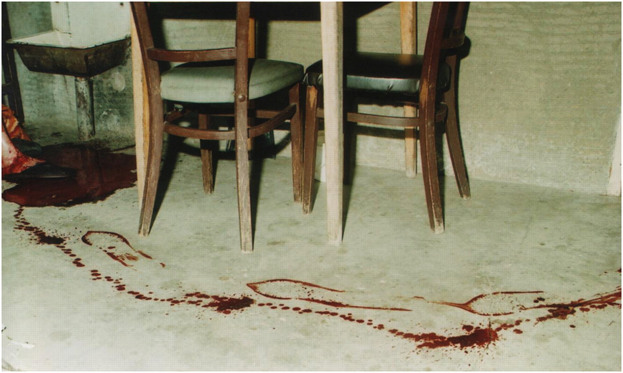
Death scene: noticeable bloodstain pattern on the porch floor in the shape of right foot steps and gushing. Feet of the deceased in the pool of blood can be seen in the upper left corner of the picture
According to Sauvageau et al. 7 only four cases of pulsating haemorrhage from varicose vein rupture have been reported in the literature, including their own case. 8,9 In two of those cases, a tricuspid insufficiency was noticed. It was proposed that tricuspid regurgitation could lead to severe chronic elevation of venous pressure, and transmission of the central venous pulse to the superficial veins.
Veins in the lower limbs contain valves that regulate the one-way flow of blood towards the heart. Backflow in these veins is stopped as the flaps of the valves fill with blood and block the vessel. The position of the valves also facilitates blood flow in the proper direction during venous compression. When a person stands up, contraction of the skeletal muscles of the legs compresses the deep veins of the legs and assists the flow of blood towards the heart. 10
The pulsating gushing haemorrhage from varicose veins rupture is produced by muscle pump: each muscle contraction of the calf during walking compresses the deep veins of the legs and pushes the blood from varicose vein rupture in the form of gushing, mimicking arterial haemorrhage, especially in cases of anatomic or functional valve insufficiency of anastomoses between superficial and deep leg veins. In our case, each time the right leg was on the floor during walking and where the right foot blood print was found, the calf muscles were contracting, supporting the body and pushing blood forward and out from the varicose vein rupture, leaving a gushing bloodstain pattern near the inside part of the blood print. Between each right foot blood print, the line of solitary blood drops could be seen on the porch floor. In those moments, the body was supported by the left leg, the right leg was lifted, the muscles of the right calf were in state of relaxation, and the muscle pump was not pushing the blood from veins forward, so the blood from the varicose vein rupture was just leaking out, leaving on the floor blood stains in the form of a line of solitary blood drops. Thus, tricuspid regurgitation, consequent chronic elevation of venous pressure and transmission of the central venous pulse to the superficial vein were probably not the cause of the pulsating gushing haemorrhage from varicose vein rupture.
The characteristic rhythmical, pulsating, gushing haemorrhage from varicose vein rupture is not produced by tricuspid regurgitation: this rhythmical blood gushing does not correspond with heart beat rhythm and tricuspid regurgitation. On the other hand, it corresponds with the rhythm of one's walking and the rhythmical action of the calf muscle pump, and tricuspid regurgitation and elevation of venous pressure could be considered as a necessary, but not sufficient, condition for gushing.
Footnotes
ACKNOWLEDGEMENT
This study was supported by the Ministry of Science and Technological Development of the Republic of Serbia, Grant no. 45005.