
Abstract
Aims
The aims of this study were (A) to investigate whether the number of years of forensic experience affected the accuracy with which forensic experts (FEs) were able to age bruises and (B) to identify the properties and colours of a bruise that were utilized by FEs in their assessment of bruise age. The study then investigated the possibility of using a more objective technique. It was decided to use readily available digital photography and software to objectively assess changes in bruise colouration and to investigate if this can be used to age bruises.
Methods
Twenty-three FEs were shown 25 photographs of bruises of varying but known ages and asked to estimate the ages. In part two of the study, bruises were inflicted on volunteers using a vacuum pump and photographs taken of the bruise daily from infliction to resolution. The images were analysed using Adobe Photoshop®. Red, green and blue (RGB) values were recorded for each bruise and analyses carried out comparing the values over time between subjects and within subjects.
Results
This study both enhanced and supported a previous conclusion that visual assessment of photographs is an unreliable method for ageing bruises. Additionally, it found that the degree of forensic experience had no effect on accuracy. It also identified that colour (particularly yellow, red and purple) and intensity of colour were the most commonly used properties of a bruise in the assessment of its age. The RGB method proved to be a reliable technique with which to measure bruise colour, but its validity in the assessment of bruise age was poor.
Conclusions
Visual assessment of bruises is unreliable and the accuracy of ageing was not improved by the degree of forensic experience. The RGB method gave highly reproducible results, but did not accurately assess bruise age. However, results within subjects suggested that there may be individual variation in haemoglobin metabolism.
Introduction
Physicians and surgeons are frequently asked to assess bruises and give an opinion as to when the injury was inflicted. Non-accidental injuries, especially in children, and injuries allegedly inflicted while in police custody, are two main areas of interest when assessing bruise age due to the legal implications in such cases. Research into whether an accurate assessment of bruise age can be made is therefore of particular importance to forensic medicine, as the accuracy of such evidence in a court of law could potentially be used to convict an innocent suspect, or indeed release a guilty suspect back into society.
A bruise or contusion is the release of blood into the skin or subcutaneous tissues due to a blunt impact that does not break the skin. 1,2 The force of the impact tears the blood vessel and as the blood leaks into the surrounding tissue the body mounts an inflammatory response that allows for the degradation and removal of the blood. 3 Macrophages are recruited to the site of injury where they break down the oxygenated (red) haemoglobin into two degradation products, biliverdin (green) and bilirubin (red/orange or yellow depending on whether it appears as powder or a biological solution). The visible colour change in bruises is attributable to the formation over time of these breakdown products. Forensic textbooks often describe the order in which colours appear in a bruise, and the time frame in which these colours will appear and disappear. There are, however, inconsistencies concerning the time at which each of these colours will appear, and even in the order of the colour change.
Many studies have been carried out investigating the ability of forensic or medical experts to accurately assess the age of a bruise visually, but the consensus is that this is a highly inaccurate method and should therefore not be relied upon in court. 4–7 A study by Langlois and Gresham 8 provided one of the only concrete pieces of evidence regarding the colour change of bruises over time. It concluded that if a bruise contained any yellow colour it must be older than 18 hours, although the absence of the colour yellow did not necessarily dictate that a bruise would be younger than 18 hours. However, further studies showed that there was considerable inter- and intra-observer variation in the ability to perceive colour and in the description of colour. 9,10 Therefore, it would seem sensible to focus on the objective assessment of bruise age, although this area is still poorly covered in terms of scientific research. A study by Thornton and Jolly 11 looked at the histopathological data from ovine bruises, and found that they could place the bruises with an acceptable degree of accuracy within either 1–20 hours or 24–72 hours. However, this information would be of limited use from a legal perspective, and it would be preferable to find a less invasive technique. Spectrophotometry is a possible option for non-invasive objective assessment of bruise age, and pilot studies into the use of reflectance spectrophotometry have produced encouraging results in demonstrating the degradation of haemoglobin in bruises. 12 A recent paper used a spectrophotometer to examine 86 accidental bruises in children: the time at which each of these bruises occurred was known and could therefore be compared with the spectrophotometry results. 13 A characteristic pattern was observed in 21 of the bruises, but there was a very high degree of variability. It therefore seems unlikely that this technique will be useful in the accurate assessment of individual bruises, although further research using this method may provide more accurate assessment of bruise age in the future. However, the spectrophotometry technique involves expensive specialist equipment that may not be readily or widely available, and is therefore not ideally suited for general and widespread use. As previously mentioned, haemoglobin and its degradation products appear to be responsible for the colour change over time of a bruise, and it would therefore seem appropriate to investigate whether we could identify other, less technically complex, methods that focus on an objective assessment of this change in colour, and which could be easily and routinely used in general forensic practice.
Previous studies have been carried out into the accuracy of visual assessment of bruise age, and it is generally agreed upon that this is an unreliable method of ageing bruises. Variables such as the age and sex of the forensic experts (FEs) have been taken into account, 14 but have been found to be irrelevant parameters in the ability of the expert to assess bruise age. The level of forensic experience, however, has not been explored in previous studies, and the first part of this study was therefore dedicated to assessing the accuracy of visual assessment of bruise age, focusing on forensic experience as a variable with the potential to alter accuracy. In addition, the study investigated the colours and properties of a bruise that are considered useful to FEs in the assessment of bruise age. We also investigated the possibility of identifying a more objective method of assessing bruise age. By identifying a technique that uses inexpensive and widely available equipment, e.g. digital cameras and common imaging software, this study aimed to provide a method for ageing bruises with the potential for widespread and routine use in general forensic practice.
Materials and methods
Visual assessment of bruise age by FEs
A group of 28 FEs was recruited from three groups: (1) qualified clinicians, nurses and paramedics attending the Diploma of Forensic Medical Sciences course at Barts and the London School of Medicine and Dentistry; (2) forensic medical examiners and police involved in forensic work attending the Human Identification course at Hendon Metropolitan Police Training Centre; and (3) members of the Essex Medical and Forensic Services (EMFS), an organization providing medical and clinical forensic services to the police. The participants, who agreed to take part in the study, were provided with a data collection form and shown photographs of bruises of known but varying ages. They were then asked to estimate the age of each bruise. The participants were asked further questions in which they identified the properties and colours of the bruise that were useful to them in ageing bruises, and they were then asked to rank these properties in order of relevance to the assessment of bruise age.
Objective assessment of bruise age using digital photography and Adobe Photoshop®
A vacuum pump attached to a 60 mL syringe barrel was used to produce bruises on the outside of the upper arm of white Caucasian volunteers using the method described by Pilling et al. 14 The participants applied the syringe nozzle to the area of skin and the pressure was reduced over five minutes to −600 mmHg. The participant held the nozzle in place for a further 10 minutes so that suction was applied for a total of 15 minutes to create the bruise. There was a resting period of 15 minutes to allow for any immediate swelling to subside and a photograph was then taken of the bruise under standardized conditions using a Canon EOS 350D digital SLR camera (settings: exposure time 1/200 seconds; ISO speed ISO-100; aperture f/8; focal length 35 mm) with a ring flash. The camera was mounted on a tripod and photographs were taken in an area away from any window to reduce the effect of natural light on the bruise colour. A second bruise was produced on the contralateral arm of the participant at the same time to allow for intra person comparison. The participants were asked to return to the laboratory on each of the following days until complete resolution of the bruise. On each day, a photograph of the bruise was taken under the same standard conditions. All photographs included a photographic colour scale (large greyscale and colour separation guide, product code DANE002, Colour Confidence Birmingham, UK) that was customized so that each of the eight colours was 1 cm in length, to allow for size and colour comparison. The images were downloaded both as JPEG and RAW (TIFF) files with a view to assessing the variability between the raw and processed images (Figure 1). A decision was later made not to use the raw data as these files were considered too large for most speedy analysis using Photoshop and the available PC. Images used for forensic purposes are commonly stored and downloaded as JPEG files and the aim of the study was to investigate a method that could potentially be used in routine forensic practice. The images were standardized by applying black-and-white set points according to the black-and-white blocks on the colour chart. However, when visually compared with one particular image (chosen randomly to be the standard image for comparison), the colour scales on many images were sometimes not identical. The images were then matched using Adobe Photoshop CS4 to alter the brightness of the image until it was deemed to be identical to that of the chosen standard image. Once standardized, an outline was drawn around the bruise using the magnetic ‘lasso’ tool and the width, height and number of pixels within this outline were recorded to help ensure all photographs of the same bruise had an outline with approximately the same dimensions and area. The red, green and blue (RGB) values of each bruise were then recorded. Values for the width, height and number of pixels were reported in the ‘info’ box of the software and the RGB values were in the ‘histogram’ section under the heading ‘channel’.
Statistical analysis
All data were recorded and analysed using StatsDirect (
Results
Visual assessment of bruise age
Twenty-eight FEs were recruited for this study, but five were excluded from further analysis. In two cases this was due to the participant failing to estimate the age of one of the bruises, while the other three participants were excluded for giving non-specific estimations of bruise age, i.e. ‘<18 hours’ or ‘>18 hours’. The results from the remaining 23 participants were analysed.
All 23 participants identified the colour of the bruise as the most important factor in their assessment of bruise age: 17 said that they considered the intensity of the bruise colour a useful property while six identified the size of the bruise as a useful indication of the age of the bruise. Six participants identified other factors as being useful to them in the assessment of bruise age including swelling and pitting, texture of the skin, surrounding erythema, a distinct edge to the bruise and having a ‘gut impression’. The most important colour in the assessment of bruise age was identified as yellow, followed by red and then purple. Blue, green and brown were considered to be relatively less useful to the FEs in their assessment of bruise age.
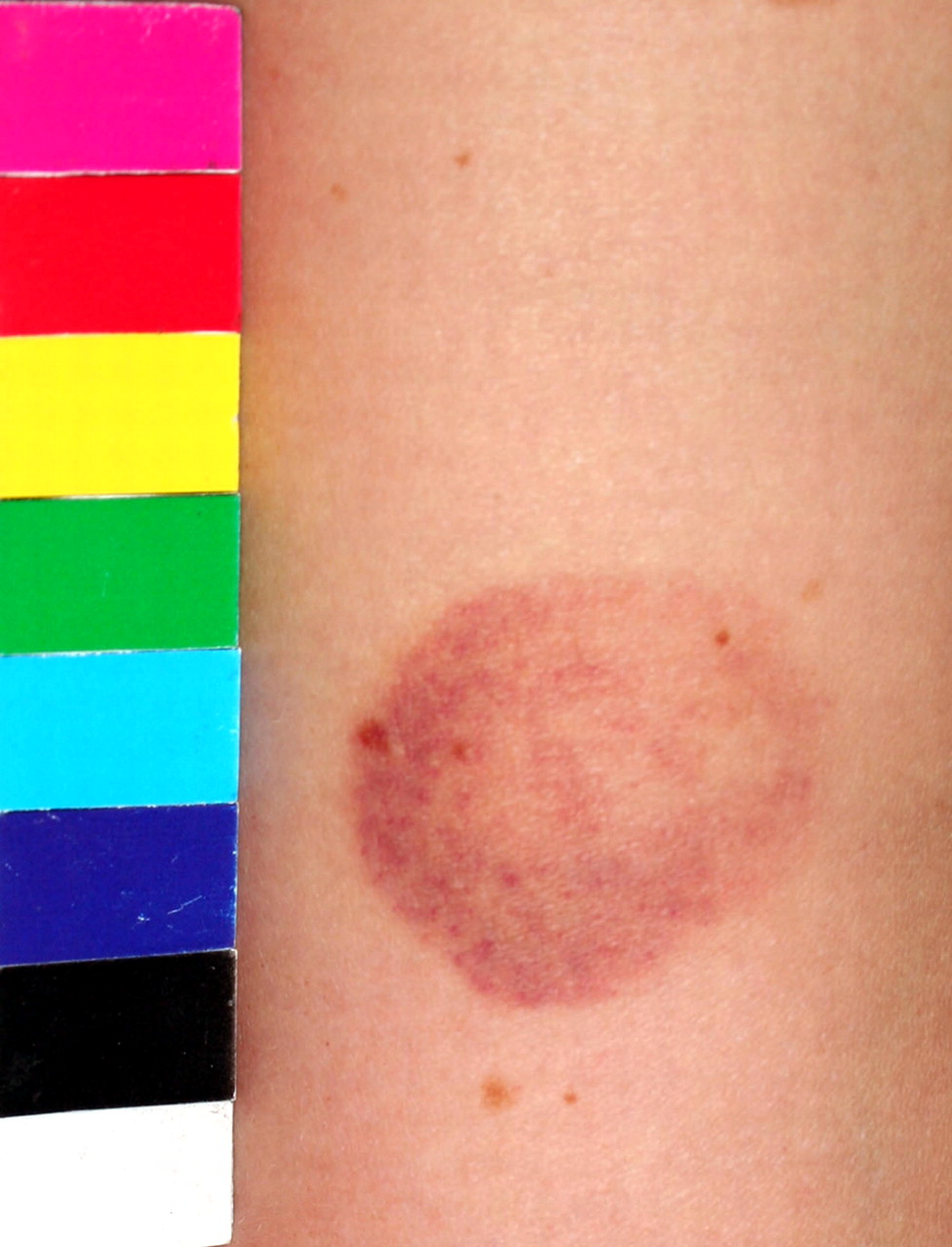
Image of a bruise once it has been cropped and rotated
The accuracy with which FEs were able to age bruises was analysed by calculating the median absolute difference (absolute difference = real age − estimated age) for each participant, N.B. this does not take into account whether it is an over or underestimate. The data were compared using box and Whisker charts (Figure 2). The results show that of all the estimations, 251 were underestimates, 320 were overestimates and only four were correct. Figure 2 demonstrates that although the interquartile range and outliers differ between participants, the medians are very similar.
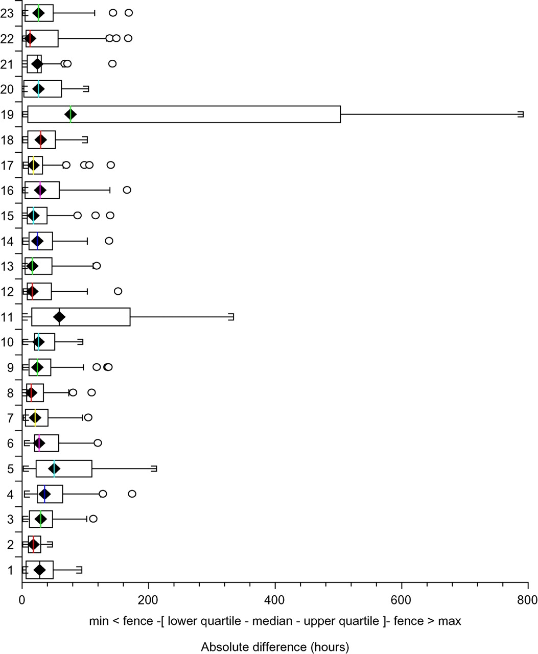
Box and whisker plot comparing the accuracy with which FEs can age bruises
The FEs showed very little accuracy in their ability to assess the age of a bruise from the photographs, which is consistent with results from several other studies that have shown visual assessment of bruise age to be unreliable. However, we also considered that the level of forensic experience may affect an expert's ability to age bruises. Figure 3 shows how the mean absolute difference for each FE varied according to their number of years of forensic experience. The r value of 0.28 suggests that there is very little correlation between the median absolute difference and the number of years of forensic experience.
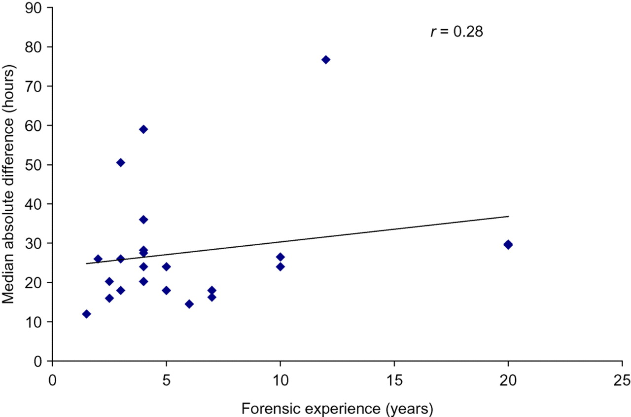
Scatter graph to investigate the relationship between the median absolute difference and the number of years of forensic experience
The years of forensic experience were grouped so that participants fell into one of three categories; 0–2 years, 3–5 years and >5 years (Figure 4). The standard deviation for each of the three groups was 43, 51 and 126, respectively, and the groups were therefore compared using the Kruskal–Wallis test instead of the one-way ANOVA as the data were not normally distributed. The results indicate that an increased level of forensic experience does not improve the participants' ability to age bruises (P = 0.91).
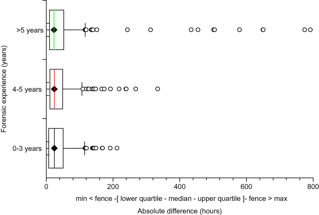
Box and Whisker plot comparing the level of forensic experience with the absolute difference for all bruises
Objective assessment of bruise age using Adobe Photoshop®
RGB values were recorded for each bruise using Adobe Photoshop. In addition, the width, height and number of pixels contained within the outlined bruise were measured, although these were to ensure consistency in the size of each set of photographs of the same bruise. Before bruising, a photograph was taken of the area of skin on which the bruise was inflicted with a view to removing the effect of the underlying skin colour from the colour of the bruise. However, during the course of this study this did not prove useful as simple subtraction of RGB skin values produced erratic colouration that bore little relation to the true bruise, with the result that the actual RGB values could not be directly compared between individuals.
On analysis, it was noted that the degree of photographic exposure could vary and it was found that several photographs were overexposed and a few were underexposed. While it was possible in some cases to compensate for this by altering the brightness during the standardizing process, some images could not be standardized to an acceptable level and this was noted alongside the data. All images were analysed, but for the purpose of comparison only bruises that consisted of well-standardized images were selected. A total of 16 bruises were used for comparison. The red values for each of these 16 bruises were plotted against the age of the bruise, and the same was carried out for the green and blue values. The graphs were then observed to look for any trends. The bruises were labelled by number and with either an ‘L’ or an ‘R’ representing whether the bruise was inflicted on the left or right arm.
All three graphs showed an overall increase in RGB values over time, although the overall increase in the blue value appears to be less than that of the red and green values. However, the individual trend for each bruise over time was highly variable and there was found to be no relationship between the change in RGB value and the age of the bruise. Figures 5a, b and c show how the RGB values of each bruise changes over time, respectively.
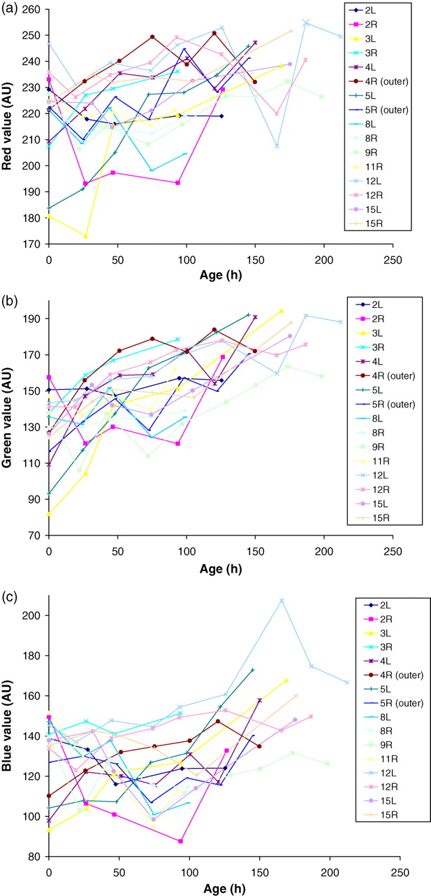
(a) Change in red value of each bruise with time. (b) Change in green value of each bruise with time. (c) Change in blue value of each bruise with time
To allow for intraperson comparison, participants were bruised on both arms. The results are shown in Figures 6a–c. There was a very close association between the RGB colour changes of the two bruises over time. There was a complete loss of association between the blue values at 165.5 hours, and the red and green values at this age also deviated further from those of bruise 12R. However, by referring to the original data we found that this image was noted to be ‘very overexposed’ and therefore unreliable.
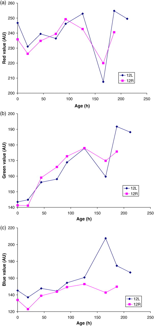
(a) The change in red value of two bruises from the same subject with time. (b) The change in green value of two bruises from the same subject with time. (c) The change in blue value of two bruises from the same subject with time. AU = arbitrary units
Discussion
Visual assessment of bruise age
The properties of a bruise that were most useful to FEs in the assessment of bruise age were identified as the colour and intensity of colour, and in particular the colours yellow, red and purple. Red was generally stated to be indicative of a fresh bruise (due to the presence of haemoglobin?) while studies have shown that the presence of yellow indicates the bruise must be at least 18 hours old. 8 In their role as FEs, it is likely that many of the participants will be aware of these facts and it is therefore not surprising that these colours were identified as relevant. A focus of this study was on the extent to which increased forensic experience might improve accuracy in visual bruise assessment by FEs. In general, there was, as previously reported, 14 very poor accuracy in the precise identification of bruise age. Participants were more accurate in ageing fresh bruises with all outliers as overestimates rather than underestimates.
The level of forensic experience was investigated as a possible factor in the FEs' ability to age bruises. Other parameters, such as age and sex of the observer, had been found to be irrelevant in a previous study. 14 The present study found that forensic experience had no bearing on the accuracy of the FEs' assessment of bruise age; the most inaccurate FE had 12 years of experience whereas the majority of participants had less than six years experience. Although this study found that more forensic experience does not improve the accuracy with which the experts were able to age bruises, it would appear that having a greater level of experience, coupled with appearance as a court expert, meant that such participants were not willing to attempt to estimate bruise age based on visual assessment, an encouraging finding considering the outcome of this study.
One problem with quantifying the level of forensic experience was that the study questionnaire did not specify the type of forensic experience. Participants recruited from EMFS specified their forensic experience as the number of years for which they had worked for EMFS. However, those from the Forensic Medical Sciences course stated their forensic experience as the number of years they had been qualified as a doctor. It would have been preferable to place participants in groups according to their job title as well as their level of FE, but this would have left too few in each group to reach statistical significance.
This study found that the most useful property in the assessment of bruise age was colour, closely followed by the intensity of colour. However, simple visual assessment is not a suitable or reliable method for the assessment of bruise age, and indeed assessment was unrelated to forensic experience; furthermore, the major impact of experience was for the FEs to be even more sceptical regarding the value of visual assessment.
In order to attempt to provide a more objective assessment, a photographic technique was validated to age bruises. The RGB values were recorded for each of the bruises and the individual RGB values then plotted to compare how one value changed over time for each subject. Over the course of the study no suitable method was identified to remove the effect of underlying skin colour, so the absolute change for each of the RGB values was compared over time across subjects. Assuming the underlying skin colour remained the same this would remove any effect of skin colour, thereby allowing direct comparison between subjects. The RGB results showed no association between the age of the bruise and the RGB value for bruises inflicted on different subjects. However, the RGB values recorded over time for bruises inflicted on a single subject (one on each arm) showed a markedly similar trend. That there is such strong intra-person association is of particular significance in view of the complete lack of association between subjects. The intra-person association suggested that the lack of inter-person association is due purely to the bruises themselves, and is not inherently an unreliable method. It also suggested that while the change of colour of a bruise may differ from one person to the next, it may be consistent within a person. This may be due to individual rates of haemoglobin degradation, resulting in the production of bilirubin and biliverdin at varying rates. If this is the case, it would indicate that further studies into the ageing of bruises using the objective assessment of colour are likely to be of little value, as the premises on which these techniques are based are not valid.
The RGB values for each bruise were plotted on one graph to demonstrate how the ratio between the three values changed over time. The results showed that the ratios between the RGB values remained fairly constant over time, and as one value changes the other two mirror this change to a similar degree. This suggests that the colour of the bruise is not affected by the ratios between the RGB values, but by the saturation or strength of the colours.
The major problem encountered during the study was the effect of the photography on the bruise colour. Several bruises were excluded from comparison due to over- or under-exposure, while many of the remaining images were altered to standardize the colour charts and in this process human error may have affected the outcome. The effect of skin colour was removed by observing the real change between values over time. However, this did not take into account the fact that skin colour does not remain constant. The filling of cutaneous and subcutaneous capillary vessels is important in determining skin colour, and this varies in response to many physical and pathological changes. 15 The most obvious of these is temperature change, which may have been a factor in this study although the studies were performed in an air-conditioned room.
The RGB method proved to be a reliable method with which to measure colour, demonstrated by inter image and intra person reproducibility studies. Both produced very low values for the coefficient of variation, independent of the age of the bruise. The validity of the method in the ageing of bruises was found to be poor, although this may be due to interpersonal variation in the metabolism of haemoglobin. If such variation exists, the only way in which a bruise age could be assessed would be to produce another bruise on the victim and observe the colour change. However, this is neither practical nor ethically acceptable in children, but could form the basis of a further study in adults.
In conclusion, these studies have confirmed the unreliability of expert visual assessment of bruise age, and that RGB analysis of JPEG images corrected for colour balance did not assist with determining the age of bruises. However, the possibility for objective assessment of colour for the purpose of ageing bruises has not been completely rejected owing to the comparable changes in bruise colour seen within subjects.
The study was supported by funding from Barts and the London School of Medicine. The authors declare that they have no competing interests.