
Abstract
Personal identification using DNA typing of formalin-fixed tissue is very important in the forensic sciences. However, few studies have been conducted to determine the detection limit of DNA typing of formalin fixation time in samples using the AmpFℓSTR® Identifiler® PCR Amplification Kit (Identifiler Kit). We collected samples from five cadavers submitted for forensic autopsies, and fixed them either in a 10% formalin solution, or in a 10% neutral-buffered formalin solution. The amount of template DNA for polymerase chain reaction (PCR) amplification and the detection limit of DNA typing for the Identifiler Kit were determined. When tissues were fixed in 10% formalin, 10 ng of DNA template was required for successful genotyping even after three-hour fixation and 100 ng was required after one-week fixation for PCR amplification. However, when tissues were fixed in 10% neutral-buffered formalin, the required amount of DNA template was 1 ng for a fixation time of three hours to three days and 125 ng for three months. Fixation time in neutral-buffered formalin was longer for successful PCR than that in formalin solution. Dropout was more common with increasing formalin fixation time. These results suggest that neutral-buffered formalin is preferred to formalin for fixation of tissues if they are to be subjected to DNA typing and that tissues fixed with neutral-buffered formalin can be used for DNA typing using the Identifiler Kit unless the fixation time exceeds one month.
Introduction
Personal identification using DNA typing of formalin-fixed or formalin-fixed, paraffin-embedded (FFPE) tissues is one of the important identification methods in the forensic sciences. We have developed a DNA identification method using FFPE tissues to prove that lymph node samples collected from patients with early gastric cancer with lymph node metastasis are not misidentified with other patients' samples. 1 In this method, we used and demonstrated the usefulness of the AmpFℓSTR® Identifiler® PCR Amplification Kit (Identifiler Kit) (Applied Biosystems, Foster City, CA, USA), which enables DNA typing of 15 short tandem repeat (STR) loci and gender determination with the amelogenin gene. In forensic medicine, formalin and buffered formalin solutions are most commonly used for the preservation of excised organs, as they provide good histological preservation. However, formalin fixation has been shown to complicate DNA testing through the formation of protein-nucleic acid cross-linking in the tissue and DNA fragmentation by formic acid resulting from the oxidation of formaldehyde. 2–4 It has also been demonstrated that the detectable size of DNA fragments decreases in proportion to the duration of formalin exposure. 5,6 Therefore, whether or not DNA testing of a formalin-fixed sample is successful depends on the duration of formalin exposure.
A kit capable of genotyping 13 STR loci, whose use was recommended by the Federal Bureau of Investigation in 1997, is currently used for DNA identification in forensic laboratories and investigative organizations around the world. 7 The Identifiler Kit, which is commonly used in Japan, allows for DNA typing of 15 STR loci and gender determination with the amelogenin gene. However, few studies have been conducted to determine the detection limit of DNA typing of formalin fixation time using the Identifiler Kit. Formalin-fixed tissues are also useful for forensic investigations where archived pathology samples are often the only reference source of DNA available for accurate identification. We have also noted that DNA typing of FFPE tissues using the Identifiler Kit requires an increased amount of DNA templates for polymerase chain reaction (PCR) than its use in the absence of formalin. 1
In the present study, we collected liver, kidney and spleen samples from five cadavers submitted for legal autopsy, fixed the samples in either 10% formalin or 10% neutral-buffered formalin solution for variable times ranging from three hours to three months, and determined, using the Identifiler Kit, the amount of template DNA for successful PCR amplification and the detection limit of DNA typing. We also examined the usefulness of column purification in some samples.
Materials and methods
Samples and formalin fixation time
Liver, kidney and spleen samples were obtained from five cadavers subjected to legal autopsy at the Department of Legal Medicine, Graduate School of Medicine, Chiba University, Japan. Samples were processed into 2-cm3 pieces and fixed either in 10% formalin solution for three hours, six hours, 12 hours, 24 hours, three days, one week, two weeks, one month or three months, or in 10% neutral-buffered formalin solution for three hours, six hours, 12 hours, 24 hours, three days, one week, two weeks, one month or three months. Samples cryopreserved at −30°C before DNA extraction were used as control samples.
Sample processing, DNA extraction from fixed samples and column clean-up
Formalin-fixed tissues were washed under running water for one day and then rinsed in phosphate-buffered saline for one day. DNA was then extracted from approximately 25 mg of each sample using the QIAamp® DNA Mini Kit (QIAGEN, Hilden, Germany). The concentrations of extracted DNA were measured using an ultraviolet spectrophotometer (BioSpec-1600, Shimadzu, Japan). DNA extracted from the spleen samples fixed in 10% neutral-buffered formalin solution for three months or in 10% formalin solution for three days or one week were further purified with BD Chroma Spin™ columns (Takara Bio Inc, Shiga, Japan) to remove degraded or degenerated DNA.
Determination of effective DNA concentration by PCR amplification of the amelogenin region
The amount of DNA template required for PCR amplification using the Identifiler Kit was determined by PCR amplification of a part of the X-Y homologous gene amelogenin. PCR primers with the following sequences were used: 5′ CCCTGGGCTCTGTAAAGAATAGTG 3′ (Amel-A) and 5′ ATCAGAGCTTAAACTGGGAAGCTG 3′ (Amel-B). 8 PCR amplification was performed in a 50 μl reaction mixture containing 1–200 ng of the extracted DNA as a template, 0.2 µL each of the primers, 0.2 mM of each dNTP and 1.25 U of AmpliTaq DNA polymerase (Applied Biosysitems), with a thermal cycler (GeneAmp PCR System 9700, Applied Biosystems) using 35 cycles of 94°C for one minute, 60°C for one minute and 72°C for one minute. PCR amplification was confirmed by 2% agarose gel electrophoresis. The amount of DNA that resulted in sufficient PCR amplification of the amelogenin region was regarded as the effective DNA template concentration for PCR amplification using the Identifiler Kit.
PCR amplification, electrophoresis and data analysis
PCR amplification was performed with the effective DNA concentration, which was determined by PCR amplification of the amelogenin region, as a template using the Amplification Kit according to the manufacturer's instructions. PCR was performed with 0.5 µL of AmpliTaq Gold DNA Polymerase, 10.5 µL of PCR reaction mix and 5.5 µL of a primer set, using a programme of 95°C for 11 minutes, followed by 28 cycles of 94°C for one minute, 59°C for one minute and 72°C for one minute, and then 60°C for 60 minutes, on the GeneAmp® PCR System 9700 Thermal Cycler. The following STR loci were amplified: D8S1179, D21S11, D7S820, CSFIPO, D3S1358, TH01, D13S317, D16S539, D2S1338, D19S433, vWA, TPOX, D18S51, D5S818, FGA and Amelogenin. PCR products were subjected to electrophoresis using an ABI PRISM 310™ Genetic analyser (Applied Biosystems, Foster City, USA). Analysis of electrophoresis results and allele analysis were performed using data analysis software Gene Mapper™ ID (version 3.2, Applied Biosystems). The threshold for allele peak height was set within a range from 150 to 6000 RFUs, and the same threshold was used for homozygotes and heterozygotes.
Results
PCR amplification and DNA typing using the Identifiler Kit with the effective DNA concentration as determined by PCR amplification of the amelogenin region revealed that the amount of template DNA required for PCR amplification increased with increasing formalin fixation time. As shown in Table 1, when liver, kidney and spleen tissues were fixed in 10% neutral-buffered formalin, the required amount of DNA template was 1 ng for a fixation time of three hours to three days, 3 ng for one week, 5 ng for two weeks, 20 ng for one month and 125 ng for three months. However, when tissues were fixed in 10% formalin, 10 ng of DNA templates was required even after three-hour fixation and 100 ng was required after one-week fixation. When tissues were fixed for two weeks or more, no PCR amplification was obtained even at higher DNA concentrations. These results indicate that fixation with neutral-buffered formalin is more suited to maintain the effective DNA concentration for PCR than formalin fixation, particularly if fixation could be prolonged. With regard to DNA typing results, regardless of organ type, the peak heights of the loci producing larger PCR products decreased faster with increasing formalin fixation time than those producing smaller PCR products, resulting in more significant drop-out. In addition, DNA typing of the loci producing larger PCR products became increasingly less with increasing fixation time (Figure 1). The detection rates of 15 STR loci and Amelogenin in the kidney after different formalin fixation times are shown in Figure 2. When tissues were fixed in 10% formalin, DNA typing was successful for all loci of all the five samples with a fixation time up to 24 hours. With a fixation time of three days or more, however, DNA typing was unsuccessful in some samples. With a fixation time of one week, only D3S1358, D19S433 and Amelogenin were successfully DNA typed in all the five samples.
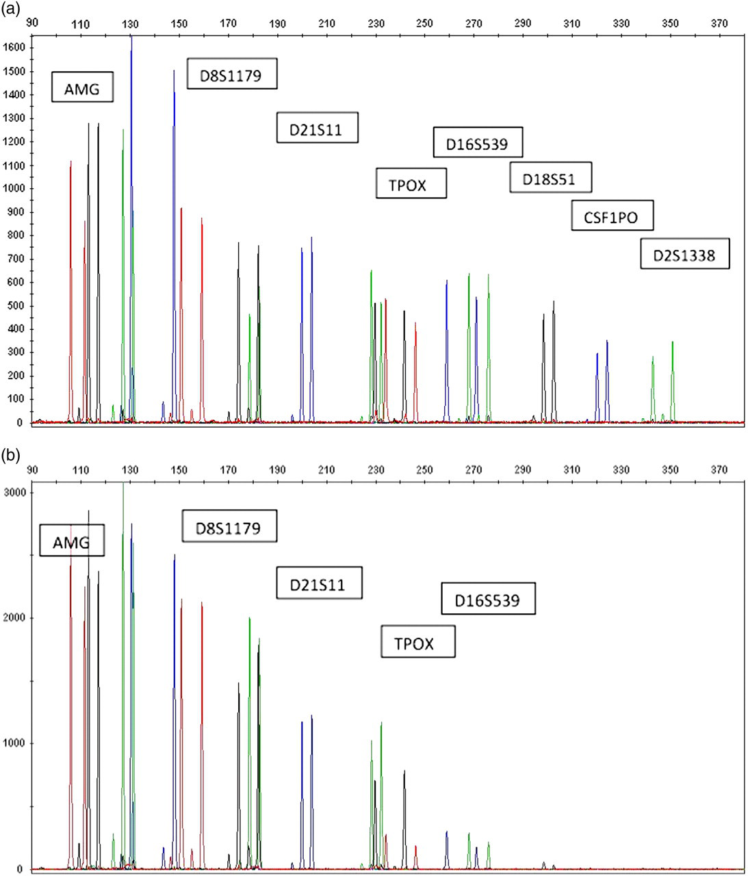
Amplification of DNA typing from (a) non-formalin and (b) three days formalin-fixed kidney samples by Identifiler Kit
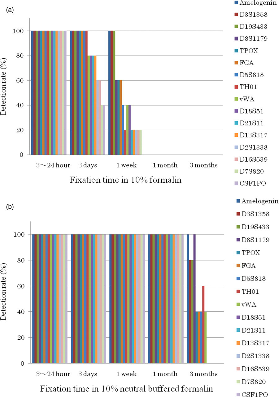
The detection rates of 15 short tandem repeat loci and Amelogenin in the kidney after different formalin fixation times in (a) 10% formalin and (b) 10% neutral-buffered formalin
Amount of template DNAs (ng) required for PCR amplification when DNAs were extracted from tissues fixed in 10% formalin or 10% neutral-buffered formalin
PCR, polymerase chain reaction
*The required amount of DNA was immeasurable because DNAs extracted from tissues fixed in 10% formalin for two weeks or more yielded no PCR amplification products even with increased amount of template DNAs
With a fixation time of two weeks or more, DNA typing was unsuccessful for all loci in all the samples. When tissues were fixed in 10% buffered formalin, as shown in Figure 2, all loci were successfully DNA typed in all the samples with a fixation time of up to one month. With a fixation time of three months, DNA typing was unsuccessful in many loci except for D8S1179 and Amelogenin.
When comparing detection rates between organs, the liver was associated with lower detection rates with shorter fixation times than the kidney and spleen.
Column clean-up of DNA extracted from spleen samples resulted in an increased number of detectable loci, with typing results consistent with those of control samples. Column clean-up of DNA extracted from spleen samples fixed in 10% neutral-buffered formalin resulted in the successful DNA typing of seven loci, namely, D21S11, D3S1358, D13S317, D16S539, TPOX, D5S818 and FGA, compared with five loci before clean-up, namely, D8S1179, TH01, D19S433, vWA and Amelogenin (data not shown).
Discussion
DNA typing of formalin-fixed tissues using the Identifiler Kit was possible with a fixation time of up to one week in 10% formalin or up to one month in 10% neutral-buffered formalin.
The effects of formalin fixation on DNA include degeneration and degradation by formic acid produced from formalin, resulting in a decrease in PCR-amplifiable DNA size. The size of the PCR products generated by the Identifiler Kit ranges from approximately 100 to 400 bps. We thus hypothesized that such DNA may not be affected by formalin if fixation time is short and can thus be DNA typed.
Samples fixed in a 10% neutral-buffered formalin solution contained more loci that could be DNA typed than those fixed in 10% formalin. This suggests that buffered formalin, a fixative resistant to pH drop, is more effective in preserving DNAs than non-buffered formalin. 9
Formalin-based fixatives supplemented with EDTA or buffer agents to prevent DNA degradation have recently been developed to allow for DNA testing of fixed tissues. 10 Reducing the size of the PCR products of the DNA regions of interest for improved detection efficiency has also been studied. 11,12 Studies have also been conducted to improve DNA purification efficiency via the improvement or modification of conventional DNA extraction methods and thereby obtain an effective DNA concentration for PCR amplification. 13–15 Column clean-up is also recognized as an effective method. In the present study, column clean-up of extracted DNAs efficiently yielded PCR products with a size of approximately 100–400 bp, which served as effective template DNAs for the Identifiler Kit, and thus increased the number of loci identifiable by DNA typing. This finding indicates that the use of column clean-up is an effective approach to improve the rate of DNA-type identification.
The Identifiler Kit is a DNA-typing kit for identification purposes that is commonly used around the world, including Japan. This kit is used for DNA typing of various kinds of samples, including fresh blood, old body fluid stains and hard tissues such as bones and teeth. Another commercially available kit of this kind is the AmpFℓSTR® MiniFiler™ PCR Amplification Kit (Applied Biosystems, Foster City, USA), which detects small PCR products of approximately 100–300 bps. This kit generates smaller PCR products from nine STR loci, namely, D13S317, D7S820, D2S1338, D21S11, D16S539, D18S51, CSFIPO, FGA and Amelogenin, than in the Identifiler Kit. In addition, while the minimum amount of template DNA required for PCR amplification using the Identifiler Kit is 1 ng, the MiniFiler kit only requires a minimum of 125 pg of template DNAs for amplification. This kit overcomes the inhibitory effects of heme and humic acid commonly contained in forensic samples and allows for the PCR amplification of degraded DNA samples. These features of the kit may allow for the detection of the STR loci that could not be DNA typed in formalin-fixed samples in the present study. Although the MiniFiler kit is very expensive and currently not as commonly used as the Identifiler Kit, future studies will need to evaluate the usefulness of this kit.
In conclusion, the results of the present study suggest that neutral-buffered formalin is preferred to formalin for the fixation of tissues to be subjected to DNA typing. Tissues fixed with neutral-buffered formalin were sufficiently available for DNA typing using the Identifiler Kit unless the fixation time exceeded one month.