
Abstract
The skeletal remains of five individuals with an unusual postmortem course were discovered in a house. According to the explanation of the putative bereaved family, the postmortem interval of the five remains was between five and 20 years. They also explained to the police that they and the dead family members believed that the dead can be resurrected, and they had kept the bodies indoors, so the bodies had followed an unusual postmortem course. The five dead were identified by kinship analysis using DNA typing. For DNA extraction, we used the DNA extraction method with ultrafiltration and a silica-based DNA extraction kit. As a result, complete amplification STR profiles were obtained from DNA from bone samples of all five skeletons and their identity was proven by kinship testing.
Introduction
The identification of unknown remains of missing persons or victims of mass fatalities 1 is one of the most important roles of forensic science. Traditional techniques, such as fingerprinting 2 and odontology, 3 have contributed to identifying remains. In addition, individual identification methods applying genetic polymorphism in DNA are currently used as very powerful tools, and samples are analysed to compare with DNA profiles from direct reference samples, such as toothbrushes and razors, and/or to carry out kinship analysis with close relatives. 1 The identification of skeletal or highly decomposed remains is carried out by performing short tandem repeat (STR) analysis from hard tissue, such as bones and teeth. 4,5 Various DNA extraction methods from these hard tissues have been reported, and we used the DNA extraction method with ultrafiltration and a silica-based DNA extraction kit.
The skeletal remains of five individuals with an unusual postmortem course were discovered in a house. In order to identify them and their relationship to the three survivors suspected family members, kinship analysis using DNA typing was carried out.
Case report
A social worker reported to the public office that there were several elderly family members that had been missing for a long time. A official and the social worker visited their house, but there was no response. Since they were not able to contact the family after that, they consulted with the police. A few days later, a police officer went into the house and found the skeletal remains of five individuals there. At the time of entry, the house was locked up and the shutters and the windows were closed. The remains were already skeletonized, and were located separately, face up, on Japanese futon mattress. The investigators designated them A, B, C, D and E, respectively, based on the position of their bodies (Figure 1).

A schematic diagram of the house where the five dead people were found
The police investigation revealed that the five skeletal remains were from one family: two parents and three children. Three other children were alive: the first son, and the third and fourth daughters of the family, who lived in another house. They explained to the police that they and their dead family members believed that the dead can be resurrected, and they had kept the bodies indoors, so the bodies had followed an unusual postmortem course. According to their explanation, it was supposed that person A was the family's first daughter; person B, the father; person C, the second son; person D, the mother; and person E, the second daughter. Furthermore, they explained that the postmortem interval (PMI) of person A was 17 years; person B, 20 years; person C, five years; person D, 10 years; and person E, eight years. The family pedigree is presumed as shown in Figure 2.
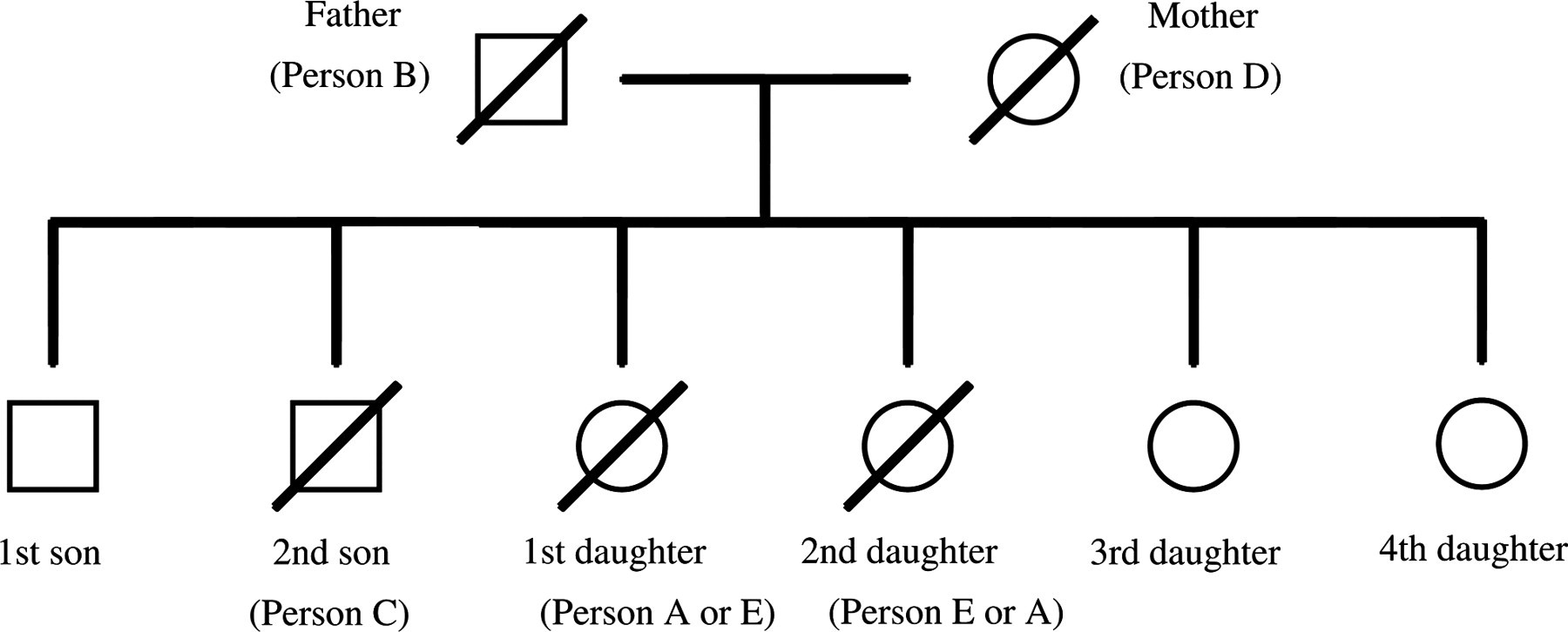
Genealogical tree of the family who underwent kinship analysis in this study. □: ♂; ○: ♀; /: deceased
Autopsy findings revealed no marked injuries to any of the five skeletal remains so their cause of death remained unknown. Morphological examination revealed their suspected gender, age, height and so on. It was also suspected that the time after death was three years or more.
Materials and methods
STR analysis
Samples
Oral mucous swab from the three living children and femur bone samples from each skeleton were collected to use as DNA sources.
DNA extraction from bone samples
The diaphysis of each femur bone was cut to about 2 cm × 4 cm with an autopsy saw, No. 810 (Stryker, Kalamazoo, MI, USA). The bone fragment was ground away to remove the external layer and bone marrow with PAL, a grinder for dental technicians (Morita Tokyo Manufacturing Corporation, Tokyo, Japan) and washed well using detergent. After drying, the bone fragment was roughly crushed with a hammer, and then pulverized with a multi-beads shocker (Yasui Kikai Corporation, Osaka, Japan).
About 1 g of bone powder was decalcified with 0.5 mol/L ethylenediaminetetraacetic acid (EDTA) solution, pH 8.0, and gently agitated in a shaking incubator at 56°C overnight, and for another eight hours with fresh EDTA solution. Following the removal of EDTA solution, and rinsing the decalcified bone powder twice with distilled water, it was rinsed once with TNE buffer (10 mmol/L Tris-HCl, pH 8.0, 1 mmol/L EDTA, pH 8.0 and 10 mmol/L NaCl). Following the removal of the TNE buffer, extraction buffer (3 mL TNE, 150 µL Qiagen Proteinase K [Qiagen, Hilden, Germany], and 150 µL of 10% sodium dodecyl sulphate) was added and incubated at 56°C overnight in a shaking incubator with gentle agitation. An equivalent volume of phenol was added to the digested bone sample and rotated slowly for an hour. After separation of the water layer (ca. 4 ml) and phenol layer by centrifugation, the water layer was transferred to an Amicon Ultra-4 Centrifugal Filter Device (Millipore, Billerica, MA, USA) and concentrated to 200 µL by centrifugation following the manufacturer's recommendations. 6 DNA isolation was carried out with a procedure for dried blood spots using a QIAamp DNA Mini Kit (Qiagen) as described by the manufacturer. The DNA was eluted with 50 µL of distilled water.
DNA amplification and fragment analysis
DNA amplification was conducted using an AmpflSTR Identifiler Kit (Applied Biosystems, Foster City, CA, USA) on a GeneAmp PCR System 9700 (Applied Biosystems) using the following conditions: 95°C for 11 minutes; 28 cycles at 94°C for one minute, 59°C for one minute, 72°C for one minute and a final extension at 60°C for 60 minutes. The reaction volume was 25 µL, and 10 µL DNA extract was added to the polymerase chain reaction mixture. The amplified STR fragments were separated by capillary electrophoresis on a 3130xl Genetic Analyzer (Applied Biosystems). STR typing was performed using GeneMapper ID version 3.2 software (Applied Biosystems).
Kinship analysis with suspected parents and statistical calculations
According to the explanation of the alleged relatives, the five dead were a father, mother and three of their children. Among the five dead, one whose amelogenin locus was ‘XY’ was considered to be the father, and one whose amelogenin locus was ‘X’ was considered to be the mother.
Likelihood ratio (LR) values were calculated with Patcan v.1.2 7 software using Japanese population data of 15 STR loci, as described by Yoshida et al. 8
Results and discussion
STR analysis
The STR profiles of the three living people believed to be children of the family are shown in Table 1. STR analysis was also performed using DNA from all five skeletal remains. We were able to obtain a full profile from all extracts, including the DNA from person B, who had been lying in the house for a suspected 20 years. The electropherogram of the amplified STR product obtained from person B showed a marked tendency for the peak height of the long amplicon loci to be markedly lower than that of the short amplicon loci. This phenomenon indicated that the DNA from the bone had degenerated as a result of postmortem changes.
STR profiles obtained from bones of five dead and three living people
In the present case report, we used the DNA extraction method reported by Imaizumi et al. 9 that we had used routinely, with a slight change in the order to gain the high DNA yield and shorten the time for DNA extraction. Various methods such as an organic-based and a magnetic particle-based DNA extraction method were reported, 10 and from among them, we selected a silica-based DNA purification technique with a QIAamp DNA Mini Kit. For applying a QIAamp DNA Mini Kit to our method, we included an additional step using an Amicon Ultra-4 column for ultrafiltration in order to reduce the volume of DNA extract from 4 ml to 200 µL, an appropriate volume for the subsequent DNA isolation step. By replacing the standard phenol/chloroform extraction method with a silica-based DNA extraction, the operation for DNA extraction could be simplified and the loss of DNA yield, which we often faced when performing the phase separation, could be kept to a minimum. It has been established that larger amounts of DNA were obtained with the silica-based DNA extraction method than with the organic-based method; 11 therefore, it is suggested that the silica-based method is more efficient in respect of quantity and speed than the organic-based method for DNA extraction from bone samples. The PMI of the most recent remains was five years and that of the oldest was 20 years; therefore, the remains analysed in this study were relatively aged. DNA degradation is one of the factors making DNA typing difficult; 12 however, we were able to obtain complete STR profiles from all five samples, even the 20-year-old bone specimen with the DNA extraction method presented here, so it is suggested that our method is suitable for aged bone.
Kinship analysis
Considering the results of the amelogenin sex-typing concerning five dead people, there were six possible combinations of parents: person B–person A; person B–person D; person B–person E; person C–person A; person C–person D; person C–person E; the former being the putative father and the latter was the putative mother, and 12 possible combinations of family. In the six possible combinations of parents, the combination of person B–person D was the only one that did not produce any inconsistency at all STR loci with the three living people as their children. Moreover, among the five decedents, person B–person D was also the only combination that did not produce any inconsistency at all STR loci with the three dead people, namely person A, person C and person E, as their children. There was no contradiction between these results of the kinship analysis using DNA typing and the three living people's explanation, indicating that their explanation was reliable. Therefore, the parents of this family were believed to be person B as father and person D as mother. Considering that the men missing from this family were the father and the second son, and that the father was already confirmed to be person B, person C was believed to be the second son. Whether person A or person E was the first or second daughter could not be confirmed by DNA typing.
To verify maternity, we calculated the LR for person D and each child being related as mother–child with genotyping data obtained from eight people of the family (Table 2). The LRs for verifying maternity ranged from 2084 to 52,797. Similarly, to verify paternity, we calculated the LR for person B and each child being related as father–child (Table 2). The LRs for verifying paternity ranged from 673 to 81,330. These LR values strongly supported that person D was the mother and person B was the father of this family. Moreover, the genotyping data obtained from STR analysis and the statistical assessments described above suggested that all eight people were related by birth.
Likelihood ratio values to verify maternity and paternity
LR, likelihood ratio
In this case report, the family believed that the dead could be resurrected so they kept their dead family members in their house. A high number of corpses, the skeletal remains of five individuals, therefore had to be identified. With the help of kinship testing, we demonstrated that all eight people were related. In addition to the morphological findings of skeletal remains, we could define the most probable pedigree consisting of two parents and their six children (Figure 2) by amplifying 15 autosomal STRs and amelogenin.
Footnotes
Acknowledgement
We thank Dr Christa Augustin and Dr Mandy Larsen, Institute of Legal Medicine, University Hospital Hamburg-Eppendorf, Germany, for helpful scientific advice in making this report.