
Abstract
The distinctive morphology of the human innominate bone (os coxae) and its clear sexual dimorphism make it of interest from anatomical, anthropological and forensic points of view. The features of the greater sciatic notch of the coxae are characteristic and are commonly used to determine sex in unknown individuals. In this study, several measurements of the greater sciatic notch, e.g. width (AB), depth (OC) and width of the posterior segment (OB) were taken and indices I and II were calculated in 64 adult (32 men and 32 women) and side (right: left) coxae radiography (A-P view). Results indicated that out of all the parameters studied, width of the notch (right and left) (P < 0.001), posterior segment width (right and left) (P < 0.001), right (P = 0.036) and left (P = 0.008) index II of notch were found to be significantly greater in women as compared with men. Discriminant function analysis showed that the accuracy of sex determination varied from 100% in the men and 40% in the women groups to 70% for the total group. These results can be used as an aid to the identification of human skeletal remains in Iranian people.
Introduction
The human skeleton usually demonstrates its gender. The distinctive morphology of the human innominate bone (os coxae) and its clear sexual dimorphism make it of interest from anatomical, bioarcheological and forensic points of view.1,2 This bone is an ideal bone for sex determination because it not only reflects the general differences between the two sexes but also the special adaptation of female bone for child bearing. Those authors who have studied this bone by osteometric methods have paid attention either to features relating to its total size or to those of various components, such as its inferior and superior border, the greater sciatic notch, the symphysial surface, the acetabulum, the obturator foramen, the arcuate line or the distance between defined morphological points on its borders. 3 12
There are metric and non-metric differences in skeletal components among populations and these variations are related to genetic and environmental factors. 1 Environmental factors such as activity, socioeconomic status influence trait expression in human skeleton. Sex characteristics secondarily vary depending on hormones and nutrition and both of these are influenced by the cultural formation of gender roles. All of these together will influence activity patterns and labour roles. 13 Thus each group should have specific standards to optimize the accuracy of identification.
While the pelvis is probably the most accurate bone from which to determine sex, 14 no research has been done on the Iranian pelvis.
The features of the greater sciatic notch of the innominate bone are commonly used to determine the sex in unknown individuals. In this radiographical study, several measurements of the greater sciatic notch, e.g. width, depth and width of the posterior segment were taken in A-P view and indices I and II were calculated.
Therefore, the aim of this paper was to collect metric data from Iranian pelvis to develop standards that can be used to determine the sex of unknown adults.
Materials and methods
The materials used in this research work included 64 radiographs of people belonging to different age groups (19-78) from the Department of Radiology, Loghlman Hakim Hospital. Out of these radiographs 32 were men and 32 were women. This study was approved by the ethical committee of the Medical Ethics committee of Hamadan University of Medical Sciences.
Pelvic radiographs were obtained using the standardized protocol: in 15-30° of internal rotation of the hip in the supine position. All films were taken at the object-film distance of 5 cm and focal film distance of 92 cm in the antero-posterior view, and a magnification correction factor of 2.86% was applied (9).
The radiographs were used to measure the width, depth and posterior segment of the greater sciatic notch.
The definitions of the measurements were taken from the literature and were selected on the basis of their being good discriminators in previous studies. These are clearly defined in the available literature.1,2,14-17
The piriform tubercle was taken as the posterior point (B) and the tip of the ischial spine was taken as the anterior point (A) of the width (AB). Maximum depth (OC) was determined by a perpendicular line between the baseline (AB) and the deepest point (C) of the greater sciatic notch. Also, (OB) was designated as the posterior segment (Figure 1). Index I: Maximal depth (OC) × 100/maximal width (AB) and index II: posterior segment of the width (OB) × 100/maximal width (AB) were also calculated.
Measurement of greater sciatic notch from the antero-posterior X-Rays photographs of the pelvis, (a): tip of the ischial spine (b): piriform tubercle. AB: baseline, OC: Maximum depth, C: deepest point of the greater sciatic notch. OB: posterior segment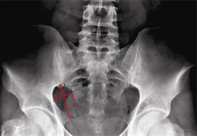
Each variable was measured by the same observer. All linear measurements were in centimeters for each parameter.
To test the results, the mentioned parameters were being measured in a blind trial by another observer. Discriminant analysis was conducted on this data.
Statistical analysis
Multivariate analysis was used to compare the mean vector of variables by sex. To determine which variable causes difference in mean vector, an independent sample t-test was used and a significance level of 0.05 was corrected according to Bonfferoni criteria.
It was accepted that the confidence interval of the difference was to be 99.58% and P values of 0.05 or higher were not significant. The SPSS version 16 program was used for descriptive analysis.
Results
Age and sex of sample used in study

Comparison of various parameters of the greater sciatic notch in both sexes
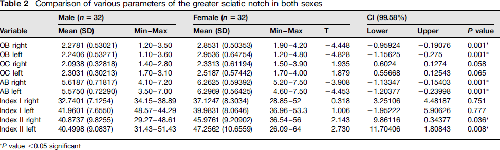
P value <0.05 significant
Comparison of various parameters of the greater sciatic notch on both sides in men and women
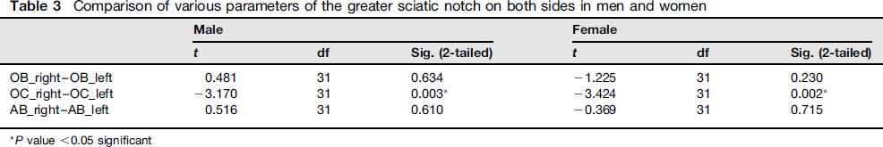
P value <0.05 significant
Correlation of age and all variables in women
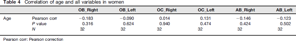
Correlation of age and variables in women

Results of discriminant analysis showed that correct classification of sexing of innominate bone using statistically significant different parameters is 100% for men 40% for women and 70% for total sample.
Using receiver operator characteristic curves, the optimum cut-off point of the index II (47.3) for the correct classification of cases in relation to sex was 90% for men, 70% for women and 80% for all.
Discussion
In this study, several measurements of the greater sciatic notch, e.g. width (AB), depth (OC) and width of the posterior segment (OB) were taken and Indices I and II were calculated in 64 adult (32 men and 32 women) and side (right: left) of the innominate bone radiography (a-p view). Out of all these parameters, width of the notch (right and left) (P < 0.001), posterior segment width (right and left) (P < 0.001), right (P = 0.036) and left (P = 0.008) index II of notch were found to be significantly greater in women as compared with men (Table 2).
Multivariate analysis of comparing mean vector of right OB, left OB, right OC, left OC, right AB, left AB right and left index I and II by gender showed that there is significant difference between mean vectors (Wilks Lambda value = 0.564, df (6,57) and P < 0.001). To determine which variable causes difference in mean vector, independent sample t-test was used which significance level of 0.05 was corrected according to Bonfferoni criteria. The result show the difference is due to right OB, left OB, right AB, left AB and right and left index II (P < 0.001).
Comparison of variables of greater sciatic notch in different studies
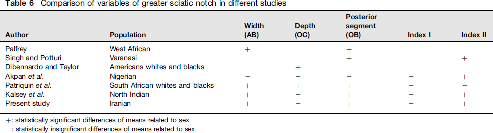
Akpan et al. used X-ray films (a-p view) of the pelvis of adult Nigerians to measure the width, depth and posterior width of the greater sciatic notch. They reported that the width, depth of the greater sciatic notch and Index I were insignificant criteria but that Index II was the most useful criterion in sex determination. 20 Patriquin et al. 21 23 determined the maximal width, maximal depth and posterior width of the greater sciatic notch in whites and blacks. They reported that the width of the greater sciatic notch is larger in women but deeper in men and that there are significant sex differences among South African men and women and whites and blacks.22,23 Steyn et al. 24 used geometric and morphometric analysis of the greater sciatic notch and reported that this feature may not be so reliable, especially in South African white males. Kalsey et al. determined the maximal width, maximal depth and posterior width of the greater sciatic notch in North Indian population. They showed that width, posterior segment width and index II of notch were found to be significantly greater in women as compared with men. 25
Our study is compatible with Palfrey, Patriquin et al, Singh and Potturi and Kalsey et al. Their results show that posterior segment width is a good indicator to detect sex (Table 6).
Singh and Potturi, Akpan et al. and Kalsey et al. as our result also show that Index II has significant sex differences among both men and women (Table 6).
In contrast to almost all the studies mentioned above, the notch was found to be deeper in women as compared to with men. In the present study, however, this difference between the two sexes was statistically not significant (P = 0.058 for right and P = 0.067 for left). Only Kalsey found it to be deeper in women of north India, however his results were not statistically significant.
When all variables were compared with respect to the side, the difference was statistically insignificant in both sexes for width and posterior segment. The depth of the left and right greater sciatic notch, as given in Table 3, was deeper on the left side and showed significant difference in both sexes. In Kalsey's 25 study the difference was significant only in women.
The mean age of the study population was 42.9 years (range, 19-78 years). Correlation between age and sciatic notch variables were analysed statistically. There was no significant difference between age and various parameters in men and women. Walker tested the correlation between age and the greater sciatic notch morphology. He showed that, for both sexes, there is a strong relationship between age and sciatic notch scores. 26 Young people tend to have wider, more feminine-appearing sciatic notches than people of greater longevity. There are also significant population differences. Environmental influences on skeletal development (vitamin D deficiency) appear to provide the most likely explanation for these population differences.
While the accuracy of the measurements of the greater sciatic notch is high, all collections used in these studies are from different populations and one cannot assume the methodology would yield equally high success rates in all of them. Therefore, this study, of data from Iranian collections ensures this technique's applicability to the Iranian population.
Footnotes
Acknowledgements
This research was funded by the Vice Chancellor for research and technology of Hamadan University of Medical Sciences and Health Services.