
Abstract
Femur bone sections from a single donor were exposed for six months to (i) outdoor conditions (exposure to sun, rain, etc.); (ii) water-vapour saturated environment favourable to mould proliferation and (iii) humic-garden soil. Following these treatments, DNA was extracted and yields were compared with that of a control bone fragment kept under optimal laboratory storage conditions. Our results demonstrate that both mould and soil are very detrimental to bone DNA conservation since more than 97% of the bone DNA was lost in these samples as compared with the control condition. Outdoor exposure gives an intermediate result with 30% of the DNA still present in the bone. Thus, environments favourable to microorganisms proliferation appear detrimental to bone DNA conservation and are a bad prognostic should bone remains be used for genetic identification purpose. Comparatively, open-air exposure is much more favourable to bone DNA analysis.
Introduction
Bone DNA analysis is often the last possibility left for identifying remains. The highly mineralized structure of bone, together with its collagen matrix, offers osteocytes a protective environment. The DNA of these cells can remain encased in bone long after death, as the only genetic witness of long bone individuals. For example, nucleotide sequences from bones tens of thousands of years old have been reported. 1 However, it is not rare that forensic or anthropology laboratories fail to extract DNA from bones, even recent ones. Several studies reported the crucial importance of bone conservation conditions on the success or failure of DNA extraction.2,3 The most striking demonstration of the impact of the conservation conditions is the side-by-side comparison of DNA extracted from two fragments from the same exact bone, one having been excavated 60 years ago and conserved in a museum since, while the other fragment had been freshly excavated from the original archaeological site. 4 The authors showed that as much DNA had been lost in the 60 years of museum ‘conservation’ as during the 3200 years of burial. We decided to compare in a strict and controlled manner the influence of multiple classical environments from which bones could be retrieved for DNA analysis. Our results strongly suggest that the conditions associated with biological contamination have the most deleterious effect on bone DNA conservation, since only about 1-2% of the initial DNA is conserved. Open-air exposure also results in a loss of DNA, but to a much lower extent, since 30% of the DNA is still present.
Methods
Bone preparation
A human femur was prepared from a deceased donor. Soft tissues were removed by two six-hour preparations in boiling water, 5 and the bone was further cleansed by brushing under running water. Bone was dried overnight on the bench and sawn transversely to obtain ±4 cm-thick cortical ring fragments.
Bone exposure conditions
Bone fragments were kept for six months, from the June solstice to the December solstice, under the four following conditions:
Control: a bone fragment was kept in the laboratory on paper tissue, in a well ventilated wooden drawer, with no direct exposure to light. The relative humidity of the room was between 40% and 50% and the temperature between 20 and 25°C;
Saturated humidity: a bone fragment was kept in the laboratory, in a plastic container with vents allowing the circulation air and spores present in the atmosphere. A water-saturated atmosphere was maintained by the constant presence of liquid water in the container, but without direct contact with the bone. The temperature of the room was between 20 and 25°C. The container was kept away from direct exposure to light;
Open air: a bone fragment was attached to the east-facing wall of a tower building, 30 m above the ground, behind a protective 4×4 cm nylon net. The cumulative rainfall during the study was 544 mm. The one-month average maximal temperature (July) was 28.9°C, with an absolute maximum of 34.3°C, while the one month average minimal temperature (December) was 5.3°C, with an absolute minimum at 0.3°C. Total sun exposure during this six month period was 1400 hours;
Buried: a bone fragment was buried under 30 cm of well drained garden loam (17% humidity measured at the end of the experiment). This top soil is composed of 35.1% sand, 52.6% silt and 12.3% clay. Its pH is 6.5, and contains a very rich and varied microorganism population. 6 A total of 544 mm of rain was recorded during this period. The one month average maximal temperature (July) was 28.9°C, with an absolute maximum of 34.3°C, while the one-month average minimal temperature (December) was 5.3°C, with an absolute minimum at 0.3°C. The soil temperature at this depth remains very close to the daily average air temperature. 7 The accumulated degree days for the six-month period (calculated as the daily average temperatures added together) is 3415°C.
Bone powder preparation and DNA extraction
The method will be described elsewhere in detail. Briefly, after decontamination by abrasion, the bone was pulverized with a Dremel bit. Powder was collected by aspiration and 200 mg was solubilized by slow rotation overnight at 55°C in an ethylenediaminetetraacetic acid (EDTA) based solution (0.25 mol/L EDTA pH 8.0, 1% Triton-×100, 10 mmol/L 5,5'-Dithiobis(2-nitrobenzoic acid), 250 μg/mL Proteinase K). DNA was then bound to laboratory-prepared silica powder, as described in ref., 8 extensively washed and eluted in water. An isopropanol precipitation was performed to remove ploymerase chain reaction (PCR)-inhibitory activities. All steps were performed under strictly controlled conditions to limit risks of exogenous DNA contamination. Purified DNA was further authenticated by sequencing mitochondrial hypervariable region 1 to rule out any contamination during the extraction process.
PCR conditions
Amplifications were performed on an ABI7000 system, in a final reaction volume of 20 μL. Reactions were as follow: 160 ng/μL bovine serum albumin, 10 μL Eurogentec MESA GREEN qPCR MasterMix 2×, 200 nmol/L final of each primer and 5 μL of diluted template. Cycling conditions compatible with all the primer sets used in this work are: five minutes 95°C (hot start) + (30 seconds 95°C - 60 seconds 64°C) × 45 + denaturation cycle.
DNA quantification
Control DNA extracted from human blood cells was purified and quantitated by spectrophotometry at 260 nm. Several dilutions were measured to precisely determine concentration. Dilutions of DNA were then compared by qPCR using Alu primer set (see below).
Dilutions (5×, 40× and 320×) of bone-extracted DNAs were prepared in water, and 5 μL of each dilution was used for qPCR in a final reaction volume of 20 μL. Our known human reference DNA was prepared at 100, 10 and 1 pg/5 μL in water. Primers for the conserved mammalian mitochondrial 12S sequence, presented in ref., 9 are: Forward: gccaccgcggtcatacgatt. Reverse: gggtatctaatcccagtttgggtcttagc. Amplicon size: 198 bp.
Human Alu primer sequences used are: Forward: gaccatcccggctaaaacg. Reverse: cgggttcacgccattctc. Average amplicon size: Alu being a highly repeated sequence, amplicons are represented by a Gaussian centred on 127 bp.
The potential presence of PCR inhibitors in our prepared DNA was monitored by amplifications on serial dilutions. Linear responses obtained with these dilutions indicated that the samples were devoid of inhibitors.
DNA identification
Human primer sequences used for mitochondrial hypervariable region 1 analysis: Forward (as well as sequencing primer): ccaccatcagcacccaaagct. Reverse: tggccctgaagtaagaaccag. Amplicon size: 517 bp.
Results
The cortical shaft of a freshly prepared human femur bone was separated in sections of similar length (±4 cm, Figure 1). These fragments were exposed for a six-month period between the June and December solstices to the four following environments: (1) control condition (temperature- and humidity-controlled laboratory atmosphere, away from light); (2) mould (temperate and humid); (3) open air (exposed to rain and sun) and (4) garden soil. Details of these conditions are provided in the Methods section. After exposure, the fragments were cleaned by brushing off any visible contamination under running water, rinsed and dried. As seen in Figure 1, the mould exposed bone appeared normal (image a) and comparable to the control (not shown). The open air-exposed fragment was discoloured (image b), while the buried fragment was stained, particularly in the periphery of the medullary cavity, which had been filled with garden loam (image c). DNA was prepared in parallel from all bone samples as described in the Methods section. To validate the identity of the purified DNA, amplification of the mitochondrial hypervariable 1 sequences was performed. The four amplicons obtained were sequenced and compared with each other as well as to the corresponding sequences obtained from the laboratory members (data not shown). The alignment unambiguously demonstrated that: (i) the DNA from the four bone fragments are indistinguishable from each others and (ii) the extracted DNAs are distinct from those of the laboratory members. Together, these results demonstrate that the bone-extracted DNA were not contaminated by exogenous human DNAs.
Visualization of the shaft and section of the three exposed bone fragments. (a) kept for six months in a water-saturated atmosphere with mould proliferation; (b) kept exposed to the elements for six months; (c) kept buried in humic-garden loam for six months. The bone fragments where thoroughly washed before being photographed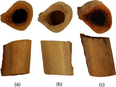
These bone fragments DNA were then quantified by qPCR using a human Alu primer set
10
by comparison to a known human DNA standard. For this quantification, three independent bone pulverizations and DNA extractions were performed from the control fragment, and seven independent pulverizations and DNA extractions from each of the three other bone fragments. Each preparation was quantified in triplicate. The box-plot presented in Figure 2 synthesizes the results obtained. The average amount of DNA extracted from the control bone was arbitrarily set to 100 to facilitate comparison, The average amounts of DNA extracted from the different bone samples were: 6098 ng/g for ‘Control’, 98 ng/g for ‘Mould’, 1797 ng/g for ‘Air’ and 134 ng/g for ‘Loam’. For each sample, the median value is represented as a wide horizontal bar, the quartile (25th-75th percentile) as a grey box, and the min/max values as narrow horizontal bars.
Box-plot representation of the amount of DNA extracted from the four bone fragments. As a reference, the amount of DNA isolated from the control was set to 100. Median values are represented by the widest horizontal bars, the quartile (25th-75th percentile) by the grey boxes, and the min/max values by the narrower horizontal bars. Statistical significance was assessed by Kruskal and Wallis non-parametric test. All values are significantly different from each others (P > 0.05), with the exception of the Mould and Loam pair
Non-parametric statistical analysis of pair-wise comparison (Kruskal and Wallis) indicates that the differences between the three-assayed conditions are significantly different from the control condition, and that the DNA quantification from the ‘Air’ sample is significantly different from that of both ‘Mould’ and ‘Loam’ samples (P > 0.05). We measured a more than 70% decrease of DNA in the ‘Air’ sample as compared with the Control sample, while the ‘Loam’ and ‘Mould’ samples represented only 2.2% and 1.6%, respectively, of the control DNA quantity, which corresponds to a 50-fold decrease in DNA amount after six months in their respective environment.
Discussion
Bone is known to be the best shelter for DNA long after death.11,12 Nevertheless, DNA extraction/analysis from bone, particularly from ancient bone, is not always successful. It has been shown that the length of the postmortem period is not the only parameter affecting bone quality, and that conditions in which the bones are kept are critical for their physical structure conservation. 13 More recent studies similarly pointed to the predominance of bone environment on the quality of extractable DNA.13,14 However, to our knowledge, no direct side-by-side comparison has been performed to quantify the variation of DNA obtained from the same exact bone submitted to different conservation conditions. In order to investigate this point, we compared the DNA yields from four bone fragments prepared from the same human femur but kept in different environments for six months. We selected four conditions covering the most common situations of bone discoveries: (1) a controlled atmosphere, which is similar to what can be found in a normally maintained building, and that we arbitrarily considered as the reference condition; (2) an open air condition, which reflects a situation where a skeleton would be found in the open, with no protection from the weather; (3) a garden-loam condition, which mimics a situation where the skeleton would be found buried; and finally (4) a water-saturated condition similar to what a skeleton exposed to a warm and humid environment could meet. Each of these environments corresponds to very specific parameters, whose influence on DNA conservation could be critical.
Our studies demonstrate that conditions favourable to microorganisms proliferation are unambiguously deleterious to bone DNA stability. Loam is a very biologically active and complex medium, 6 while a temperate water saturated condition yields to the rapid proliferation of mould spores present in the atmosphere. It is well-established that bones are commonly contaminated by microorganisms whose DNA can account for more than 99% of the total DNA extracted from these bones. 1 Microbial contamination results in nucleases secretions, as shown by Ye et al., 15 likely yielding to the dramatic loss of DNA reported in this work. About 50 times less human DNA was isolated in both cases as compared with the reference bone fragment.
A much higher amount of human DNA was extracted from the bone fragment exposed to the open air as compared with both loam- and mould-exposed bones. Nevertheless, more than 70% of the bone DNA was lost in this condition as compared with the reference fragment. This suggests that prolonged exposure to ultraviolet and weather does have an effect on bone DNA conservation, since less than 30% of the DNA found in the control bone fragment could still be isolated.
The four conditions selected in this work evidently do not cover all possible situations. However, they were chosen to be as representative as possible. Humic-garden loam is known to be one of the most biologically active soil. 6 As such, it may have a higher influence on bone DNA degradation than for example a sandy soil that will drain water faster, and will be less favourable to microbial growth. Similarly, the water vapour-saturated condition used here is likely to be more extreme than a classical humid atmosphere, in which mould growth may not be as active. Finally, the exterior condition was such that the bone fragment was maximally exposed to elements, with no freezing period that could have protected the DNA.
Our results are in good agreements with earlier reports that did not specifically focus on bone DNA analysis. 16 We focused in this work on the effect of the environment on bone DNA conservation. To exclude the influence of flesh decomposition, all soft tissue was removed from the bone in the laboratory prior to the experiment. Our results thus solely reflect the relation between bone, DNA and environment. The quality of bone DNA will be highly dependent upon the environment(s) the bone met during its postmortem existence. As a general rule, bones kept under dry conditions will be more likely to still contain usable DNA than bones found in environments favourable to microorganisms proliferation.
Footnotes
Acknowledgements
This work was supported by the Centre National de la Recherche Scientifique (CNRS). Bone samples were obtained from the ‘Don du Corps’ of the Laboratoire d'Anatomie de Nice, under the responsibility of Pr F De Peretti, with the authorization of the French Ethic Committee.