
Abstract
Background
In this retrospective autopsy study, we aimed to review the anatomopathological findings observed in cases of hanging death for a five year period and to evaluate the role of contributing factors such as age, sex, type of hanging and localization of the ligature knot.
Methods
Autopsy reports of 102 hanging cases performed by the Department of Forensic Medicine, Faculty of Medicine of Pamukkale University, between January 2007 and September 2011, were retrospectively reviewed.
Results
In the 102 hanging cases 73 of the victims were males (71.6%) and 29 (28.4%) were females, with a mean age of 40.97 ± 17.41 years. All cases were suicidal hanging. Fifty four cases (52.9%) were typical hanging, with the ligature knot located posteriorly. There were petechial hemorrhage on the face and eye lids in 46 (45.1%), ecchymoses of the cervicale muscles in 43 (42.2%), and fractures of the neck structure(s) in 69 cases (67.6%).
Conclusions
The incidence of neck structure fractures increased with age. In addition, there was no correlation between the incidence of neck structure fractures and sex or type of hanging.
Introduction
Hanging is a form of ligature strangulation in which the force applied to the neck is derived from the gravitational drag of the weight of the body or part of body. 1 Death is caused by compression of the blood vessels of the neck that causes an insufficient amount of oxygenated blood to the brain. Obstruction of the airway can also occur, either through compression of the trachea or, when the noose is above the larynx, elevation and posterior displacement of the tongue and floor of the mouth. 2 Another mechanism of death is pressure on the baroreceptors situated in the carotid sinuses, the carotid sheaths and the carotid body, called as ‘reflex cardiac arrest’, ‘vasovagal shock’ or ‘reflex cardiac arrest’, this reflex can cause immediate cardiac arrest. 1 Hanging is a common method of suicide. Suicidal hangings are most common while accidental hangings are rare, and homicidal hangings are even rarer. 2 Hanging has been classified as typical or atypical depending upon the position of the knot in the noose. If a knot is present on the posterior region of the neck it is termed as typical hanging or otherwise as the atypical hanging.1,2
Detection of ecchymosis in the cervical soft tissues and muscles, fractures of the hyoid bone and thyroid cartilage, and petechiae are critically important to determine the cause of death as hanging. Therefore, neck structures need to be thoroughly examined during medicolegal autopsies of hanging cases. 3
The larynx projects ventrally between the great vessels of the neck and is covered anteriorly by the skin, fasciae and muscles. The thyroid cartilage is the largest of the laryngeal cartilages. It consists of two quadrilateral laminae. Posteriorly the laminae diverge, and their posterior borders are prolonged as slender horns, the superior and inferior cornua. The cricoid cartilage attached below to the trachea, and articulates with the thyroid cartilage. It forms a complete ring around the airway, the only laryngeal cartilage to do so. 4 The U-shaped hyoid bone is attached to the larynx: it has a body, two greater and two lesser horns. 5 The hyoid often has natural joints that lie at the junction of the body with the greater horns. There are two lesser horns on the upper surface of the body that have no forensic anatomical significance. 1 In middle and later life the hyoid and thyroid horns calcify and become more brittle. The cricoid cartilage also become partly calcified as age increases. 1
The aim of the present study is to review the anatomopathological findings of hanging, to evaluate the role of contributing factors, such as age, sex, type of hanging and location of the ligature knot and to discuss the literature.
Materials and method
Autopsy reports of 102 hanging cases performed by the Department of Forensic Medicine, Faculty of Medicine of Pamukkale University, between January 2007 and September 2011, were retrospectively reviewed. Cases were analysed for age, sex, type of hanging (typical- atypical), localization of the ligature knot, presence of fracture of the hyoid bone and thyroid cartilage, cervical intramuscular haemorrhage, carotid artery injury and external petechiae (facial or conjunctival petechiae). Hyoid bone and thyroid cartilage fractures were identified with naked eye examination and manually. Proof of antemortem fractures obtained by demonstrating macroscopic haemorrhage at the fracture site. 1 Statistical analyses were performed by using SPSS statistics 10.0 software (Pamukkale University, Denizli, Turkey). Chi-squared test was used to test the statistical significance of the results. A P value of less than 0.05 was considered statistically significant. This study was approved by the Ethics Committee of Pamukkale University.
Results
Age and gender distribution of hanging
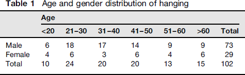
Of the 102 cases, typical hanging comprised 52.9% (n = 54) while atypical hanging comprised 47.1% (n = 48). The location of the knot was on the posterior region in 54 cases (52.9%); on the right side of the neck in 19 cases (18.6%); on the left side of the neck in 22 cases (21.6%) and on the anterior region in seven cases (6.9%).
Neck structure injuries according to different types of hanging and localization of ligature
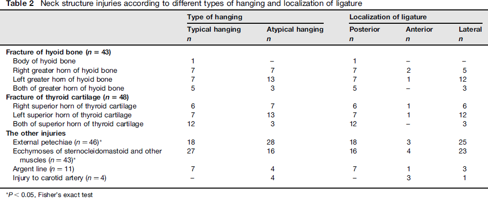
P < 0.05, Fisher's exact test
Ecchymoses occurred in the sternocleidomastoid and/or the other neck muscles in 43 cases (42.2%). Of the 43 cases that have ecchymoses, the localization of the ligature knot was on the lateral sides (right or left) in 22 (53.5%); on the posterior region in 16 (37.2%) and on the anterior region in four (9.3%) (χ 2 = 8.494; P = 0.037). In atypical hangings, haemorrhage into neck muscles was significantly more common compared with typical hangings (62.8%; n = 27 versus 37.2%; n = 16, Fisher's exact test; P = 0.009; Table 2).
The argent line, formed by the condensation of the subcutaneous adipose tissue beneath the hanging mark, was observed in 11 cases (10.8%; Table 2). The occurrence of the argent line was not correlated with each of age, sex, ligature knot localization and type of hanging.
In four cases, Amussat's sign, the laceration of the intimal layer of carotid arteries, was observed (Table 2). All of these cases were atypical hanging and in three cases, the ligature knot was anteriorly located.
Fractures of the neck structures occurred in a total of 67.6% cases (n = 69); 20.6% (n = 21) had isolated hyoid bone fracture while 25.5% (n = 26) had isolated fracture of the thyroid cartilage and 21.6% (n = 22) had fracture of the thyro-hyoid complex (Figure 1). Fracture of the hyoid bone was most commonly at its left greater horn, followed by right greater horn. In the thyroid cartilage, left superior horn fracture was the most common fracture (Table 2). The cervical vertebra was fractured in one case while fracture of the crycoid cartilage was not observed.
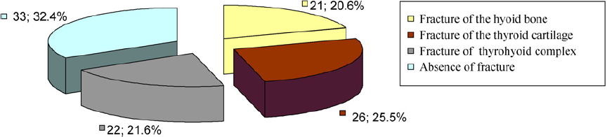
The distribution of fractures of the hyoid bone and the thyroid cartilage in hanging
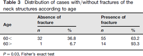
Distribution of cases with/without fractures of the neck structures according to age
Cases with fractures of neck structures comprised of 47 men (68.1%) and 22 women (31.9%). Sex did not correlate significantly with the incidence of fractures (Fisher's exact test; P = 0.35).
Distribution of cases with/without fractures of the neck structures according to the type of hanging and the position of the knot
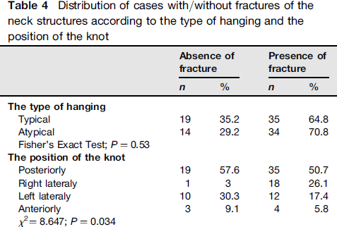
We found a significant correlation between the incidence of fractures of neck structures and location of the ligature knot (Table 4). Incidence of fracture was significantly higher when the ligature knot was posteriorly located (50.7%; χ22 = 8.647; P = 0.034).
Discussion
Among the 102 cases of mortality due to hanging, 73 (71.6%) were men and 29 (28.4%) were women. There are studies in the literature, which showed that mortality due to hanging was more common among men. In studies by Azmak, Suárez-Peñaranda et al. Cantürk et al. Ege et al. Uzün et al. and Balci, 83.9%, 77.8%, 72%, 72.63%, 70.56% and 64.3%, respectively, of the subjects were males.6-10 There were twice as many male subjects as females in the Sharma et al.‘s 11 study. On the other hand, Goren et al. 12 reported that the majority of the subjects were women (57.7% as opposed to 42.3%) and the authors argued that there could be regional differences.
With 24 cases, 21-30 year age group was the largest group in the present study. Gören et al. Ege et al. Azmak, Uzün et al. and Sharma et al. reported that mortality due to hanging was most common in the young age groups, 11-20, 20-29, 25-30, 20-29 and 21-30, respectively.3,6,9,11,12
Hanging is classified as typical or atypical according to the location of the ligature knot. The frequencies of typical and atypical hanging vary in the literature. In the present study, 52.9% of the cases were typical and 47.1% were atypical hanging. Goren et al. 12 reported the frequencies of typical and atypical hanging as 60.7% and 39.3%, respectively, while Cantürk et al. 8 reported as 87.4% and 11.8%, respectively. On the other hand, with a frequency of 78.78%, a typical hanging was found to be more common by Talukder et al. 13 Atypical hanging was found to be more common by Sharma et al. 11 as well, with only eight cases of typical hanging among 66 hanging cases.
In the present study, the ligature knot was most commonly located on the posterior region (52.9%). This finding is in accordance with previous studies. The incidence for this localization reported as 66% and 61.71% by Azmak 6 and Nikolic et al. 14 respectively.
Petechiae are classical findings of asphyxia and are commonly observed in hanging. It is caused by extravasation of erythrocytes as a result of capillary endothelial damage due to increase in venous pressure and hypoxia.1,2 Authors also agree that petechiae were correctly interpreted as products of vascular rupture due to obstructed venous return, rather than hypoxia. 15 We noted external petechiae (facial or conjunctival petechiae) in 45.1% of our cases, 65.2% of which were atypical hanging cases. This was in accordance with the literature. Sharma et al. 11 reported petechiae in 42% (n = 28) of 66 hanging cases, 18 of which were incomplete atypical, seven were complete atypical and three were incomplete typical hanging. Załuski et al. 16 observed petechia on the face and eyelids in 33.5% of their cases. Clément et al. reported that petechiae were more common in incomplete hanging. 17
We found haemorrhage into neck muscles in 43 cases and atypical hanging comprised 62.8% of these cases. Azmak, 6 Suárez-Peñaranda et al. 7 and Załuski et al. 16 reported intramuscular haemorrhage in 51.7%, 55.8% and 18.4% of their hanging cases, respectively., Sharma et al. 11 suggested haemorrhage into the cervical muscles as a general sign of typical and complete hanging.
The argent line caused by the condensation of the subcutaneous adipose tissue beneath the hanging mark is one of the general findings. Suárez-Peñaranda et al. 7 reported the incidence of this finding as 76.8% though we found this in only 10.8% of our cases. In accordance with the literature, the occurrence of the argent line was not correlated with each of age, sex, ligature knot localization and type of hanging. 7
Tears that appear on the intima of the carotid arteries are termed as Amussat's sign. We found Amussat's sign in 4 cases (3.9%). This incidence was 9.1% and 16.1% in the studies by Sharma et al. 11 and Petr, 18 respectively. Amussat's sign has been reported to increase with age but we did not find a significant correlation confirming this. 18
We noted fracture of the neck structures in 67.6% of cases. The incidence of the fracture of the neck structure has been reported as 76.8%, 68%, 57.3%, 47.5%, 28% and 23.4% by Azmak, Nikolic et al. (2003), Nikolić et al. (2011), Green et al., Clément et al. and Godin et al. respectively.6,14,17,19-21 Hanging can be associated with either isolated fractures of the hyoid bone or thyroid cartilage or fracture of the combined thyro-hyoid complex. In the present study, isolated thyroid cartilage fracture ranked first, with a frequency of 25.5%. Isolated fracture of the thyroid cartilage was the most common fracture in other studies as well, with an incidence of 26%, 22.5%, 22% and 18.6% by Nikolić et al. (2011), Green et al. Clément et al. and Godin et al. respectively.17,19-21 On the other hand, Azmak 6 reported isolated hyoid bone fracture most commonly and followed by isolated thyroid cartilage fracture, with a frequency of 17.8%. Incidence of isolated hyoid bone fracture has been reported as 46.4% 15.1%, 10%, 2.6% and 1% by Azmak, Nikolić et al. (2011), Green et al., Godin et al. and Clément et al. respectively.6,17,19-21 In the present study, incidence of isolated hyoid bone fracture was found to be 20.6%. The incidence of combined hyoid bone and thyroid cartilage fractures varies between 2.2% and 16.2% in the literature.6,17,19-21 In our study, this incidence was 21.6%, higher than those reported in the literature. Similar to studies by Clément et al. 17 and Godin et al. 21 we did not find fracture of the cricoid cartilage in any of our cases.
In this study, we found that incidence of the fracture of neck structures increased with age. This finding is in accordence with results of numerous studies in the literature.3,11,14,17,22 Fusion of greater horn to the body of the hyoid bone and ossification of the thyroid cartilage increase with age.23,24 Miller et al. 25 reported that bilateral fusion of the greater horn to the body of the hyoid bone might occur in the age groups 20-30 years, while Gupta et al. 26 reported this age group as 30-40 years. Gupta et al. 26 also reported that frequency of hyoid bone fusion rose sharply after 50 years of age and all the hyoid bones in the present study were bilaterally fused after 60 years of age. Therefore, neck structures become more fragile. We also found that the incidence of fracture was higher after the age of 60. Increase in the incidence of fracture with age might be due to calcification and ossification as well as age-related mineral loss in the bones.
We did not find a significant correlation between the incidence of fracture of the neck structures and sex. Feigin 22 and Suárez-Peñaranda et al. 7 reported a significant correlation between fracture incidence and sex. However, some studies reported a higher incidence in men11,17,20,21 while other reported a contradictory result. 27
Significant correlation between fracture of the neck structures and the location of the ligature knot have not been reported in the literature.7,14,17 Results of the present study showed that fracture was more common when the knot was posteriorly located.
In conclusion, the incidence of the neck structure fractures was found to be 67.6% and age is the most important factor that contributes to the occurence of neck structure fractures in hanging.