
Abstract
Endometrial stromal sarcoma (ESS) rarely infiltrates the great vessels. We report a successful surgical resection of the inferior vena cava (IVC) after extensive infiltration with metastatic low-grade ESS. A case of presumed recurrence of low-grade ESS demonstrated complete IVC occlusion from tumour thrombus with extensive local disease. Radical resection of the tumour and caval reconstruction was performed. The IVC graft was thrombosed at short-term follow-up. Curative resection of extensive caval infiltration with metastatic low-grade ESS can be achieved. Caval reconstructive procedures may be redundant in the presence of an adequate collateral circulation.
Introduction
Endometrial stromal sarcomas (ESSs) are rare malignant tumours that constitute approximately 10% of all uterine sarcomas, but only around 0.2% of all uterine malignancies. 1
Low-grade ESSs (LGESSs) are a subset of these tumours which demonstrate indolent growth. Owing to the infiltrating margin of LGESS and its tendency to be angio-invasive, 1 however, late recurrence of up to 10–20 years and distant metastases can occur. Despite this, infiltration of the great vessels due to tumour recurrence is a rare event.
Report
We report a case of curative surgical resection and reconstruction of the inferior vena cava (IVC) after extensive infiltration with metastatic LGESS.
A 60-year-old woman was investigated for right flank pain and gross haematuria. She had previously undergone a radical hysterectomy and bilateral salpingo-oophorectomy for endometrial sarcoma confirmed histologically as LGESS 20 years ago. Physical examination and laboratory tests were unremarkable.
Contrast phase computed tomography (CT)-angiogram and CT-venogram revealed complete occlusion of the IVC from a soft tissue mass with extension into both renal veins. Planning magnetic resonance imaging revealed mixed signals from the lesion consistent with tumour rather than tumour thrombus. The proximal extent of the tumour was just below the hepatic veins in the supra-renal cava (Figure 1). There was no evidence of metastasis to other organs. Given the extensive nature of the disease, radical resection of the tumour and caval reconstruction was performed.

Preoperative venous phase abdominal computed tomography scan demonstrating tumour thrombus occluding the inferior vena cava and tumour infiltration into the right renal vein
The patient was positioned in left lateral position with the table jack-knifed and right arm over. A retroperitoneal approach was used with an incision at the base of the costal margin extending towards the umbilicus. The rectus sheath muscles were not divided due to the high incidence of incisional hernia at this site. This provided exposure to mobilize the liver and access the hepatic veins. The IVC was dissected from the liver to bifurcation. Division of the inferior hepatic vein was required to achieve macroscopic tumour clearance. The right renal vein contained an occlusive tumour and was excised into the hilum of the kidney as it branched into its tributaries, hence it was not possible to reconstruct. The left renal vein contained a non-adherent tumour and remained patent; it was therefore excised proximally until the healthy vein was reached. The distal resection was to the origin of the common iliac veins that were found to be tumour-free. Thrombus was not found to extend past the margins of proximal or distal resection. Caval reconstruction was undertaken using a 20-mm polytetrafluorethylene (PTFE) prosthetic graft. The left renal vein was re-attached to the main body of the graft using an 8-mm PTFE graft.
Therapeutic anticoagulation was commenced postoperatively. The patient was discharged on warfarin with normal renal and liver function tests.
Histopathology confirmed recurrence of LGESS and all the margins of the excision (proximal IVC, distal IVC and ends of both renal veins) were tumour-free (Figure 2).
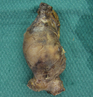
Surgical specimen photo showing resection of inferior vena cava and tumour from the iliac confluence to hepatic veins
Follow-up imaging one month postoperatively revealed a patent left renal vein graft but complete thrombosis of the IVC graft distal to this. Despite this, the patient remained asymptomatic and is currently playing tennis without evidence of venous hypertension or claudication. Twelve-month disease-free survival was demonstrated on a CT scan.
Discussion
Extension of LGESS into the large vessels is rare. To date, there have been only 19 reported cases of metastatic LGESS with invasion of the great vessels and IVC tumour thrombus. 1 In only three cases was a partial/segmental resection of the IVC wall required due to tumour infiltration. In these cases, the IVC was reconstructed with xenopericardium or a PTFE prosthesis. In a further case, the IVC was resected without reconstruction. 1
Our case is unique given the extensive infiltration of a tumour requiring partial resection and reconstruction of the renal veins and common iliac veins in addition to the IVC to the level of the hepatic veins.
The decision to reconstruct the IVC after resection is debatable. There is evidence supporting reconstruction only for patients with poorly developed collateral circulation on preoperative evaluation or those with intraoperative oliguria and haemodynamic instability. 2 In our case, the IVC was chronically occluded with a tumour allowing development of a significant venous collateral circulation. Preoperatively this enabled sufficient drainage of lower limb venous return; however, the extensive dissection required ligation of a significant number of these collaterals, increasing the risk of postoperative limb swelling and venous claudication. Reconstruction of the cava was therefore undertaken. This decision was supported by previous work demonstrating a reduction in swelling associated with delayed graft occlusion compared with non-reconstruction at the time of surgery where swelling is significant. 3 Given our intention of reconstruction was to minimize venous hypertension, PTFE graft was used to avoid lower limb oedema and the morbidity associated with a panelled great saphenous vein or femoral vein graft. The possibility of adjuvant radiotherapy was also considered and if required was felt likely to induce radiation injury to an autogenous vein graft resulting in early graft occlusion or sclerosis.
Venous grafts are more prone to occlusion than arterial grafts given the slow venous flow against a hydrostatic pressure gradient, low intraluminal pressure and often competitive flow from a collateral circulation. 4 In a study of 10 patients who underwent caval reconstruction for chronic iliocaval thrombosis, the patency ranged from immediate postoperative occlusion for three grafts to a maximum of 3.3 years. 5 Our case is consistent with these findings and suggests that complex caval reconstructive procedures may be redundant in the presence of adequate collateral circulation. The creation of arterio-venous fistula has been used to successfully overcome such problems in lower limb reconstructive procedures. 3 Owing to the high flow expected in the cava, this was considered unnecessary in our case. In retrospect, we would not have reconstructed the cava.
In summary, metastatic LGESS involving the great vessels is rare. We have shown that in symptomatic patients with extensive disease curative resection can be achieved with return to normal quality of life.