
Abstract
Objective
To assess the presence of pulmonary embolism and inflammation after polidocanol foam injection into the peripheral veins of rabbits.
Method
The animals were treated with polidocanol foam (1 or 3 mg/kg) or vehicle. Early (15 minutes) and late (30 days) animals were evaluated by perfusional lung scintigraphy and histopathological examination.
Results
In the control group no alterations were found. After polidocanol foam injection it was observed that an important reduction of pulmonary perfusion in the early periods, was mainly in the left lung (P < 0.001), with consequent embolism in the histological evaluation. In late periods it was observed that the presence of thrombus was with fibrin in small veins, compatible with chronic thrombus and the presence of chronic pulmonary inflammation.
Conclusions
The injection of polidocanol foam in experimental animals can induce venous embolism and chronic inflammatory infiltration.
Introduction
The medical treatment of chronic venous failure includes changes in the lifestyle, compressing with elastic socks and medicament therapy. However, most of the patients, at some point of the evolution of the disease, will need invasive therapy, such as conventional surgery and physical methods with laser or chemical methods with the use of sclerosing substances. 1 Among these sclerosing agents polidocanol is one of the most used. 2 Although rare, there are some systemic effects after the injection of polidocanol foam, some patients complain of auto-limited chest pain, cough and visual alterations. 3 In addition, the long-term effects of polidocanol foam in lung function were not assessed. The main objective of our study was to assess the occurrence of pulmonary embolism and long-term inflammatory activation after polidocanol foam injection in the peripheral veins of rabbits.
Methods
The investigation was approved (7 September 2011) by the animal ethics committee of the university. Domestic rabbits, of both sexes, weighing around 2 kg were used.
The animals were divided into two groups, the control group (injection of saline solution, n = 4) and the experimental group (injection of polidocanol foam 1 and 3 mg/kg, n = 4 each dose), administered in the lateral superficial auricular vein. Polidocanol foam was prepared using a 3% solution. It was mixed with 1 mL of 3% polidocanol solution with 4 mL of air to obtain polidocanol foam solution. For the 1 mg/kg group it was injected with 0.3 mL of this solution and for the 3 mg/kg it was injected with 1.0 mL of this solution. Polidocanol foam was injected as a slow bolus over one minute.
Lung scintigraphy was realized 15 minutes after polidocanol foam with technetium (Tc99m) conjugated with albumin. The interpretation of the images was done by a board-certified doctor blinded to the study group. After scintigraphy the rabbits were killed and the lungs were removed for histopathological evaluation. A separated group of animals was reserved for late histopathological evaluation (30 days after injection). The pathological exam was realized by a board-certified pathologist blinded to the study group.
The differences in the extension of lung scintigraphy alterations between the right and left lung within groups were analysed by chi-square-Fisher exact test. The extension of lung scintigraphy alterations between the groups were compared by ANOVA's test followed by Tukey's post hoc. Statistical significance was considered when P < 0.05.
Results
In the control group no alterations were found (Figures 1a and d). When the pulmonary perfusion was evaluated after the injection of 1 and 3 mg/kg polidocanol foam it was observed that an important reduction of pulmonary perfusion occurred, but without statistical significance when compared with the two polidocanol foam groups (Figures 1b-d).

Lung scintigraphy findings after polidocanol foam administration. Lung scintigraphy was performed 15 minutes after saline or polidocanol foam administration in domestic rabbits. (a) Representative scan from a saline-injected animal; (b) representative scan from polidocanol foam (1 mg/kg) injected animal; (c) representative scan from polidocanol foam (3 mg/kg) injected animal; (d) quantification of lung perfusion in saline- and polidocanol foam-injected animals, a = P < 0.05 compared with the control (saline) of ipsilateral lung; b = P < 0.05 compared with the left lung in the same group; RL, right lung; LL, left lung
The pathological exam of the control group did not show alterations (Figure 2a). The pathological exam at earlier times after the injection of polidocanol foam shows the presence of platelet-fibrin clots mainly in the small pulmonary veins, independent of the dose administered (Figures 2b and c). In addition, it was observed that the occasional presence of droplets can correspond to fat embolism (Figure 2b) and an acute inflammatory response (Figure 2c). In later times after polidocanol foam injection (30 days), it was observed that the occasional presence of thrombus with fibrin, is suggestive of chronic thrombus in small veins (Figure 2d) and the presence of droplets that can correspond to fat collection (Figure 2e). It was also observed that the mixed chronic inflammatory response, can be sparse (Figure 2d) or more limited (Figure 2f).
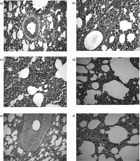
Lung histology findings of early and late times after polidocanol foam administration. Lung histology was performed immediately after lung scintigraphy (early) or 30 days (late) after polidocanol foam injection in domestic rabbits. (a) Representative image from a saline-injected animal; (b,c) representative image from polidocanol foam (1 and 3 mg/kg) injected animal, acute phase; (d-f) representative image from polidocanol foam (1 and 3 mg/kg) injected animal, late phase
Discussion
This work shows that the injection of polidocanol foam in experimental animals leads to acute and chronic alterations in pulmonary perfusion and lung inflammation.
Unfortunately, we cannot find in the literature similar studies to compare with our results. The medication prescription information (http://www.asclera.com/downloads/Asclera_FULL_PRESCRIBING_INFORMATION.pdf, accessed on 17 May 2012) reveals that doses between 4 and 10 mg/kg lead to a possible damage of the fetus, but stresses that there is no evidence of muscular and visceral damage. The acute alterations observed in these animals can only be the normal dynamic of polidocanol foam bubbles, as was previously demonstrated, even in humans, that bubble emboli entered the heart early after foam injection. 4 This is reinforced by the fact that there are no differences between the lower and higher polidocanol foam doses. Thus, the acute alterations can have no meaning from the lesion point of view.
In addition, we found the development of mixed chronic inflammatory infiltration in the lung parenchyma at later points after polidocanol foam administration. As any other detergent, the presence of polidocanol can induce an inflammatory response. 5 Again, there is no animal model study or long-term follow-up study in humans that determines the parameters of pulmonary inflammation or alteration of pulmonary function related with chronic inflammation. This appears to be an effect not expected and not explored since most of the patients submitting to foam injection, in general, did not show acute alterations of the pulmonary function. But, if few amounts of polidocanol foam can be deposited into the lung (in low quantity, not causing significant pulmonary embolism) it can induce inflammation in the pulmonary parenchyma. However, the lack of experimental or clinical data and the limited follow-up in our study did not allow us to determine if the inflammatory response observed for 30 days can persist or evolve to chronic forms of pulmonary diseases, such as fibrosis.
Some limitation must be mentioned. First, the use of rabbits does not allow us to use manoeuvres that, in humans, can reduce the incidence of embolism such as leg elevation before or after the injection and immobility post treatment. In addition, the veins used to administere polidocanol foam in the rabbits cannot reflect exactly the veins used in humans. Rabbit ear could be similar to a reticular varix, but the distance between the rabbit ear and the lungs does not match the distance between the lower limbs and lungs in humans. Since it is known that albumin can deactivate polidocanol 6 it is possible that the time elapsing for blood flows from the rabbit ear to its lung is lower than the time needed to inactivate polidocanol. Thus, the results presented here must be considered with caution.
We conclude that the injection of polidocanol foam in experimental animals can induce venous embolism and chronic inflammatory infiltration. Longer follow-up and human studies must be performed to assess the long-term effects of polidocanol foam on lung inflammation.
Footnotes
Acknowledgement
We thank Dr Rocha who analysed lung perfusion scintigraphy. This work was supported by UNESC.