
Abstract
The aim of this paper is to demonstrate the location of the venous foot pump using an anatomical study. Four hundred cadaveric feet were injected with green neoprene latex followed by a dissection. A coloured segmentation of the venous system was achieved. The Lejars’ concept of the venous sole of the foot is incorrect: the true blood venous reservoir of the foot is located deeply in the plantar veins, between the plantar muscles. The medial and mostly lateral plantar veins converge into the plexus shaped calcaneal crossroad, where the blood is ejected upwards into the two posterior tibial veins. In addition, the several medial perforators of the foot directly connect the deep system (medial plantar veins) to the superficial venous system (medial marginal vein). This forms a true ‘medial functional unit’ which is unique in the limb given its directional flow is from deep to superficial. In conclusion, the plantar veins play an important role in the physiology of the venous return since a venous reservoir of 25 mL of blood is mobilized upwards with each step during walking. Therefore, the impairment of the foot pump by a static foot disorder should be considered as an important risk factor for chronic venous disease, and should be evaluated and corrected in any patient with venous insufficiency.
Introduction
Historical timeline
In 1861, Sucquet observed small channels in the thickness of the skin going from a precapillary arteriole to a postcapillary venule. This structure shunts the capillary blood and facilitates passage into the arteriovenous anastomoses. These channels are observed in areas of high pressure in the sole of the foot and the palm of the hand.
In 1885, Bourceret 1 demonstrated a fine plexus of dermal and subdermal veins along the entire plantar surface of the foot. This venous network drains directly into the medial and lateral marginal veins, and into the medial and lateral plantar veins via fine perforators in fatty tissue.
In 1889, Braune 2 observed an arch, the anterior part of the plantar venous network by the interdigital veins that opens into the dorsal veins. He confirmed the existence of the fine plexus described by Bourceret.
In 1890, F Lejars 3 was the first to describe a venous pump activated by walking: the plantar venous pump. He described large superficial vessels that form a true plantar reservoir. But these large superficial veins were observed as a result of an arterial injection technique done under high pressure and thus Lejars’ observation is a technical artefact.
In 1992, J H Scurr 4 recorded changes in the volume using plethysmography and estimated the quantity of blood ejected from the sole of the foot during contraction to be 20–30 mL.
In 1993, Gardner and Fox 5 proposed the hypothesis that the stretching of the medial and lateral plantar veins, with each step, pushes the blood into the saphenous veins and the deep venous network. The foot pump and the calf pump are shown to function sequentially (Figure 1).
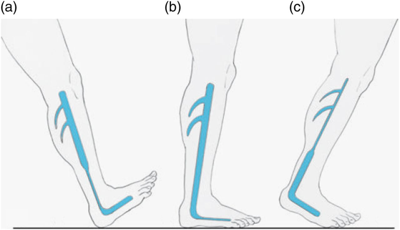
Venomuscular foot and calf pumps during walking (from Gardner and Fox, 1993). (a) Activation of the distal calf pump. (b) Activation of the foot pump. (c) Activation of the proximal calf pump
Materials and methods
We used 200 non-selected, non-embalmed cadaveric subjects (mean age of 84).
Technique of injection
After exposing the medial marginal vein, a no. 19 butterfly venous catheter was inserted and directed towards the toes (countercurrent to blood flow).
A tube was inserted into the common femoral vein to perform lavage-irrigation with soapy water (Figure 2), and was repeated several times. Massage of the muscles was performed until a clear liquid was obtained. Then, after ligation of the femoral vein, green neoprene latex was injected (about 120–150 mL per limb), over 30 minutes (Figure 3). Dissection was started the next day. A coloured segmentation was achieved by painting of the veins for a more comprehensive identification.
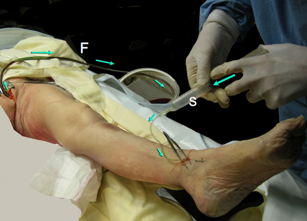
Irrigation and washing of the venous network before latex injection. Irrigation with soapy water is done by the medial marginal vein towards the toes with a 20 cm3 syringe (S), with outflow via a tube inserted in the common femoral vein (F). This is repeated several times, until a clear liquid is obtained
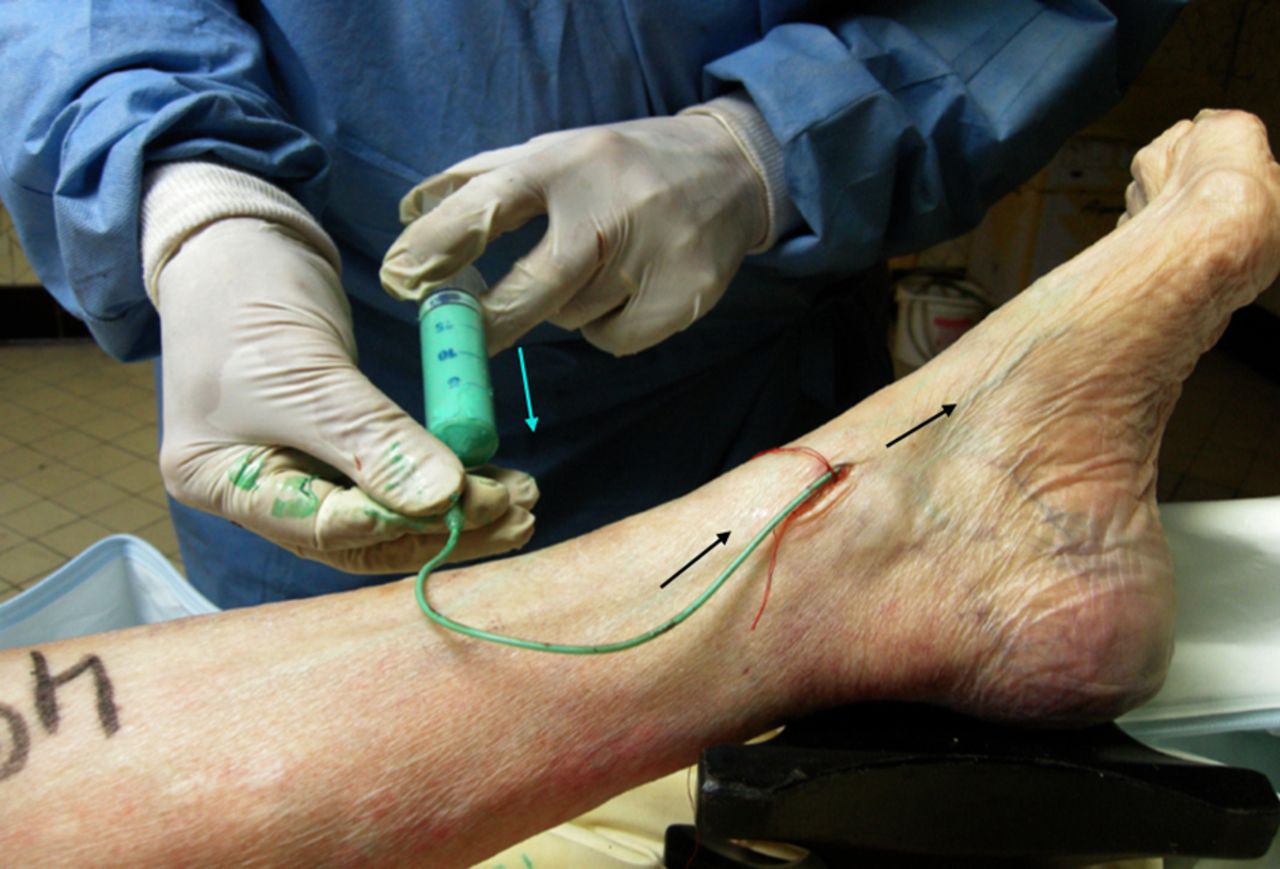
Injection of green latex. Undiluted green latex with neoprene is slowly injected towards the toes with a 20 cm3 syringe, the total amount being about 120–150 mL for one limb
Results
Anatomical description of the veins in the foot
Superficial venous network
Veins in the sole of the foot
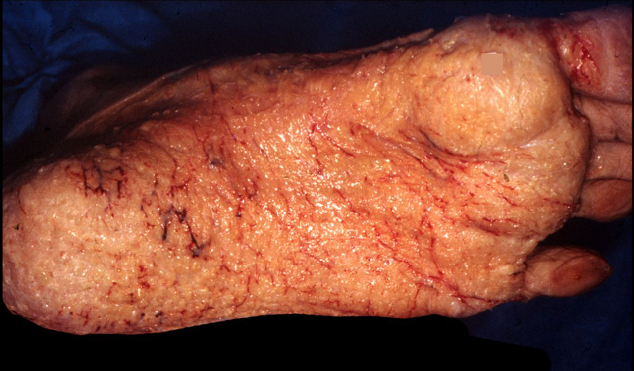
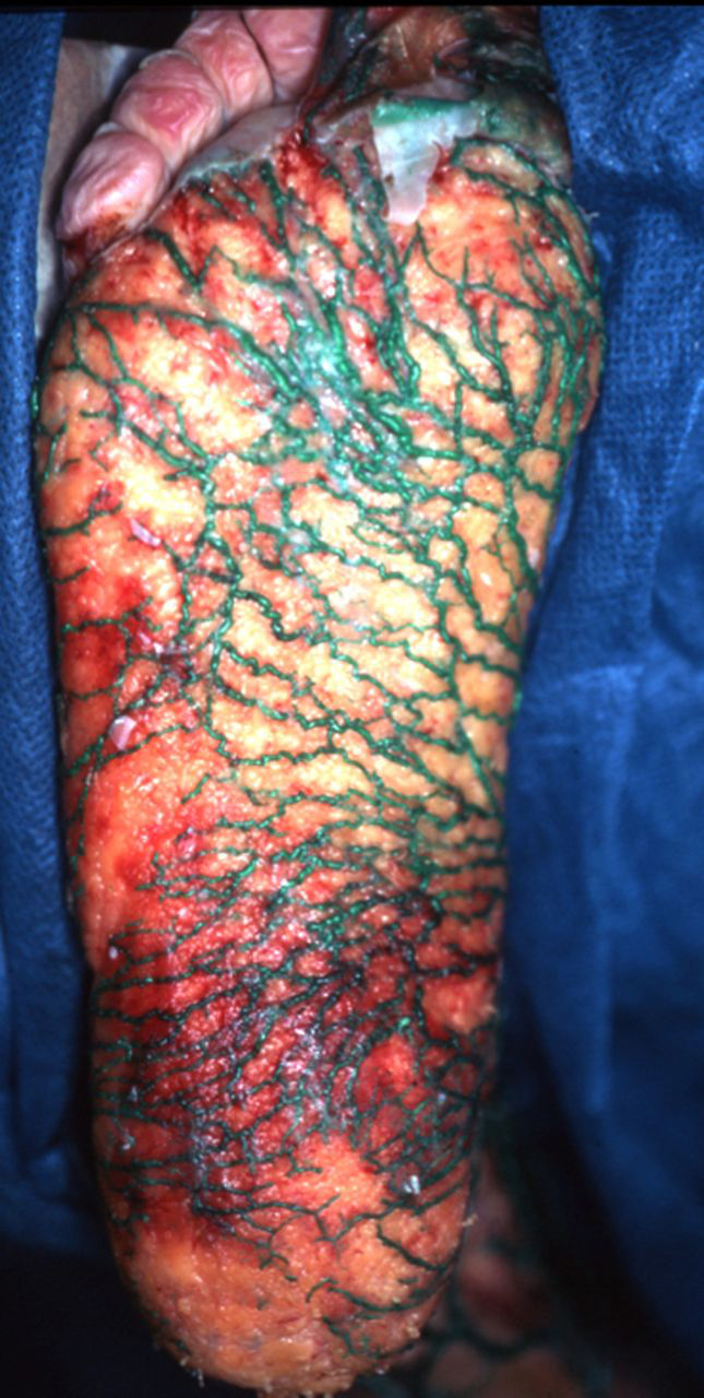
The superficial veins in the sole of the foot formed a fine network that drained into the marginal veins via small, valve-bearing veins. These valves prevent the passage of blood into the superficial area (Figures 4 and 5). In the event of total or partial absence of valves in these superficial plantar veins, moderate dilation of the plantar venous network was observed (the so-called ‘Bourceret sole’, Figures 6–8). In contrast, in the event of abnormal reflux, major dilation of this network occurred, leading to ‘Lejars’ plantar venous sole’ (Figure 9). This venous dilation was produced by severe venous stasis, most often by a superficial and/or deep vein reflux.
Dissection of the sole venules (sole of Bourceret). Note that none of the superficial veins of the sole are injected due to the expected presence of competent valves. On can see the small caliber of these venules filled with blood
Competence of the terminal collectors of the foot. The cutaneous veins of the sole (white arrows) are not injected with latex. In green, the terminal part of the small venules of the sole: the reflux is stopped at the level of the competent valves (blue dots). In red, the communicating veins and their perforation of the fascia (yellow dots)

Injected sole of Bourceret. Non-valvular veins of the cutaneous network are visible throughout the sole. Reflux of the small terminal collector veins (red lakes): it is a ‘Bourceret venous sole’, plexus of small veins, 1–2 mm in diameter
Another example of sole of Bourceret
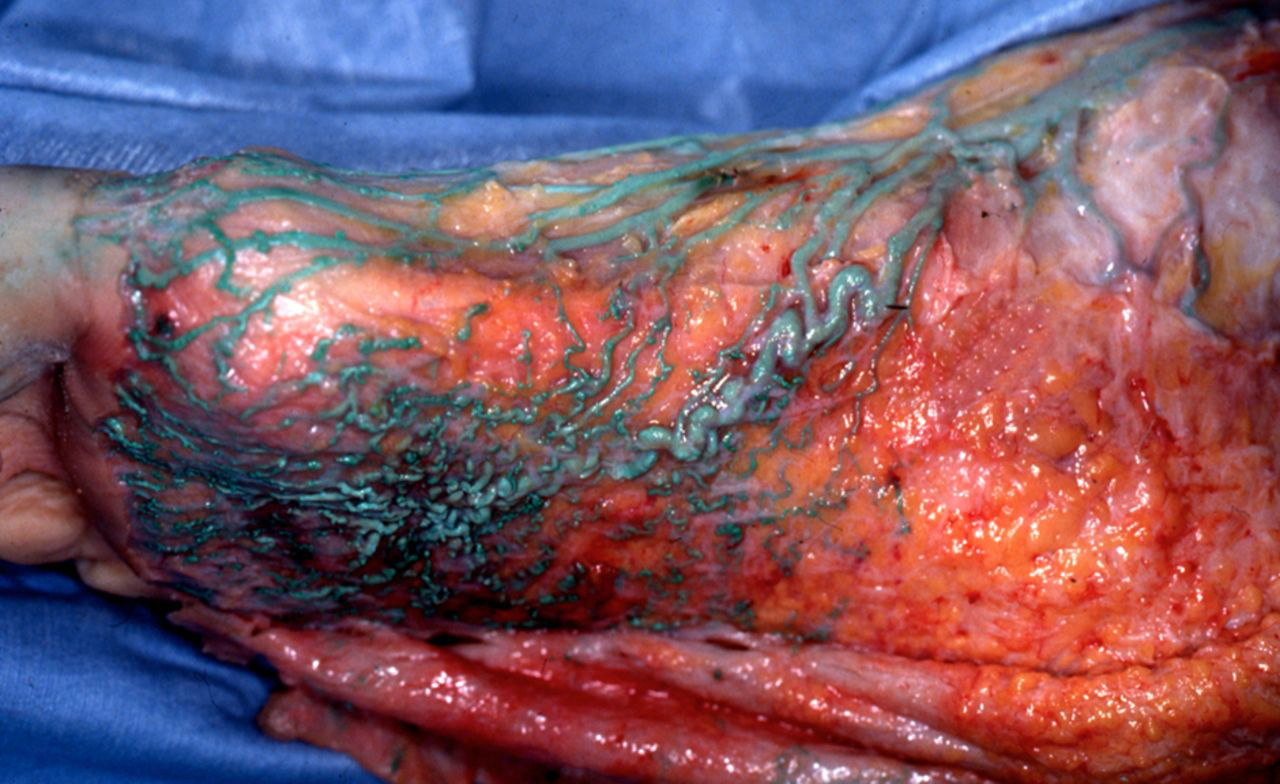
A case of plantar superficial reflux, with a rare varix of the sole. Major dilation of the anterior part of the plantar cutaneous veins, which originates from a severe reflux of the great saphenous trunk down to the ankle and foot

Sole of Lejars. Very important dilation of all the veins of the sole due to a severe venous stasis and reflux
(2) Marginal veins (Figure 10)
The superficial venous network also comprised of medial and lateral marginal veins (Figures 11 and 12). The medial marginal vein arises from the perforator of the first metatarsal interspace and continued giving rise to the great saphenous vein (GSV). This vein formed a functional unit with the medial plantar veins that we will discuss later. The lateral marginal vein also arises from the perforator of the first metatarsal interspace and ends in the small saphenous vein (SSV).
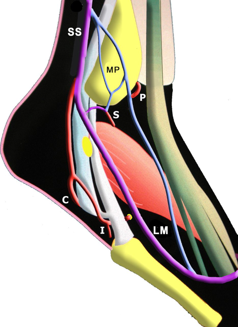
Medial longitudinal section of the foot showing the muscle topography of the plantar veins. 1: lateral plantar veins, intermuscular, located in the slit that separates the fleshy body of the plantar quadratrus (PQ) from that of the abductor hallucis (A): produces effective muscular action; 2: medial plantar veins: fibrous and tendinous relations: tendon-long flexor of the hallux (F) and deep fascia of the abductor muscle: rigid structure with no great direct action on the veins. N: navicular perforator; AP: perforator in fatty tissue; MM: medial marginal vein; LM: lateral marginal vein. Some superficial veins of the sole (S) are connected to the deep veins by adipose perforators (AP). D: dorsal arterio-venous pedicle; TE: tendons of the extensor digitorum longus
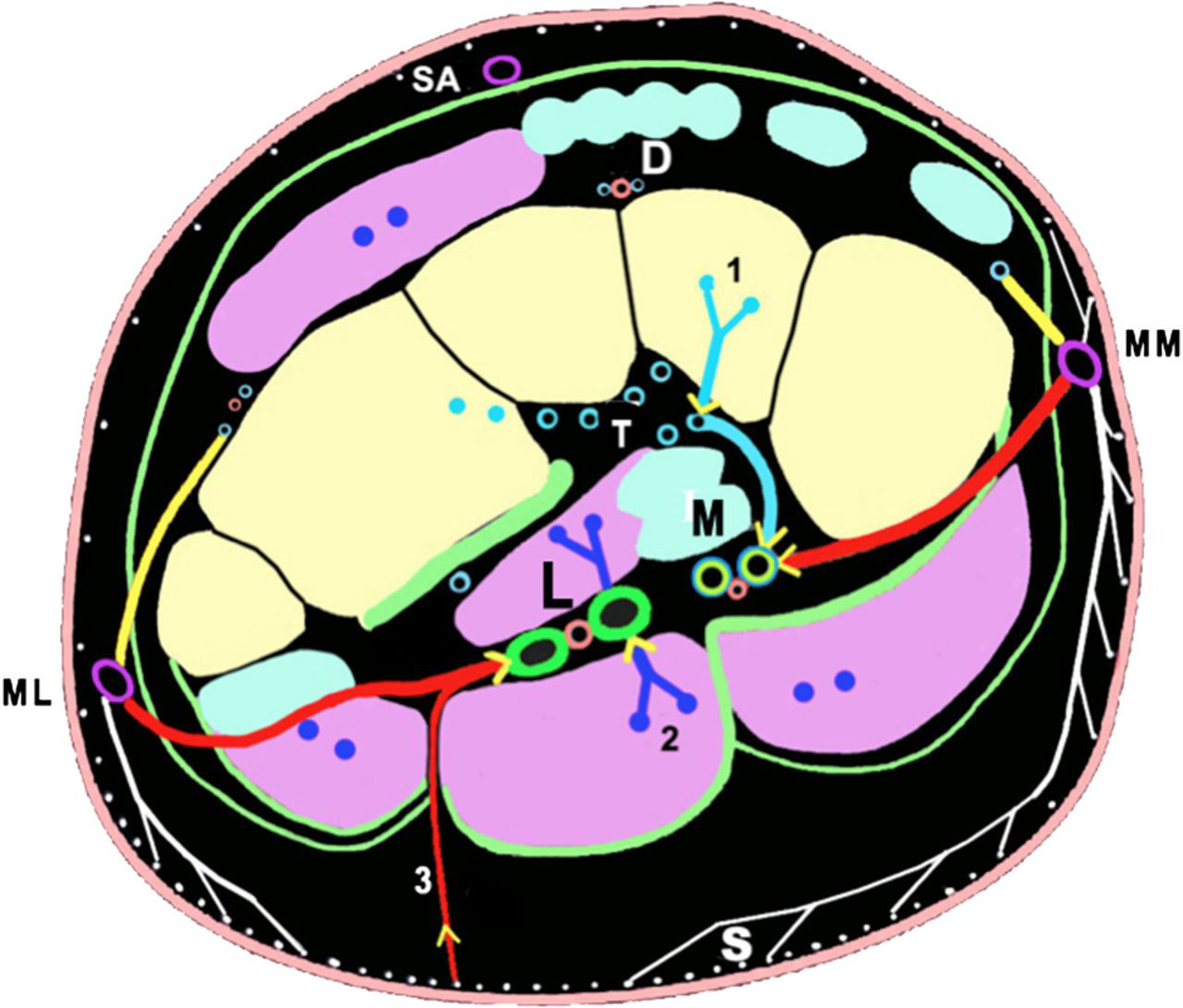
Frontal section of the tarsus showing the three layers of the foot veins. Three layers of veins are clearly visible: S: superficial, subcutaneous of the sole. Deep veins of the muscular layer: (M) medial plantar near tendons, (L) lateral plantar, between the muscles deep veins of the bony layer: T (veins of the tarsus fossa). 1: drainage of the bone veins; 2: drainage of the muscular veins; 3: small ‘adipose perforators’. MM: medial marginal vein; LM: lateral marginal vein; D: dorsal arterio-venous pedicle
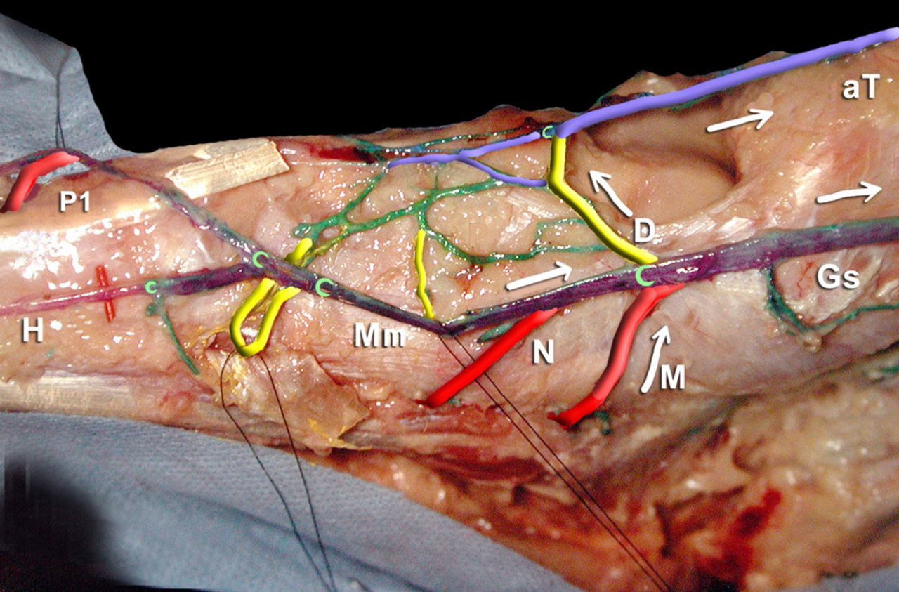
The medial perforators of the foot. The dorsal and plantar medial perforators are connected to the medial marginal (Mm) vein at the origin of the GSV (Gs). Note the three-root origin of the GSV: it arises from the medial marginal vein Mm, the dorsal communicator which opens into the anterior tibial veins aT, and the malleolar communicating vein connected to the calcaneal plexus. Note the large diameter of the communicating vein of the first metatarsal interspace P1. Note also the abrupt change in diameter of the dorsal vein of the foot after receiving the dorsal perforator vein D, thus becomes the anterior tibial vein. Mm: Medial marginal (or internal) vein Gs: GSV. H: dorsal vein of the hallux aT: anterior tibial veins. The dorsal perforator veins are shown in yellow. D: dorsal perforator vein which communicates anteriorly with the anterior tibial veins (aT). The plantar perforator veins are shown in red, anteriorly to posteriorly. P1: perforator of the first metatarsal interspace; N: navicular and M: malleolar (or perforator of the talus)

Frontal anatomical slice of the tarsus. The three layers of the foot veins: skin, muscle and bone are shown: (1) The white arrows show the superficial veins of the sole; (2) the blue arrows show the veins of the muscular layer: the lateral and the medial plantar veins (in red the companion arteries) and (3) yellow arrow shows the bone plexus draining the spongy bone
(3) The interdigital veins drained into Braune's arch from the dorsal veins
The deep venous network of the foot
This network comprised of two layers of veins as seen in this frontal section (Figures 10–12).
The deep ‘bony’ veins, located in contact with the tarsal bones in the concavity of the bony arch, which drains the cancellous bone.
The network of large collecting veins, the musculotendinous veins (Figures 16–18).
This network comprised of the medial and lateral plantar pedicles, which joined together posteriorly to form the calcaneal confluence of the plantar veins.
The medial (or internal) plantar pedicle was short, about 5 cm in length, and relatively rectilinear. It occupied only the posterior part of the sole of the foot, behind the tendon of the lateral fibular vein. It comprised of two veins and in some cases was plexiform:
It extended along the medial border of the foot and received the perforators of the medial marginal vein; Laterally, it received blood from the adjacent muscles: the abductor hallucis, the flexor digitorum brevis and the quadratus plantae muscles. These two veins were small and projected on tendons and so were ineffective for the plantar venous pump.
The lateral (or external) plantar pedicle was longer (12 cm), curved and larger because it was located between the two muscle layers of the sole of the foot, and thus was compressed during contraction. It arises opposite the first metatarsal interspace from the venous arch of the first interspace.
It emerged laterally, and then was rectilinear and emptied into the calcaneal confluent vein where it joined with the medial plantar veins. This plexus joins the posterior tibial veins. The plantar pedicle generally was formed of two veins that parallel the artery, but sometimes only one collecting vein exists along part of its pathway. Along their pathway, the lateral plantar veins sometimes present fusiform dilations, the plantar sinuses, comparable to those of the medial head of the gastrocnemius muscle and the soleus muscle. This is evidence in support of a venous pump.
The lateral plantar pedicle received perforators from the lateral marginal vein, perforators in fatty tissue, the inter-metatarsal veins (in particular from the first and fourth metatarsal interspaces), the calcaneal veins and the veins in the adjacent large plantar muscles.
(3) The calcaneal confluence of the plantar veins lied in the calcaneal groove. It was formed by the medial and lateral plantar veins and appeared as a fine venous plexus that condenses to form the posterior tibial veins.
The calcaneal confluence of the plantar veins (Figures 14–18) was semi-plexiform and multi-valvular, and was connected to the GSV by the tibial malleolar perforator vein (sometimes the navicular). This calcaneal confluence lied at the distal end of the plantar muscle pump. It distributes blood both into the posterior tibial and into the GSV via the malleolar and navicular perforators.

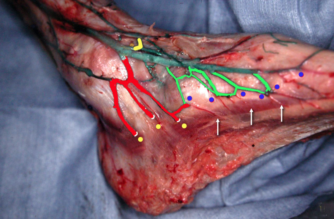
The calcaneal crossroad, convergence of the plantar veins. The convergence of the plantar veins (not visible) forms the plexus shaped calcaneal crossroad (C) that originates the two posterior tibial veins (pT). Here, the two main medial perforator veins of the foot are clearly visible, ending in the GSV: navicular (N) connected to the medial plantar veins and sub malleolar (M) to the calcaneal plexus (C). They are provided with valves (painted in yellow) allowing blood to go from the deep to the superficial system, particularity of the foot
The medial perforator veins (PV) of the foot: connection to the plantar veins. The three medial perforator veins on this dissection are connected to the medial plantar veins (1: cuneal PV and 2: scaphoid PV) or to the calcaneal convergence (3: infra malleolar PV). MM: medial marginal vein GS: GSV; PT: posterior tibial veins; D: dorsal communicating vein; M: medial plantar veins; L: lateral plantar veins; C: calcaneal convergence; 4: ankle perforator

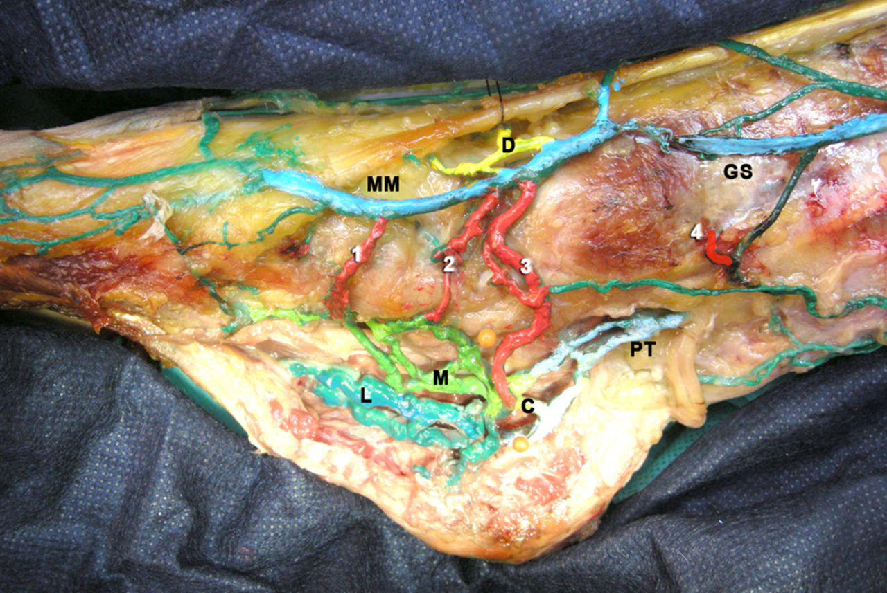
The two venous systems of the foot (the first metatarsal is partially resected). The medial system: the plantar and marginal veins communicating via many perforators, producing a true medial functional unit. The lateral system, the lateral plantar veins, a true venous pump in the foot. 1: medial marginal vein, 2: GSV; 3: perforator of the first metatarsal interspace; 4: cuboidal perforator; 5: navicular perforator; 6: malleolar perforator; 7: medial plantar veins (light green) have low capacities and communicate via the medial perforators; 8: lateral plantar veins (dark green), with their large diameter and length form a true blood reservoir in the plantar venous pump; 9: calcaneal plexus; 10: posterior tibial veins
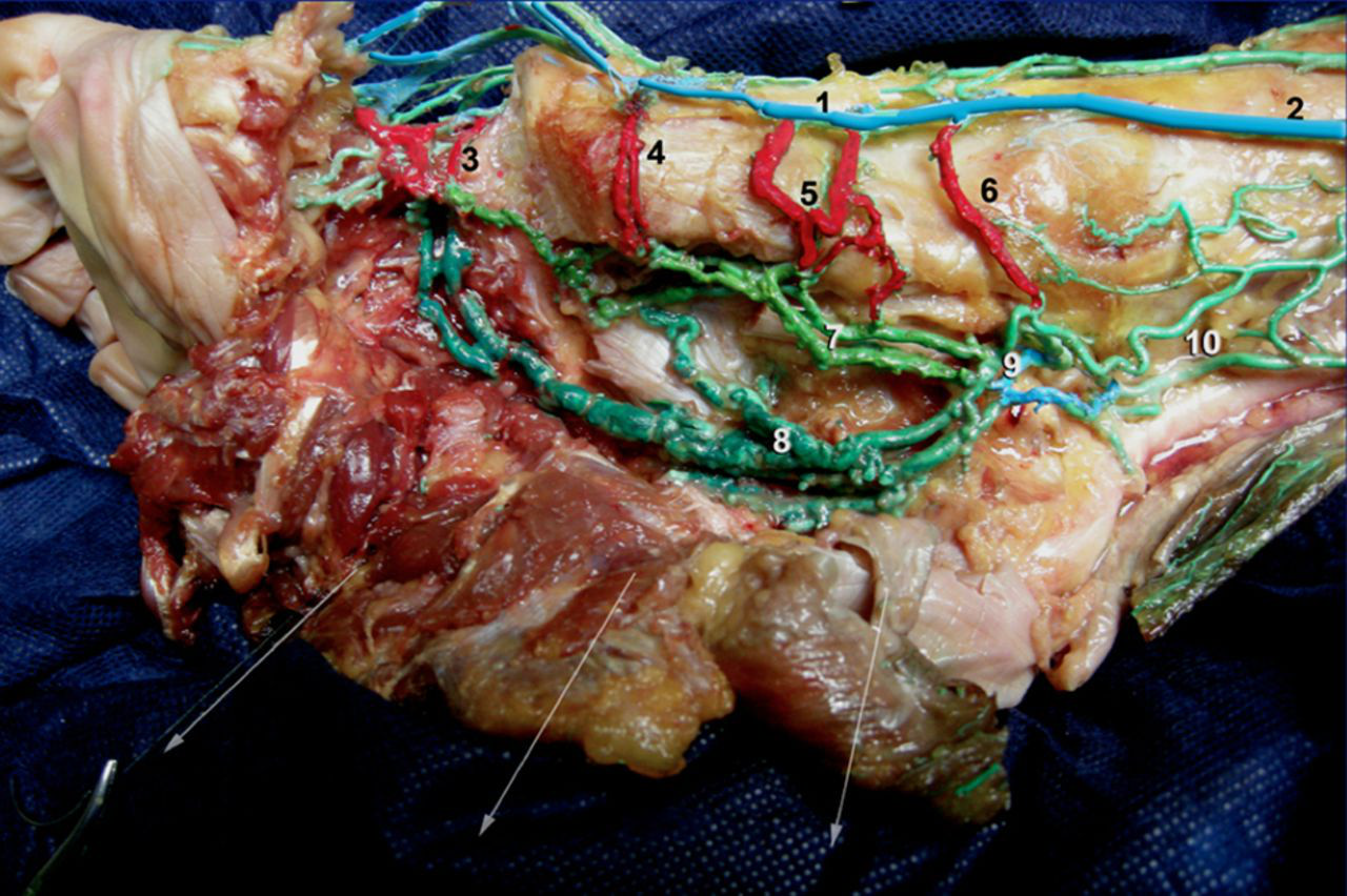
The two plantar veins (medial view after resection of the first metatarsial bone). 1: medial marginal vein; 2: GSV; 3: perforator of the first metatarsal interspace; 4: cuboidal perforator; 5: navicular perforator; 6: malleolar perforator; 7: medial plantar veins (light green); 8: lateral plantar veins (dark green); 9: calcaneal plexus; 10: posterior tibial veins
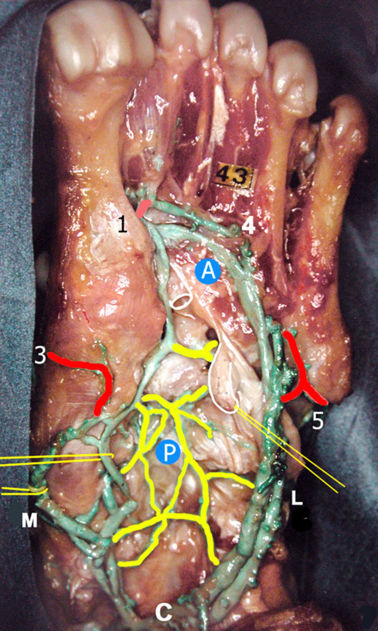
The deep muscular veins of the foot pump (sole view, the muscles have been resected). L: large lateral plantar veins; M: medial planter veins (pulled medially); P:plexus shaped anastomosis between L and M; A: plantar Arcade, origin of lateral plantar veins connected with the metatarsal perforators; 1: of the first space; 4: of the fourth space; 3: cuneal PV; 5: cuboidal PV; C: calcaneal confluence or crossroad
The terminal valves of the medial and lateral plantar veins were competent in normal subjects.
The perforator veins of the foot
The perforator of the first metatarsal interspace, generally of large diameter (Figure 16), was a relay because it was connected to the venous arch from the first interspace, which was the starting point of all venous networks in the foot, superficial and deep. The anterior tibial veins, the lateral plantar pedicle and the medial plantar pedicle arise from it. This arch was situated in relation to the superficial dorsal arch that joined the medial marginal vein and the lateral marginal vein;
The medial marginal perforator veins (Figures 13–16) open into the medial marginal vein and provide the three-root origin of the GSV. They differentiate into plantar and dorsal veins.
There are three plantar perforator veins (Figures 16 and 17):
the malleolar (or talus) perforator vein: close to the malleolus, it joins the confluent of the plantar veins; the navicular perforator vein: it is close to the tubercle of the scaphoid bone; the cuneiform perforator vein: it crosses the first cuneiform bone.
The dorsal medial perforator veins (shown in yellow).
The dorsal perforator vein (D) (shown in Figures 13, 15 and 16) communicated in front with the anterior tibial veins (Ta).
A: projection of the plantar venous axes. The plantar compression maneuver shows that while ejection is very effective in the plantar arch on which the venous axes project, normally it is zero in the weight bearing area. This also contradicts Lejars’ theory. The principal medial perforators. C: cuboidal; N: navicular; M: malleolar; 1: perforator of the first metatarsal interspace; 3: perforator of the third interspace; LP: lateral plantar veins; MP: medial plantar veins; B: relations with the weight-bearing area. Red circular patch:weight-bearing area on the ground. Blue circular patch best zones for manual compression area of the plantar veins. Purple circular patch: extremities of the foot reservoir (lateral plantar veins)
(3) The lateral marginal perforator veins (Figure 20): there were two – the calcaneal and the cuboidal which joined the lateral marginal vein. They crossed the lateral fibular tendons (inter-tendinous and subtendinous perforator).
The lateral perforator veins of the foot. Origin of the main portion of the small saphenous vein (SS): the lateral marginal (LM) vein is not a constant finding, but is often large; and the lateral malleolar plexus MP, on the contrary, is a constant finding. It gives rise to perforator veins. P: premalleolar; S: submalleolar. One can see a common vessel of the lateral perforators of the foot (C), which is a true third root of the SSV; it crosses the fibular long flexor tendons: the intertendinous (I), and cuboidal (Cu) perforator veins
Summary of this anatomical study
The overall organization of the veins in the foot traditionally differentiates the superficial veins, mainly the dorsal and marginal veins, and the deep veins, which are the medial and lateral plantar veins.
We propose a revision of the anatomical classification of these veins.
In fact, two very different venous compartments can be differentiated, with functional implications, as clearly shown in Figures 16, 17 and 21.
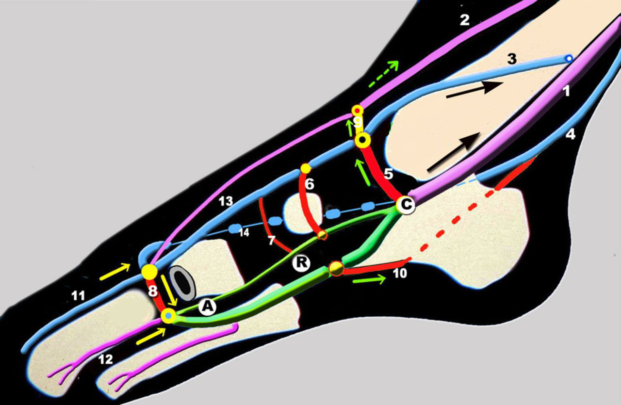
Hypotheses on venous foot pump function. The pump (shown in green) comprising the plantar veins is polarized and contains three parts going from front to back: A suction pole (A), a reservoir (R) in the plantar veins, and an ejection pole (C) the calcaneal confluence. Anteriorly, the distal pole (directed towards the toes) or aspiration A: blood enters the pump during raising of the foot in plantar flexion by relaxation of the plantar muscles. The pump is supplied by the highly vascularized toes and the highly developed metatarsal muscles. It is also supplied with superficial blood carried by the communicator vein of the first metatarsal interspace (8). The body of the pump or reservoir (R) is enhanced by the bony and muscular veins, but also by superficial blood by the medial and lateral perforator veins of the foot (in red). The distal or ejection pole is represented by the calcaneal confluence or calcaneal cross-road (C). Blood is supplied to the posterior tibial veins (1), but also the GSV (3), by the submalleolar perforator vein (5), and to the anterior tibial veins (2) by the dorsal perforator vein (9). 1: posterior tibial veins; 2: anterior tibial veins; 3: GSV; 4: SSV; 5: submalleolar perforator; 6: navicular vein; 7: cuneiform perforator vein; 8: perforator vein of first metatarsal interspace; 9: dorsal perforator vein; 10: calcaneal perforator vein; 11: dorsal vein of the hallux; 12: intermetatarsal vein; 13: medial marginal vein; 14: Lateral marginal vein
First, a medial compartment which includes the medial marginal vein and the medial plantar veins. They are well connected by three or four well-developed medial perforator veins, making a true functional medial unit. Here there is something unique to the foot: the directional flow of the blood from deep to superficial.
Second, a lateral compartment that consists practically of a single lateral pedicle, comprising the lateral plantar veins. They are long and of large diameter, with few perforators and thus without a major connection with the lateral marginal superficial venous system.
According to this classification, the venous pump is mainly deep and intermuscular, confined to the lateral plantar veins that supply the posterior tibial blood flow. Only the perforator of the first metatarsal interspace is common to the two venous compartments of the foot.
Three-dimensional modelling of the venous system of the foot by computed tomography venography
The anatomical findings of this study are supported by the investigation of our patients by computed tomography venography (CTV), performed in some complex patients or for recurrence after venous surgery. Our CTV acquisition and reconstruction protocol and results have been described previously. 6–9 It gives an accurate depiction of the foot veins, with interactive three-dimensional assessment, as shown in Figures 22–25.
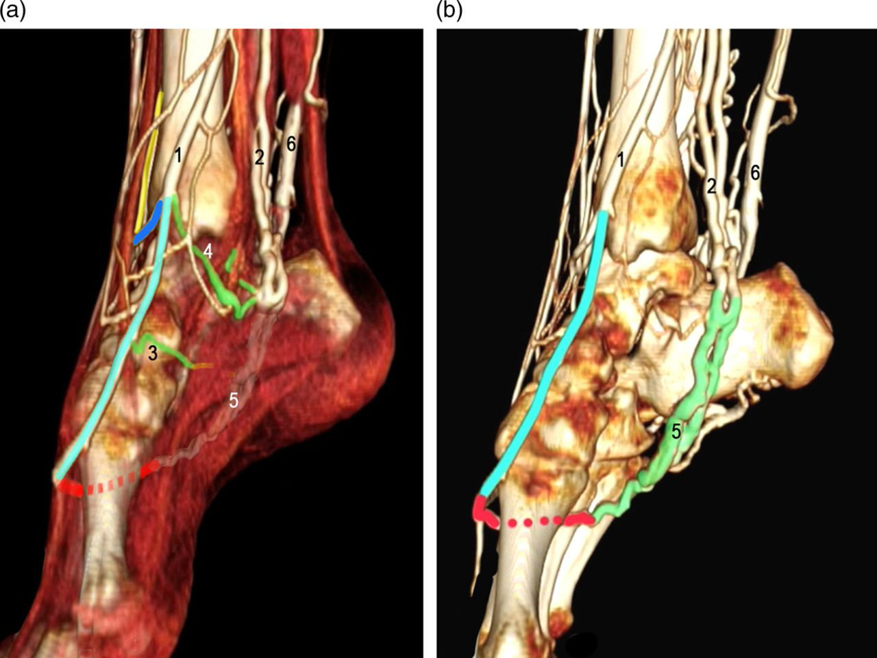
Three-dimensional reconstruction of the roots of the great saphenous vein (GSV), medial perforators and plantar veins by CTV. On the left (a), the medial marginal vein is coloured in light blue, the anterior tibial veins in yellow, the dorsal communicating vein in dark blue, and the medial perforators in green (3,4), and the perforator vein of first metatarsal interspace in red. 1: GSV; 2: posterior tibial veins; 3: scaphoid perforator; 4: infra malleolar perforator vein. The lateral plantar veins (5) are seen in transparency through the muscles. 6: SSV (dilated). On the right (b) the tendons and muscles have been removed. The lateral plantar veins (5, in green) and first metatarsal interspace (in red) are better seen

Three-dimensional reconstruction of the sole view of the plantar veins and calcaneal confluent. 1: lateral plantar veins; 2: medial plantar veins; C: calcaneal crossroad (confluence) of the plantar veins; T: the two tibial posterior veins

Three-dimensional (3D) reconstruction of the medial aspect of a Lejars’ sole. This 3D modelling in a recurrence after surgery shows a very important venous stasis with large dilatation of the veins of the sole (white arrows) and dilated lateral planter veins (marked 1). The perforator vein of first metatarsal interspace is marked 2

Three-dimensional reconstruction of the same patient. These images better show, coloured in blue, the pathological dilation of the veins of the sole (type Lejars) corresponding to a distal reflux with severe venous stasis
These new imaging tools also give access to virtual reality techniques, 6 which is mainly the virtual dissection of the limb. It can be clearly shown how it is possible to modify the transparency of the tissues to study interactively the superficial anatomy (Figure 22a) or the deep plane (Figure 22b).
Physiology of the foot pump
Clinical demonstration of the plantar venous pump
The existence of a plantar venous pump is an unquestionable reality. It is confirmed every day by phlebologists who carry out what they erroneously refer with the patient's foot immobile, manual compression exerted on the plantar venous arch triggers frank acceleration of blood flow in the posterior tibial veins, revealed in the ankle by Doppler scanning. Therefore, this acceleration of deep blood flow is immediate, intense, and repeated. This is a deep blood reservoir, mobilized by manual pressure exerted on the sole of the foot, corresponding to the lateral (and medial) plantar veins included between two fleshy muscles.
Proposal of physiology of the plantar pump in lower limb venous return
At rest, the venous pump is not active, as we shall subsequently see, therefore the system is necessary and sufficient in itself to provide specific continuity of venous return;
When a subject goes from the seated to the standing position, under the influence of gravity, the weight of the column of blood exerts a pressure of about 90 mm of mercury;
After a certain number of steps (about 10–20), ankle pressure falls to 30 mm of mercury. This decrease is related to mobilization of the volume of blood, due to activation of the different venous pumps in the lower limb during walking. A well-known mechanism in the calf and in the thigh, where the intramuscular veins of the triceps and of the quadriceps act as a blood reservoir, and in particular those of the soleus muscle and the medial head of the gastrocnemius muscle, and those of the vastus lateralis muscle. Contraction ejects the volume of blood and muscle relaxation allows filling of this reservoir. A series of valves channels the blood propelled to the root of the limb and prevents any reflux. 4,5,10–16
Where is the plantar venous pump located?
Several lines of evidence confirm that the plantar venous pump is located in the deep, intermuscular space:
The plantar venous axes are connected directly to the posterior tibial veins, which are extensions of them; The direction of the valves indicates that blood flows from the deep to superficial; The volume of blood ejected by the pump is 20–30 mL, which corresponds to the capacity of the lateral and medial plantar veins. These veins are primarily inter-muscular, thus pointing to the intervention of a motor apparatus for venous ejection during walking, which adds to the pressure exerted by the foot on the ground. Anteriorly, the suctioning pole (A), is directed towards the toes. There are many supply pathways: the highly vascularized toes, the highly developed metatarsal muscles, the large metatarsal perforator vein (arising from the superficial network of the medial marginal vein); The middle portion comprises the body of the reservoir (R) of the pump, whose average volume is 15–25 mL. It is enhanced at this level by bony and muscular veins, the medial and lateral perforators. Weight bearing and the action of walking produce a massage effect on the plantar vascular area; Posteriorly, the calcaneal confluence (C), which corresponds to the ejection pole. Of course, it empties with full diameter into the posterior tibial veins; But it also goes in the inframalleolar perforator into the medial marginal vein, the origin of the GSV below the ankle; Lastly, it goes into the anterior communicating vein of the ankle, which is connected to the anterior tibial veins.
Our anatomical description of the lateral plantar veins shows the three components of this pump that determine its function (Figure 21):
The ejection of blood goes into three main axes:
This is confirmed in everyday phlebological practice by the substantial increase in posterior tibial as well as saphenous and anterior tibial blood flow after manual compression of the plantar surface, which has been confirmed by many authors.
4,12–14
In summary, three phases can be described during walking:
The weight-bearing phase: contact of the foot on the ground produces direct compression of the reservoir in the sole of the foot between weight-bearing areas; The impulse phase: weight bearing on the forefoot with flexion of the toes that fixes the foot on the ground, resulting in compression of the pump in the musculo-tendinous plane by muscle contraction; The suspension phase of the foot, lifted off the ground, allows filling of the pump.
During walking, the pump reloads cyclically when the foot is lifted up and empties when weight bearing is applied. This plantar venous pump is the only effective pump up to the level of the calf, where its action is taken over by the calf pump of the soleus muscle. Its dual action, on both the deep and the superficial saphenous vein circulation, underlines the unimpeded circulation of blood between the two vascular compartments.
In daily practice for phlebologists
The blood reservoir, mobilized by manual pressure on the sole of the foot and emptied during weight bearing during walking, is not a subcutaneous plantar, superficial one like ‘Lejars’ sole’.
It is a deep reservoir, corresponding to the lateral plantar veins between the two fleshy muscles and is compressed at each step of walking.
It is important to recognize the utility of the stimulation of the foot pump in the prevention of post-surgical deep vein thrombosis. This should be emphasized 17,18 in high-risk patients.
This can be done by simple manual massage going upwards, using intermittent pneumatic compression, or by simple elastic compression starting at the root of the toes.
A compression stocking of 22 mm of Hg at the ankle level has also been shown by magnetic resonance imaging to significantly reduce the diameter of the leg veins in standing position. 19
This anatomical study also allows us to better understand and confirm the major role played by the foot pump in the venous return of the whole limb. This pump of the human foot is the first step in venous return from the lower extremity to the heart. The calf pumping mechanism, produced by contraction of the soleus muscle and of the gastrocnemius muscle, then takes over. 4,5,12,18
Therefore, the impairment of the foot pump could have an important clinical impact in patients with chronic venous disease. This is the case with the foot static disorders such as flat or hollow foot.
These abnormalities are very common in patients with chronic venous disorders (CVD), and should be considered as an important risk factor of CVD. 20 This is the reason why, in daily practice, they should be looked for, and when found corrected with an insole.
Conclusion
This anatomical study confirms that the plantar venous pump is an undeniable reality: it is located deeply between the plantar muscles in the plantar veins (particularly the lateral ones).
Their ejection pole is at the calcaneal crossroad going straight upwards in the two posterior tibial veins, but also by the infra malleolar perforator into the anterior tibial and the GSV.
The plantar veins are the true blood reservoir of the foot, which moves upwards as the result of manual compression of the sole or weight bearing during walking. Thus the foot pump is literally the first step of venous return during walking.
For this reason it is crucial to look for foot static disorders 20 in patients with chronic venous disease. Such disorders are any foot dysmorphism (flat or hollow foot) that may reduce the efficacy of the foot pump, but which have also been proved to be responsible for an impairment of the calf pump. 21 In such cases, treatment with an insole 21 will reduce both abnormalities and thereby improve the venous insufficiency and the symptoms of the patient, which are not always of venous origin.