
Abstract
The purpose of our study was to evaluate whether total laparoscopic aortofemoral bypass can be performed routinely in patients who require surgical intervention for aortoiliac occlusive disease. In a prospective study, 68 consecutive patients underwent total laparoscopic aortofemoral bypass between 2002 and 2004. Among these patients, there were 50 men and 18 women, with a mean age of 68.4 ± 9 years. The mean operating time was 199 minutes, with a mean aortic cross-clamp time of 85.8 minutes. There were five major complications (7.3%). The mean postoperative hospital stay was 6.3 days. Most of the younger patients could be discharged on the third or fourth postoperative day. Our results show that total laparoscopic aortic surgery can be offered as a routine procedure to the majority of patients with long-segment aortoiliac occlusive disease.
Total laparoscopic aortic procedures can be performed in patients with aortoiliac occlusive disease (AIOD) or abdominal aortic aneurysm. 1–3 According to a number of publications, laparoscopic aortic surgery can only be performed via transperitoneal approach mainly in selected cases because of the technical challenges and the time required. 4 We wanted to evaluate in a prospective study whether a total laparoscopic aortic operation can be offered to patients with long-segment AIOD as a routine procedure without preoperative selection.
Patients and Methods
The patient was placed on the operating table on a vacuum bag. When tilting the table to the right, the patient could be positioned almost 70° on the right lateral decubitus position. Seven or eight ports are used for access. In all patients, a transperitoneal, left retrocolic access was used for exposure of the aorta. The left hemicolon and splenic flexure were mobilized medially (Figure 1). The apron technique, originally described by Dion and Gracia, 5 was used to separate the abdominal contents from the retroperitoneal space. Laparoscopic exposure of the aorta was initiated at the level of the left renal vein (Figure 2). The laparoscopic camera was positioned in the left upper abdomen during exposure of the aorta. Only the area for the anastomosis proximal to the origin of the inferior mesenteric artery was dissected to avoid injury of the lumbosacral nerves adjacent to the aortic bifurcation.
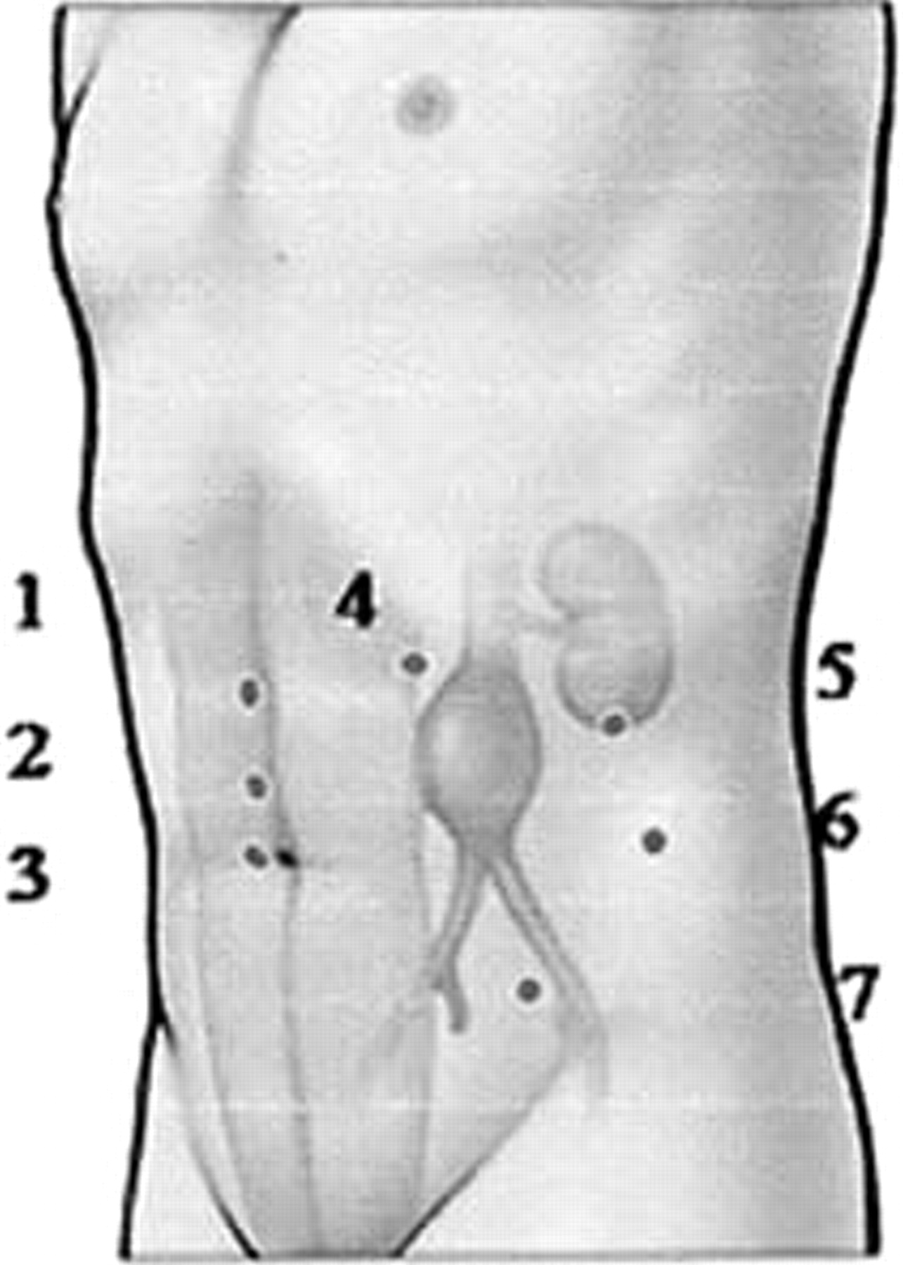
Medial mobilization of the left hemicolon and the splenic flexure to expose the aorta.

Intraoperative photograph showing the left renal vein (solid arrow) and the aortic Dacron graft (interrupted arrow).
In patients for whom an end-to-end anastomosis was planned, the infrarenal aorta was stapled with a linear, noncutting stapler. If calcification was too severe, thromboendarterectomy of the aortic stump was performed. A deployable aortic clamp (Karl Storz, Tuttlingen, Germany) was used to occlude the distal aorta. Using this novel device, only the port for the proximal aortic clamp was obstructed with an instrument.
Laparoscopic dissection and suturing were performed with the surgeon standing on the right side of the patient during the operation. 6 In patients with an end-to-end anastomosis, the aorta was completely transected to facilitate the suturing process. In patients with an end-to-side anastomosis, an aortotomy was performed, and the anastomosis was started at the heel and posteriorly with a 3-0 polypropylene suture, 10 cm in length. A second suture was taken anteriorly, and both were tied intracorporally. Time could be saved by using two 3-0 polypropylene sutures blocked with a pledget at the end, as originally described by Coggia and colleagues. 6 Tunneling was performed under laparoscopic control dorsal to the ureter after exposure of the femoral arteries in the groin.
Reasons for conversion to a minilaparotomy were outlined before surgery. According to our self-established guidelines, they included an aortic cross-clamping time of more than 2 hours and a total operating time exceeding 4 hours. In these cases, we converted to a laparoscopic hand-assist procedure, in which the anastomosis was performed under the pneumoperitoneum but with the nondominant hand of the surgeon inserted into the abdomen. 7–10 Other reasons for conversion included extensive adhesions, a severe calcified aorta, and uncontrollable blood loss. The mean value and standard deviation of the mean are given in the table below (Table 1). When appropriate, nonparametric tests were used to describe a statistical significance of p < .05.
Perioperative and Postoperative Parameters
Results
Total laparoscopic aortofemoral bypass was performed in 68 consecutive patients admitted in two hospitals from 2002 to January 2004. Among 68 patients, there were 50 men and 18 women, with a mean age of 68.4 ± 9 years. The mean body mass index (BMI) was 28 ± 1 kg/m2. Indication for surgery was disabling claudication with a walking distance of less than 30 meters in 30 patients and necrosis or gangrene in 38 patients. The majority of the patients (51 of 68) were heavy smokers until the day of the operation.
Preoperative angiographic results showed the following TransAtlantic Inter-Society Consensus (TASC) classification 11 : 4 TASC B, 7 TASC C, and 57 TASC D lesions. Among the 68 patients, 51 (75%) patients had undergone interventional procedures, including subintimal angioplasty and/or multiple stent placements, prior to their surgical intervention. Laparoscopy and suturing were performed by one of three surgeons who were trained in these procedures. In eight cases, surgery was performed without prior attempts to use angioplasty and stent placement. The mean operating time was 199 minutes, with a mean cross-clamp time of 85 minutes; the length of time depends not only on the time required for the aortic anastomosis but also on the complexity of the reconstruction in the groin (see Table 1).
There were five major complications (7.3%). One patient died after a massive myocardial infarction. Another patient with Leriche's syndrome, who underwent an end-to-end anastomosis with an aortic cross-clamp time of 34 minutes, developed paraplegia postoperatively. One patient had transient renal failure for 3 weeks after developing a compartment syndrome, which eventually resolved completely. One 82-year-old patient with forefoot gangrene remained in the intensive care unit for more than 5 days because of pneumonia. Another patient developed limb ischemia owing to graft thrombosis and underwent graft thrombectomy and profundaplasty.
Limb salvage was achieved in 68 patients. During a mean follow-up period of 47 months, additional procedures, such as distal bypass or peripheral angioplasty, were performed in 16 patients because of the large number of cases involving critical limb ischemia and requiring a staged multilevel revascularization. An end-to-end anastomosis was performed in 21 cases and an end-to-side anastomosis in 47 cases. An end-to-end anastomosis was routinely performed in patients with concomitant small aneurysms (< 5 cm) and occlusive disease and in those cases with Leriche's syndrome. Conversion to a minilaparotomy was required in three cases, in one case because of difficult exposure and in two cases because of severe calcification of the aorta, which did not permit safe clamping. In two patients, the suprarenal aortic segment was clamped, and thromboendarterectomy of the juxtarenal aorta was performed prior to completing the proximal anastomosis.
Discussion
Patients with atherosclerotic, AIOD are at high risk of postoperative complications, and a minimally invasive procedure may favorably affect their postoperative recovery. The majority of the 68 patients (75%) in our series had undergone interventional procedures prior to their surgical intervention. Our preference is to treat the occlusive lesions via endovascular means and to reserve surgical intervention for those who have failed endovascular treatment. Laparoscopic vascular surgery is another minimally invasive, feasible, safe, and effective technique for the treatment of AIOD and aneurysms. 12–15 Most of our patients stayed less than 1 day in the intensive care unit.
All operations were performed by three different surgeons. When operating and cross-clamp times were analyzed according to the individual experience of the operating surgeon, the cross-clamp time was significantly surgeon dependent. A reduction in total operative time can be accomplished only when the entire operative team has sufficient experience with laparoscopic aortic procedures. In this series of patients, the mean BMI was 28 ± 1 kg/m2; however, obesity was not a contraindication for laparoscopic aortoiliac surgery. It appears that obese patients can benefit particularly from a minimal invasive approach. Postoperative hospital stay was correlated with the age of the patient. 16 The majority of patients younger than 60 years could be discharged on the third or fourth postoperative day. Older patients, especially those in their eighties, were kept in the hospital longer, mainly for nonmedical or social reasons.
In our experience, an end-to-end anastomosis was more difficult simply because of the aortic pathology. This would require adjunctive procedures, such as a thromboendarterectomy and polytetrafluoroethylene graft reinforcement of the suture line. A major advantage of the transperitoneal, left retrocolic access is the position of the graft on the left lateral aspect of the aorta, which prevents the development of a fistula between the duodenum and the prosthesis. With this kind of access, peritoneal covering of the graft does not cause problems, as opposed to a transperitoneal approach, especially in very thin patients.
Although there is a learning curve, a laparoscopic approach can be offered routinely to most patients who require surgical intervention for AIOD. Operative times, as well as aortic cross-clamp time, were only slightly longer than for open surgery. We now have the operative technique and the instrumentation to progress to total laparoscopic aortic procedures as a routine operation for the treatment of long-segment AIOD.