




Abstract
Mucormycosis is an aggressive, opportunistic infection caused by fungi belonging to the class of Phycomycetes. Rhino-occulo-cerebral mucormycosis is the commonest anatomical presentation of mucormycosis and is a potentially fatal disease. We report here a case of a 65-year-old diabetic female who presented with a locally invasive type of mucormycosis and was treated successfully by surgical debridement along with medical treatment (Amphoterecin-B and insulin).
Case history
A 65-year-old woman presented with complaints of a low-grade fever accompanied by headache and nasal discharge of two months' duration. She had been diabetic for 4 to 5 years and had poor glycaemic control. She developed redness and swelling over her nose for which she was started on antibiotics. Her investigations revealed haemoglobin (Hb) 10.0 g%, total leucocyte count 7600/mm3, erythrocyte sedimentation rate 39 mm in the first hour and random blood sugar (RBS) 340 mg%. Kidney function tests, serum electrolytes and liver function tests were normal. She was put on intravenous fluconazole in addition to the antibiotics which were given for one week and, because there was no response, she was referred to our hospital.
On admission she was afebrile, had facial puffiness, right-sided periorbital oedema, right-sided sixth cranial nerve palsy and nasal twang because of a perforation of the hard palate (Figure 1). Investigations revealed anaemia (Hb 8.2 g%) and uncontrolled blood sugar (RBS 440 mg%) but the urine examination was negative for ketones. Potassium hydroxide smear of nasal swab revealed fungal hyphae, which was found to be aseptate hyphae of mucormycosis on histopathological examination. Computed tomography of the head revealed destruction of nasal septum and hard palate without any evidence of intracranial extension. She was started on IV Amphotericin-B 1 mg/kg/day along with insulin therapy and debridement was performed under general anaesthesia. During the course of the treatment her blood urea rose from 20 to 51 and the dosage for Amphotericin-B had to be decreased – it was later increased after the normalization of the blood urea. During her stay in the hospital she began to improve and was subsequently discharged.
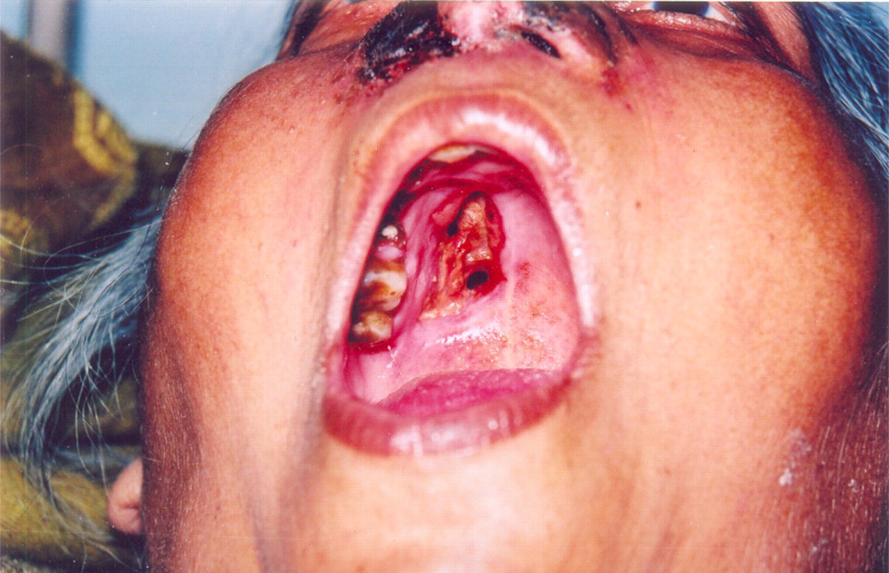
Clinical photograph showing black-coloured nasal discharge and hard palate perforation
Discussion
Mucormycosis is an opportunistic infection which spreads by direct as well as haematogenous dissemination. Based on clinical presentation and the involvement of a particular anatomic site, mucormycosis can be divided into at least six clinical categories: (i) rhinocerebral; (ii) pulmonary; (iii) cutaneous; (iv) gastrointestinal; (v) disseminated; and (vi) miscellaneous. These categories tend to occur in patients with a specific host defence defect. For example, diabetics in ketoacidosis typically develop the rhinocerebral form of the disease and, much more rarely, develop pulmonary or disseminated disease. 1 Other predisposing factors include leukaemia, lymphoma, long-term immunosuppressive therapy after bone marrow or organ transplantation and IV drug abuse.
Mucormycosis originating in the nose and para-nasal sinuses produces a characteristic clinical picture. Low grade fever, dull sinus pain, and sometimes nasal congestion or a thin bloody nasal discharge, are followed in a few days by double vision, increasing fever and obtundation. Examination reveals a unilateral generalized reduction of ocular motion, chemosis and proptosis. The nasal turbinates on the involved side may be dusky red or necrotic. A sharply delineated area of necrosis, strictly respecting the midline, may appear in the hard palate. Facial skin adjacent to paranasal sinuses may be invaded by direct extension, turning progressively red, purple and black. 2 Involvement of III, IV, VI and the ophthalmic division of the V cranial nerve is not uncommon. Clinical features often resemble those of cavernous sinus thrombosis. 3 The case described above had typical presentation. Rhino-orbito-cerebral mucormycosis in poorly-controlled diabetics carries a high mortality. Seemingly trivial symptoms, such as unilateral nasal blockage, discharge and headache, should not be ignored. 4
The successful diagnosis and treatment of mucormycosis requires: a high degree of clinical suspicion; prompt diagnosis (particularly in immunocompromised patients); the availability of senior ENT and ophthalmic surgeons, on an emergency basis, for the prompt extensive surgical debridement of the lesions; appropriate and aggressive antifungal therapy (Amphoterecin); and reversal of the underlying predisposing factors where possible. 5 The exact dose of Amphoterecin-B for effective eradication of mucormycosis is not known. Western literature mentions 1–1.5 mg/kg body weight daily until a maximum of 3 g is reached. However, our patients require a much smaller dose as their tolerance to conventional Amphoterecin-B is extremely poor. Liposomal Amphoterecin-B, which is less toxic, is beyond the reach of most of the patients as it is quite expensive. 3
Conclusion
Mucormycosis is a serious infection and can be rapidly fatal. One should always bear it in mind and investigate patients with suggestive nasal and ocular clinical manifestations, particularly those with poor glycaemic control. A high degree of clinical suspicion, early diagnosis and prompt aggressive surgical evacuation of the inflammatory material, along with Amphoterecin-B therapy, can save lives.