




Abstract
Summary
Ophthalmomyiasis externa is mainly caused by the sheep botfly Oestrus ovis; therefore, it is usually seen in rural areas. We report here a case of ophthalmomyiasis externa in a young man from Istanbul. The patient had no known history of exposure to farm animals or rural areas.
Introduction
The term ‘myiasis’ was originally coined by F W Hope in 1840 to define parasitic infestation by insect larvae. Myiasis can be classified based on the mode of parasitic behaviour (obligatory, facultative or accidental) and area of infestation (skin, wounds, intestines, urinary tract, eye, ear, etc.).
Ocular myiasis (ophthalmomyiasis) can be seen in many countries around the world, albeit rarely.1–3 There were seven reports of human cases in Turkey, between the years 1922 and 1965. No further cases have been reported since that date, except for a case of scar tissue myiasis involving the tissue surrounding the eye. In order to draw attention to this rare phenomenon, we report a case of ocular myiasis with first instar larvae of Oestrus ovis.
Case history
On a hot dry day in August 2008, a 25-year-old male medical student presented to our hospital complaining of itching, burning, pain and redness on his left eye. He explained that two hours before he had been taking a stroll in downtown Taksim (Istanbul) during the lunch hour when something flew past his eye. His complaints began after this event and after rubbing his eye he noticed small white larvae on his hand. A physical examination revealed redness in the conjunctiva, prominent blood vessels in the sclera and excessive lacrimation. Small white motile larvae were noticed under the eye lids and a total of 13 larvae were extracted using forceps (Figure 1). The conjunctival sac was washed with a saline solution. Topical steroid cream and prophylactic antibiotic treatments were applied.
One of the larvae extracted from the patient's eye. View under the light microscope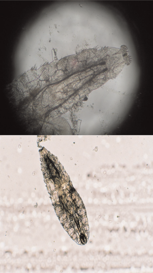
The extracted larvae were 1–2 mm long, white and oblong-shaped, with two large, black, horn-shaped mouth hooks at the anterior end. On the abdominal side there were double rows of short spines located posterior to every segment and lateral to them were groups of longer and thinner spines. The posterior spiracle consisted of two lobes with approximately 9–10 chitin-like spines on each lobe. These characteristics confirmed that these were first instar O. ovis larvae.
Discussion
Ocular myiasis is a rare phenomenon in humans and generally presents with the involvement of the conjunctiva (ophthalmomyiasis externa). Involvement of orbital tissue (ophthalmomyiasis interna) is extremely rare.
Causative agents in most cases of ocular myiasis are the sheep parasite O. ovis larvae and equine parasite Rhinoestrus purpureus larvae. Occasionally, other species such as Wohlfahrtia, Hypoderma and Chrysomyia may result in ocular myiasis. Most of the cases reported from Turkey involved O. ovis, with only one published case of R. purpureus. 4
O. ovis is a cosmopolite insect which can be found anywhere where sheep and goats are present. It is a viviparous insect laying larvae instead of eggs. In hot dry weather the insect leaves its larvae in moist areas of the host animal, such as the nose, eye or mouth. Larvae move to the paranasal sinuses causing disease symptoms of serious respiratory distress which may even lead to the death of the host. Larvae complete their developmental cycle inside the host body (this may take 1–12 months) and upon reaching the mature larval stage leave the host by falling on earth. There they pupate and emerge as adult flies within 3–4 weeks.
Although humans are not natural hosts of O. ovis, numerous cases have been reported in people who have close contact with sheep and goats. They usually cause painful conjunctivitis in humans. A typical history involves an incidence when something has flown by, after which the patient experiences pain, itching, dryness and redness in the eye. Since O. ovis larvae cannot complete their development in humans, they are only found as first instar larvae and cannot survive for more than 10 days. Therefore, symptoms usually recede by themselves within a few days.
Symptoms such as sensation of a foreign body, itching, burning, pain, photophobia and excessive lacrimation develop within a few hours after the adult female fly has squirted its larvae in the recipient's eye. Redness and oedema are seen in the conjunctiva. Larvae can only be detected by their movement upon careful examination. Sometimes they may move into the nasal cavity and cause symptoms of rhinitis. Usually 8–20, sometimes more, larvae can be found in one eye. Their small size and transparency may make it difficult to correctly diagnose the cause of conjunctivitis.
Treatment is performed by the mechanical extraction of the larvae using forceps. Local anaesthesia immobilizes the larvae and may aid the extraction process. 5