




Abstract
Caecal volvulus is an uncommon cause of acute intestinal obstruction caused by axial twisting of the caecum along with the terminal ileum and ascending colon. Early diagnosis is essential in order to reduce the high mortality rate, though the condition is rarely diagnosed correctly at the time of presentation. We report a series of four cases describing their presentation, management and subsequent outcome.
Introduction
Caecal volvulus is characterized by axial rotation in the caecum, terminal ileum and ascending colon associated with twisting of mesenteric vessels. 1 The annual incidence of caecal volvulus is reported to range from 2.8–7.1 per million people in the general population. It is responsible for 1–1.5% of all adult intestinal obstructions and 25–40% of all volvulus involving the colon. 2,3 It is generally agreed that patients with acute caecal volvulus benefit from surgical intervention for the correction of intestinal obstruction. Contemporary surgical options include manual detorsion, caecopexy, caecostomy and colectomy by open or laparoscopic approaches. 4
In this case series, we report clinical features, management and subsequent outcome of four patients presenting with caecal volvulus.
Case histories
Case 1
A 32-year-old man presented to the emergency department complaining of abdominal distention, multiple episodes of bilious vomiting and abdominal pain of six days. On examination, he was in a state of shock. An abdominal examination revealed generalized distension, board-like rigidity and absent bowel sounds. A plain X-ray of the abdomen showed multiple dilated jejunal and ileal bowel loops. After resuscitation, with the diagnosis of acute small bowel obstruction, exploratory laparotomy was done. On exploration, 3 L of faeculent fluid was aspirated and dilated small bowel loops were noted, with a twisted, hugely dilated and perforated caecum located in the left upper quadrant of the abdomen (Figure 1). Ileoceacal resection followed by ileoascending anastomosis was done. In the postoperative period, the patient developed wound infection followed by dehiscence. On the 10th postoperative period, leakage from the anastomosis was noted. He was managed expectantly with success and was discharged on the 23rd day postoperatively.
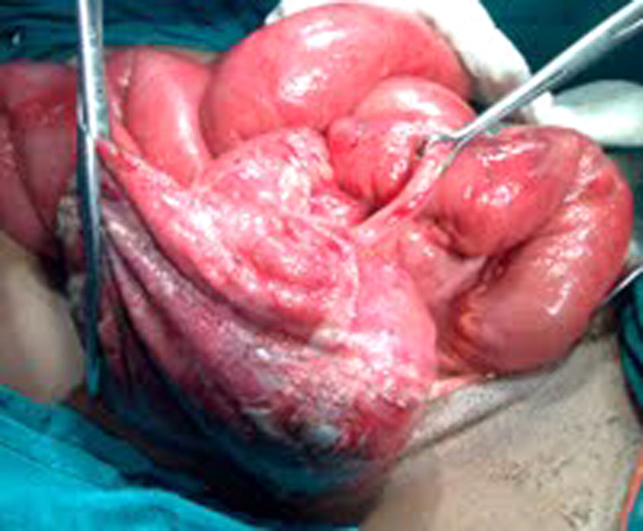
Intraoperative photo showing a hugely dilated and edematous caecum with perforation at the superior most part
Case 2
A 65-year-old man presented to the emergency department with a history of sudden onset abdominal pain, frequent vomiting and abdominal distension. On clinical examination he was in a state of shock and his abdomen was grossly distended with features of generalized peritonitis. His abdominal X-ray showed multiple dilated bowel loops with air fluid levels (Figure 2). A chest X-ray revealed bilateral pleural effusion. On analysis of the arterial blood, he had metabolic acidosis with a pH of 7.1, bicarbonate level of 8 mmol/L and pCO2 of 33 mmol/L. He had raised serum urea and creatinine. After adequate resuscitation, the patient underwent exploratory laparotomy under general anaesthesia. On exploration, he had a twisted and dilated caecum with perforation and dilated small bowel loops. Limited ileoceal resection followed by ileoascending anastomosis was performed. During the intraoperative period inotropic support was started in order to maintain his blood pressure. He was then transferred to the intensive care unit (ICU) for mechanical ventilation. His condition deteriorated further during his ICU stay. On the second postoperative day he had cardiac arrhythmias followed by a fatal cardiac arrest.

Plain X-ray of the abdomen showing multiple air fluid levels, with dilated air fluid filled caecal loops at the right lower abdomen
Case 3
A 21-year-old man presented with complaints of severe colicky abdominal pain, several episodes of bilious vomiting and gradual distension of the abdomen for a duration of three days. At presentation, he was dehydrated and had generalized abdominal distension with visible bowel loops. Plain abdominal X-rays showed multiple dilated small bowel loops. He underwent exploratory laparotomy, revealing caecal volvulus with dilated proximal bowel loops. Limited ileoceal resection, followed by ileoascending anastomosis, was performed. He had an uneventful postoperative period and was discharged on the seventh postoperative day.
Case 4
A 60-year-old man presented with a complaint of recurrent abdominal pain for one year, distension and multiple episodes of nonbilious vomiting for three days. On examination, he had a soft, nontender but distended abdomen with absent bowel sounds. His distension progressed with expectant management and he finally underwent exploratory laparotomy. On exploration he had dilated small bowel loops, a mobile and dilated caecum and a collapsed large bowel. The patient underwent caecopexy, had a smooth postoperative period and was discharged on the sixth day. He has remained asymptomatic for 18 months.
Discussion
The first report of caecal volvulus was by Rokitansky in 1837. It is characterized by axial twist of the caecum, ascending colon and terminal ileum around a mesenteric pedicle. 1 Caecal volvulus usually occurs in patients with a mobile caecum or a right colon that is not properly secured to the parietal peritoneum. Various diverse factors, such as postsurgical adhesions, early postoperative period, high fibre diet, congenital malformations, Hirshsprung's disease, increased peristalsis induced by purgatives or diarrhoea, pelvic tumour and pregnancy have been associated with the development of volvulus. 5–9 Although caecal volvulus is traditionally thought to be more common in females, all four patients in our series were male. 2,10 In our report the age of the patients ranged from 21–65 years, suggesting that it can occur in both young and old adults.
As abdominal clinical and radiological findings are frequently nonspecific, a definitive diagnosis in most patients is rarely established on the basis of the initial evaluation. 11,12 In a simple X-ray of the abdomen the usual findings are swollen loops of the small intestine with the absence of gas in the distal colon - typical symptoms of a blockage of the small intestine. An abdominal X-ray showing an unusually distended caecum pulled towards the epigastric or left hypochondrial region should arouse a suspicion of caecal volvulus. However, this typical finding was absent in our patients. 13,14
The use of colonoscopy or a barium enema for diagnosis and then reduction should be considered, as anecdotal methods have a risk of perforation and are a source of delay before surgery. The computed axial tomography scan is now being considered as the primary imaging modality. The most reliable sign of caecal volvulus is the ‘whirl sign’, which is composed of a spiralled loop of the collapsed caecum and engorged mesenteric vessels. 15
Patients with acute intestinal obstruction due to caecal volvulus usually require surgical intervention. Contemporary surgical options include manual detorsion alone, caecopexy, caecostomy and resection by open or laparoscopic approaches. 5 In the presence of a gangrenous colon, resection, usually a right hemicolectomy should be the treatment of choice. 2,4,5 Laparoscopic assisted resection has been described in the literature for uncomplicated cases. 16 The decision to perform a primary anastomosis or ileostomy depends on the patient's condition, presence or absence of perforation and the situation of the colon at the time of surgery. In retrospect, in view of our second patient (Case No. 2) we emphasize that resection of the gangrenous segment followed by ileostomy, with provision of ‘second-look surgery’ may be the safest modality of management in poor risk patients. However, the appropriate extent of the operative therapy in patients without gangrenous changes has remained undetermined. Recurrences of caecal volvulus have been reported following caecopexy. Caecostomy is associated with serious complications such as gangrene, caecal necrosis, intraperitoneal leakage of faeces, a greater mortality than with caecopexy, and recurrence. 4,5