




Abstract
Radiographic films remain the norm in developing countries. Interpreting them is easier and more reliable, using a view box, than holding them up to a light. Commercial view boxes are expensive and difficult to repair. We designed and built an inexpensive radiographic view box using commonly available materials and local craftsmen. If they break, they can be easily repaired by hospital staff. This design can be replicated anywhere in the world.
Introduction
While digital radiographic monitors proliferate in clinical settings in the most developed countries, producing radiographs on standard film remains the norm in most parts of the world. Often the lack of radiograph view boxes (light boxes) makes reading the films more difficult and less reliable than necessary.
A solution to this problem is to wait for either donations of used view boxes or the purchase of new ones; neither might occur. Improvised and less-than-optimal methods for viewing radiographs without view boxes include holding films up to a light fixture (which produces uneven and patchy image illumination) or taping films to windows (which works only during daylight hours, if a window of sufficient size is accessible and if the window design [i.e. louvers] or objects outside the window do not obscure the image).
Clinicians at the Kintampo Municipal (District) Hospital in rural Ghana often need to view radiographic images in the busy emergency department and outpatient clinics and the often full inpatient paediatric, maternity and adult wards. The only functioning view box was located adjacent to the X-ray imaging equipment, an inconvenient and time-consuming trek from most clinical areas. While the hospital had purchased two expensive commercial view boxes in the recent past, they had malfunctioned after about two months and could not be repaired.
Method
Recognizing that new view boxes were not forthcoming, we decided to build our own from local materials. To our amazement, no design for making such a device was available and so we produced a construction design based on the standard commercial view box but using locally available materials (Figure 1). The design consists of a wooden case containing fluorescent bulbs and a translucent plastic viewing screen. We used bulbs that were used in the hospital's ceiling lights and the plastic was readily available locally. The plastic slides into grooves in the sides of the box and can be easily lifted out to change the bulbs. Radiographs rest on a lip at the bottom of the box and are held in place with both the thin top wooden slat that holds the plastic sheet as well as two wires tightly strung across the front of the box between the eye bolts. A typical activation switch on the electrical socket serves to turn the light box on and off.
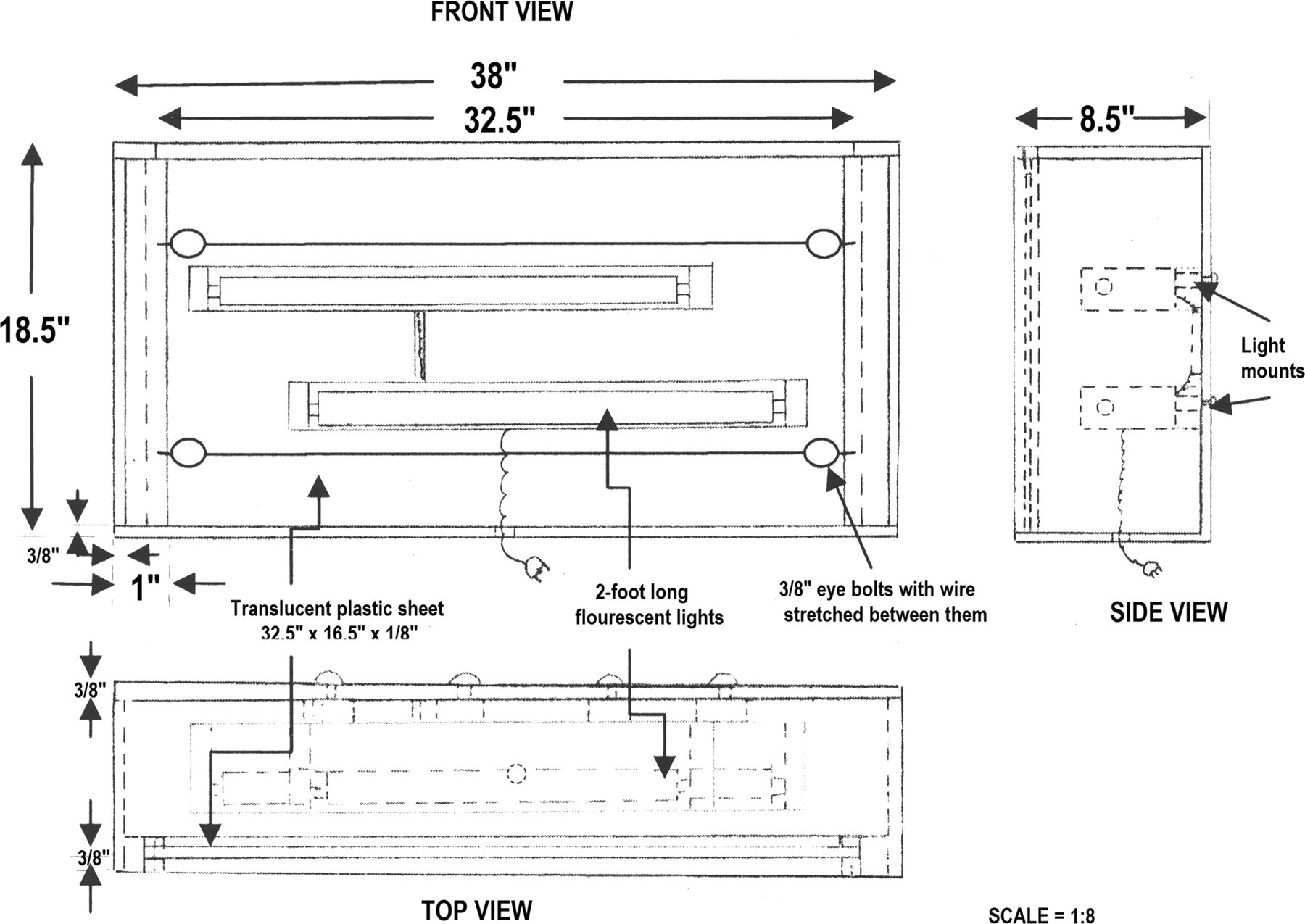
A diagram for the construction of a radiographic light box holding two films
As shown in Figure 2, the light box accommodates two full-size radiographs simultaneously. Black, fully exposed radiographs or the paper folders used to hold radiographs are used to darken the portions of the viewing surface not being used. Single-radiograph-sized view boxes can be constructed using the same design but with smaller light fixtures.
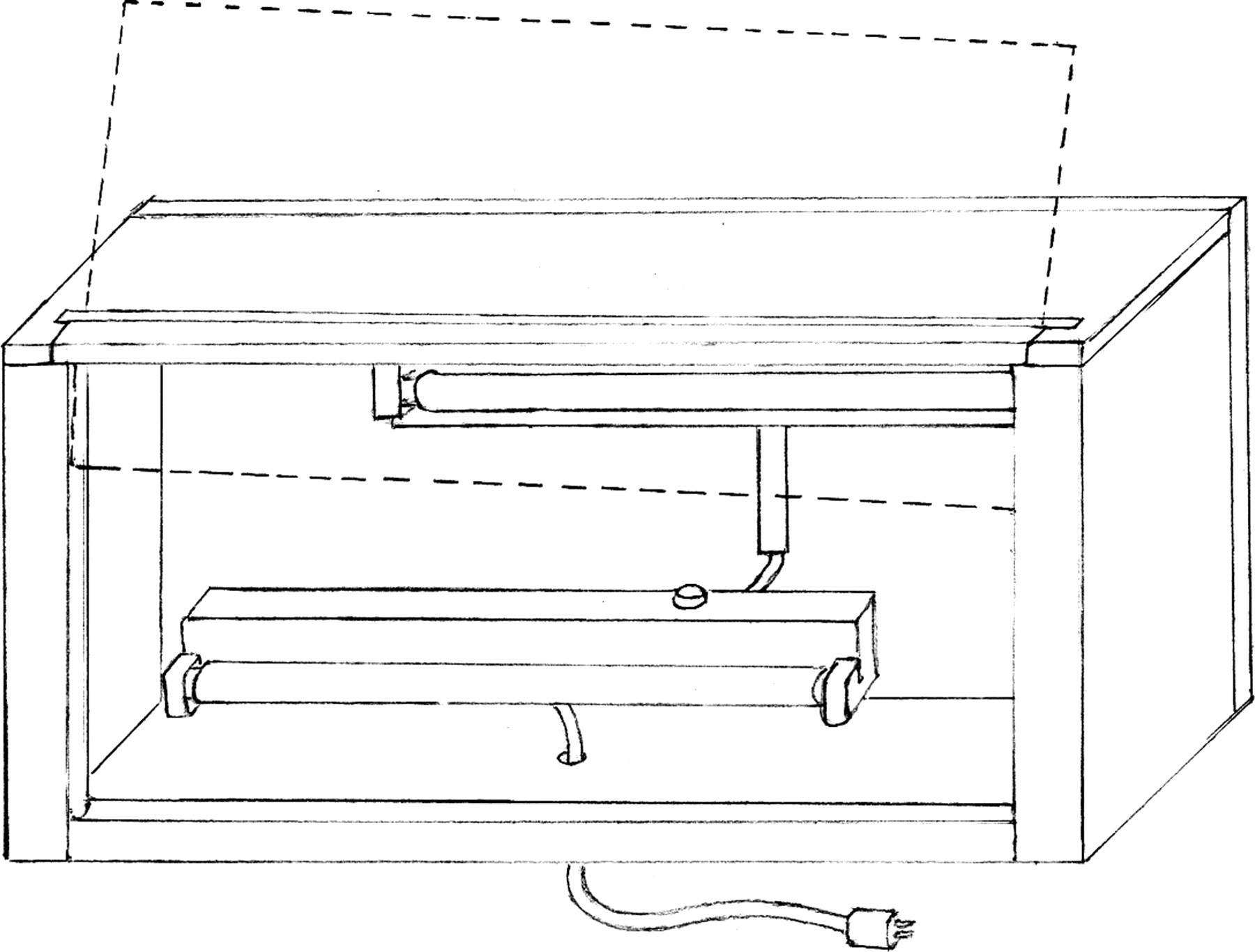
A diagram of the completed light box
Constructed by the hospital carpenter and electrician, each box took 3 ½ h to build (2 h carpentry; 1 ½ h electrical) at a cost for materials and labour of 90 Ghana Cedis (US $63). Comparable view boxes cost US $200 or more, plus shipping, taxes and duties. Not only is the box much cheaper than the commercial boxes, but also if this unit breaks, it can be easily repaired by the hospital staff.
The light box can be set on a desk or shelf or, as at our hospital, mounted on the wall (Figure 3) using standard brackets.

A photograph of the light box mounted and functioning in our emergency department
Conclusion
Clinical facilities throughout the world can easily and inexpensively produce similar radiographic view boxes. Having them available will facilitate the viewing of radiographic images and improve the accuracy of the interpretation of X-rays.