




Abstract
In this study we design and implement a novel flow chamber to assess electrostatic and streptavidin-biotin attachment mechanisms of a high-frequency ultrasound contrast agent (UCA) under high wall shear stress (WSS). The flow chamber was calibrated up to 50 Pa WSS to investigate attachment of the UCA. Attachment of the agent was assessed through measurement of the mean backscatter from targeted microbubbles attached to an agar surface within the flow chamber. The mean backscatter from microbubbles attached via a streptavidin-biotin mechanism decreased from −20 to −30 dB with increasing WSS up to 50 Pa. Mean backscatter from microbubbles attached via electrostatic attraction was indistinguishable from that of the agar surface alone beyond 0.66 Pa WSS. At 50 Pa WSS, the mean backscatter from the streptavidin-biotin attachment mechanism was 4 dB higher than from the plain agar surface. We conclude that the streptavidin-biotin attachment mechanism is ∼75 times stronger than an electrostatic mechanism. The ability of the streptavidin-biotin bond to withstand high WSS makes this attachment mechanism suitable for use as a targeted UCA for intravascular ultrasound studies and in high-resolution small animal studies, in which WSS can reach 40 Pa.
Coronary heart disease
Coronary heart disease (CHD), also known as coronary artery disease, ischaemic heart disease or atherosclerotic heart disease, is a type of cardiovascular disease affecting the heart and blood vessels of the cardiovascular system. It is the result of a build-up of atherosclerotic plaque within the arteries supplying the heart. CHD causes over 7.2 million deaths per year worldwide and 100,000 deaths each year in the UK alone, 1 and is the leading cause of mortality in the developed world. 2
Atherosclerotic plaque is a result of fatty deposits building up in the arterial walls, 3 leading to the development of either stable or unstable (vulnerable) plaques. Stable plaques consist of thick fibrous caps with hard cores, while vulnerable plaques have soft lipid cores coated by thin fibrous caps, making the latter liable to rupture. 4,5 Despite much research into the potential of elastography 6,7 and intravascular ultrasound (IVUS) 8 for plaque characterization, a reliable and accurate diagnosis of vulnerable plaque is not yet available.
A novel, lipid-based, high-frequency, microbubble ultrasound contrast agent (UCA) is currently under development in our laboratory. 9 This contrast agent is being developed to target inflammatory markers expressed by vulnerable plaques 10,11 to enable them to be more readily identified with an IVUS examination. In addition, the agent has potential applications for targeting in small animal models.
Ultrasound contrast agents
Modern UCAs consist of microbubbles with a diameter between 3 and 10 µm, which when injected into the blood pool or a cavity enhance ultrasonic signals. 12 There are currently a variety of commercial contrast agents, 13–15 which are used diagnostically in investigations of the liver, 12,16 heart and kidneys. 12,17 Improved understanding of microbubble-ultrasound interactions 18–20 has led to recent developments in the therapeutic applications of UCAs, such as drug and gene delivery. 21–24 In addition, targeting of UCAs has led to improved diagnostic approaches to ultrasound examinations. 25–28
Targeting of UCAs has been widely studied and various targeting strategies are described by Klibanov
23
and Lindner.
29
The targeting mechanisms either exploit the properties of the microbubble shell or involve the attachment of target-specific ligands or antibodies to the shell's outer layer. The microbubbles used in this study are targeted through the attachment of plaque-specific antibodies via a streptavidin-biotin bridge (Figure 1). The streptavidin-biotin bond is widely used for conjugating antibodies to microbubbles
30,31
and has been used for targeting in small animal experiments.
32–34
Attachment of antibodies to UCAs via a streptavidin-biotin bridge. UCA, ultrasonic contrast agent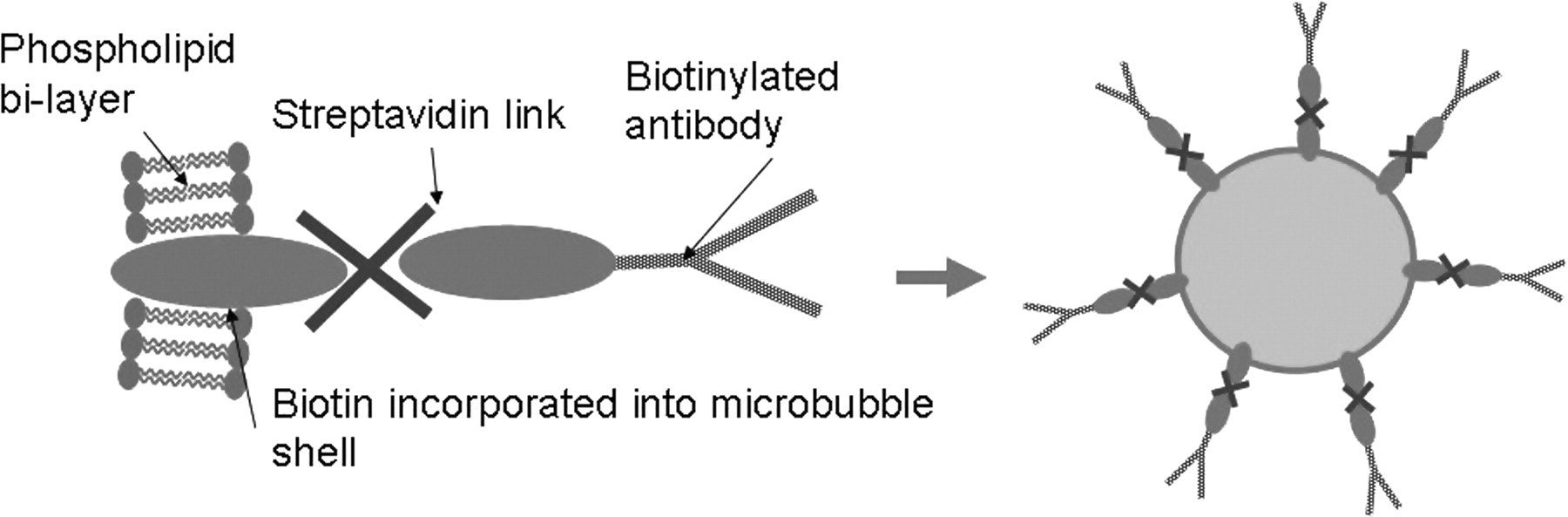
The microbubble UCA has been developed for use with high-frequency IVUS at 30 MHz, 9 and more recently at 40 MHz. Previously, a version of the agent was shown to remain attached within a flow chamber up to a wall shear stress (WSS) of 3.4 Pa. 35 Peak WSS in the human coronary arteries has been reported at 3.5 Pa. 36 However, initial trials of the agent in vivo will involve small animal studies and the peak WSS experienced in the coronary arteries of healthy mice can reach 40 Pa. 37 It is, therefore, important to assess the maximum WSS under which the agent will remain attached to a surface to ensure that it is viable for small animal studies.
In this study a novel parallel-plate flow chamber was developed and calibrated to 50 Pa WSS. The strength of a streptavidin-biotin bond was then compared with an electrostatic bond to determine the feasibility of using a streptavidin-biotin bond for targeting of the UCA in a clinical scenario. The relative strengths of these two bonds were then investigated for the UCA up to the maximum attainable WSS in the flow chamber.
Materials and methods
Flow chamber
A flow chamber was designed for the assessment of UCA attachment at very high WSS, and calibrated for WSS up to 50 Pa using laser Doppler anemometry (LDA). A diagram of the parallel-plate flow chamber developed for this study can be seen in Figure 2. The parallel-plate flow chamber was made from three panels of Perspex. The front panel (Figure 2a) incorporates a microscope slide for compatibility with LDA. The flow channel was 300 mm long with a 3 × 3 mm2 cross-section, incorporated into the central panel (Figure 2b). Inlet and outlet reservoirs, designed to stabilize the flow, were located at either end of the flow channel. Finally, the rear panel of the flow chamber (Figure 2c) included an agar well for attachment of the microbubbles, two IVUS channels for ultrasound assessment of the chamber and 8 mm wide inlet and outlet producing high flow volumes and high WSS.
Flow chamber schematic: (a) front panel; (b) central panel and (c) rear panel of parallel-plate flow chamber
The panels of the parallel-plate flow chamber were sealed with silicone grease and clamped together to ensure no leakage from the chamber. Water or a glycerol solution was then passed through the flow channel and over the agar strip. The flow volume was controlled by a pump (Micropump Inc, Vancouver, WA, USA) connected to a power supply with an analogue voltage display (Weir Lambda Electronics, Bognor Regis, UK). LDA was used to determine the WSS at volumetric flow rates of 60–350 mL/minute. Higher WSS was achieved by passing higher viscosity fluids through the flow chamber. Fluids of different viscosities were produced using glycerol (Sigma-Aldrich Inc, St Louis, MO, USA) solutions of 40%, 60% and 80% corresponding to viscosities of 0.0045, 0.0240 and 0.0599 Pa s, respectively.
Laser Doppler anemometry
LDA is a non-invasive optical technique used to measure point flow velocities within a fluid. The system used was produced by Dantec Dynamics (Copenhagen, Denmark).
38
The technique involves introducing a seeding particle to the flow and focusing two coherent laser beams at a point in the flow. Due to the fringe pattern produced at the point where the laser beams cross, a seeding particle passing through the measurement volume will scatter light back to the sensors at a frequency (f) proportional to the velocity of the particle as illustrated in Figure 3. If the fringe spacing (d
f
) is known, the particle and fluid velocity (v) can be calculated from eq. (1):
38
Measurement volume and fringe pattern at crossing point of two coherent laser beams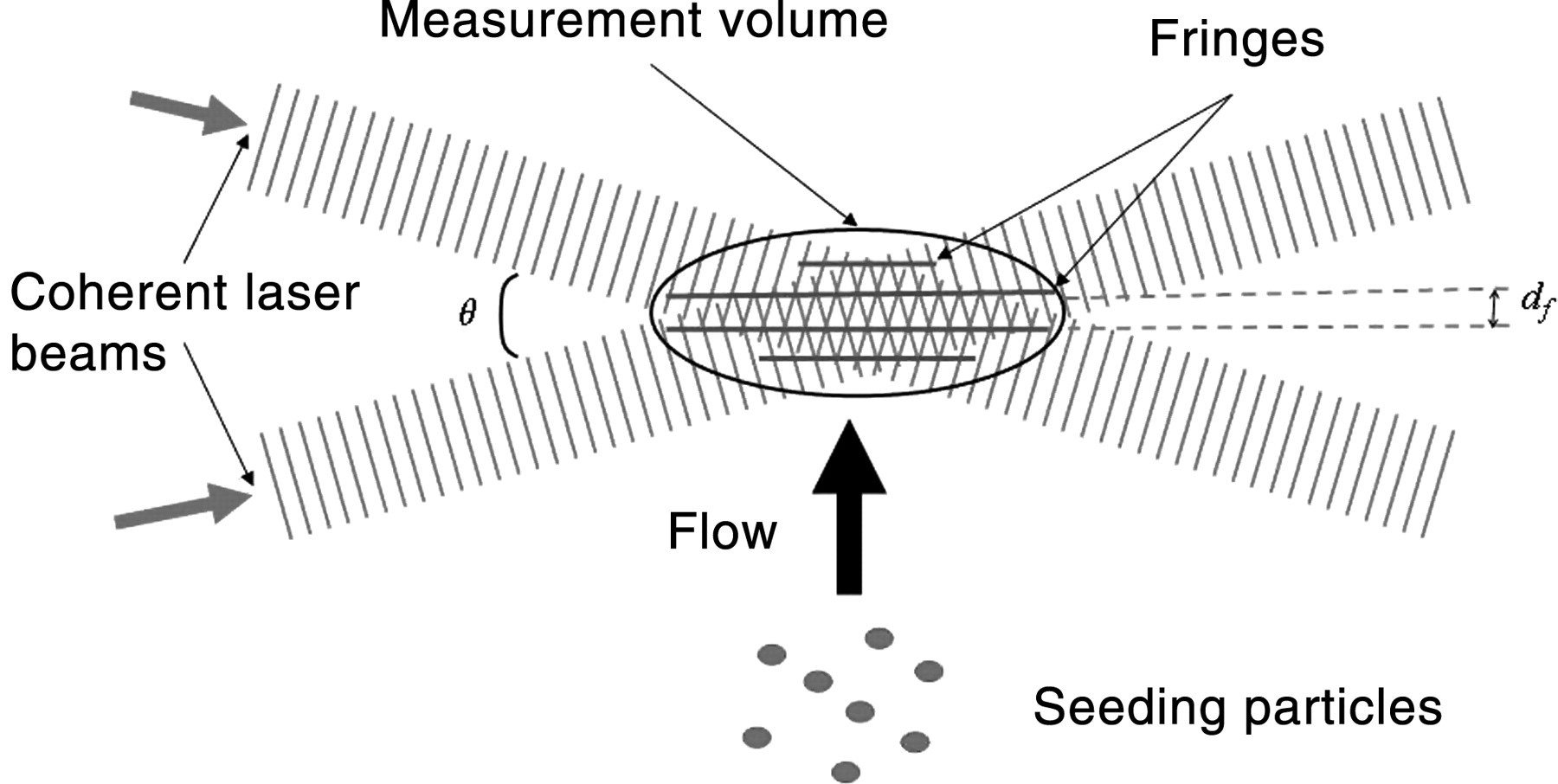

In order to determine the flow velocity of the water and glycerol solutions at varying volume flow rates within the flow chamber, the LDA measurement volume was scanned through the flow channel. Flow profiles were produced for volume flow rates of 60–180 mL/minute at 20 mL/minute increments and at 350 mL/minute for higher shear stress. The wall shear rate (dv/dx) was determined from the gradient of the flow profiles adjacent to the flow chamber wall. WSS (τ) at each flow volume were then calculated from eq. (2),
39
where μ is the viscosity of the fluid:

Microbubble production and optimization
Biotinylated lipid microbubbles were produced using a method similar to that described by Moran et al. 40,41 modified by heating and mixing of the lipid constituents, whereas the previous method involved the use of solvents. The agent was produced by combining phosphatidylcholine, cholesterol, phosphatidylethanolamine, phosphatidylglycol (PG) and biotinylated PG (Avanti Polar Lipids Inc, Alabaster, AL, USA) in a 56:30:6:3:5 ratio by mass. The lipids were mixed with saline using a high shear mixer (IKA®, Staufen, Germany) and heated to their transition temperature (65–77°C) in a water bath. Following this, the agent was rapidly agitated using a CapMix™ device (3M™ ESPE™ AG, Seefeld, Germany).
The agent was found to have a mean diameter of 5.64 ± 0.36 μm and an average size distribution as shown in Figure 4. The average concentration of the microbubble samples was 64.50 ± 0.02 × 10⁶ microbubbles/mL.
Average size distribution of UCA. UCA, ultrasonic contrast agent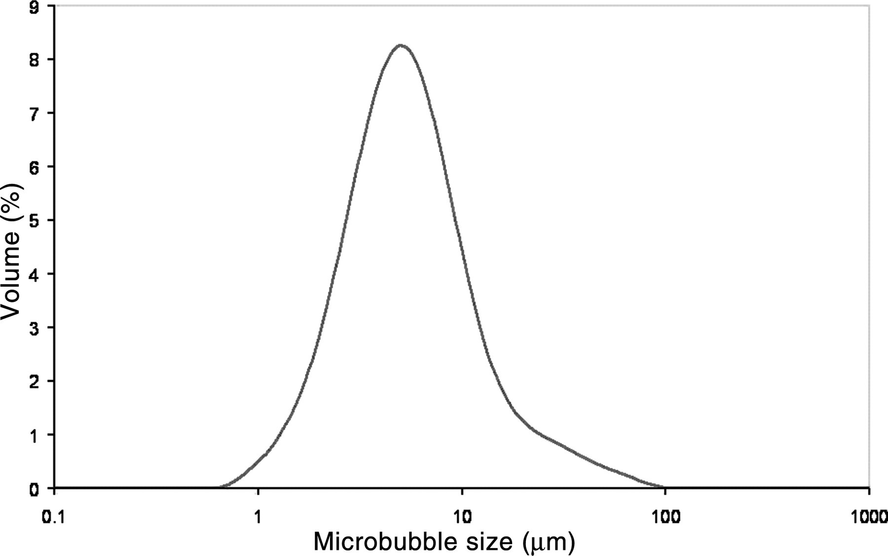
The acoustic stability of the agent was assessed over 24 hours at room temperature to ensure that the agent would remain echogenic throughout the course of our investigations. Echogenicity of the agent was determined through visual assessment of IVUS images and analysis of RF (radiofrequency) data.
Microbubble attachment
Agar was produced using a standard tissue mimicking material recipe 42 without the addition of scatterers. Strips of agar were then cut to fit into the agar well of the flow chamber (2 × 7 × 200 mm3). Agar was identified as the optimum substrate due to its acoustic transparency, making it possible to assess the surface of the agar using IVUS.
In order to assess the WSS that the streptavidin-biotin link was able to withstand, the agar sample was coated in streptavidin and biotinylated bubbles attached as illustrated in Figure 5. Attachment was achieved by coating the agar strips with a solution of streptavidin and 0.1 mmol/L sodium bicarbonate coating buffer (pH 8), in a 1:12 ratio by volume. The streptavidin was left on the agar for a minimum of 18 hours at room temperature (22°C) to optimize coating, as previously determined by Butler,
35
before being rinsed with saline to remove any excess. The microbubble suspension was then pipetted onto the agar surface and left for two hours at room temperature before being rinsed with saline to remove any unattached bubbles. Microbubble attachment has previously been shown to occur within a two-hour period.
35
As a control sample, an agar strip coated in streptavidin but with no bubbles attached was produced at the same time.
Attachment of biotinylated microbubbles to streptavidin-coated agar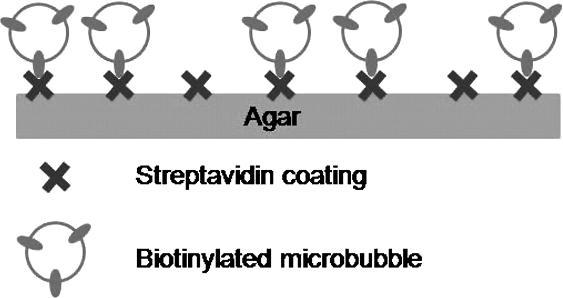
In addition, an investigation into the electrostatic attachment of the UCA was carried out. To achieve this, 1 mL of the microbubble suspension was pipetted onto an uncoated agar sample surface and left for two hours at room temperature before being rinsed with saline.
Assessment of the strength of attachment of the UCA
To assess the WSS withstood by different attachment mechanisms, agar strips with and without the UCA attached were placed inside the agar well of the flow chamber. The agar strips were then subjected to WSSs varying between 0 (no flow) and 50 Pa.
The flow volume (and therefore WSS) was increased at 20 mL/minute intervals from 60 to 180 mL/minute for the four different viscosity fluids described previously; a further result was collected at 350 and 270 mL/minute as the maximum achievable flow rate with 0–60% glycerol solutions and 80% glycerol solutions, respectively. IVUS images and RF data of the agar surface were collected at each interval for the electrostatic and streptavidin-biotin attachment methods over a 30-minute period.
A Clearview IVUS scanner (Boston Scientific, Natick, MA, USA), with a 40 MHz Atlantis™ SR Pro coronary imaging catheter, was used to collect images of the attached microbubbles. The transducer tip was placed into the IVUS channels. IVUS images were captured by a PC linked to the scanner via a video-out cable, using a Matrox Intellicam program (Matrox Imaging, Montreal, Canada).
In addition to IVUS images, RF data were captured for each image by a digitizing card with a 250 Hz sampling frequency (Gage Applied CS8500, Gage Applied Technologies, Lachine, Canada) added to the PC. The RF data were then analysed to acquire the mean backscatter power of the contrast agent attached to the agar surface using a program written in IDL5.4 (ITT Visual Information Solutions, Boulder, CO, USA). This allowed quantification of the agent's echogenicity. Selection of the appropriate echo for analysis was achieved through observation of flow through the chamber and the structures identified in Figure 5.
Echoes from plain agar and streptavidin-coated agar without microbubbles were also collected to compare the mean backscatter from these surfaces to that from the agar with attached microbubbles. This investigation was repeated four times for both attachment methods and IVUS images and RF data collected from both IVUS channels for each WSS interval. Detachment investigations were carried out using saline at room temperature (22°C).
Results
Calibration of flow chamber: LDA results
The WSS at varying flow volumes was experimentally evaluated using LDA, the peak velocities and wall shear rate for the four different viscosity fluids can be found in Table 1. These results enabled calibration of the flow chamber such that the UCA could be assessed under known WSS. Figure 6 illustrates the increase in WSS with increasing flow volumes for the four different viscosity fluids.
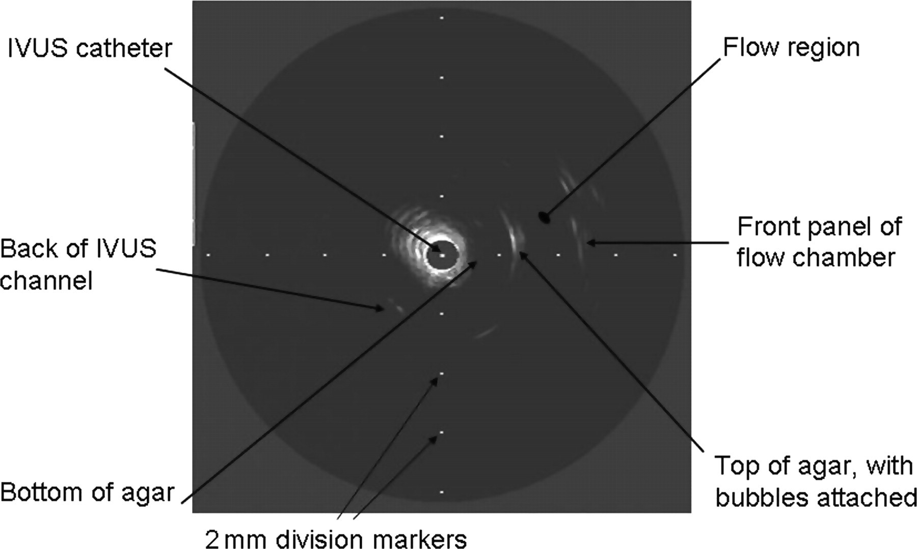
Structures observed in an IVUS image. IVUS, intravascular ultrasound
Peak velocities and shear rates of different viscosity fluids
The WSS was found to increase linearly with flow volume, with higher viscosity fluids resulting in a more rapid increase in WSS. From these results it was possible to identify the WSS under which microbubbles attached to the agar strip were subjected.
Acoustic stability of agent
The average mean backscatter of the unattached UCA over a period of 24 hours was −32.12 ± 2.06 dB. The variation in mean backscatter and average microbubble diameter can be seen in Figure 7. The mean size and mean backscatter varied by 2 dB over the initial 24-hour period confirming that the agent remains acoustically stable for the duration of the investigation. The agent has also been shown to remain acoustically stable for up to three months when stored between 2 and 5°C. However, for all experiments, the agent was used within one week of manufacture.
Mean WSS at varying flow volumes with four different viscosity fluids, showing error bars to one standard deviation. WSS, wall shear stress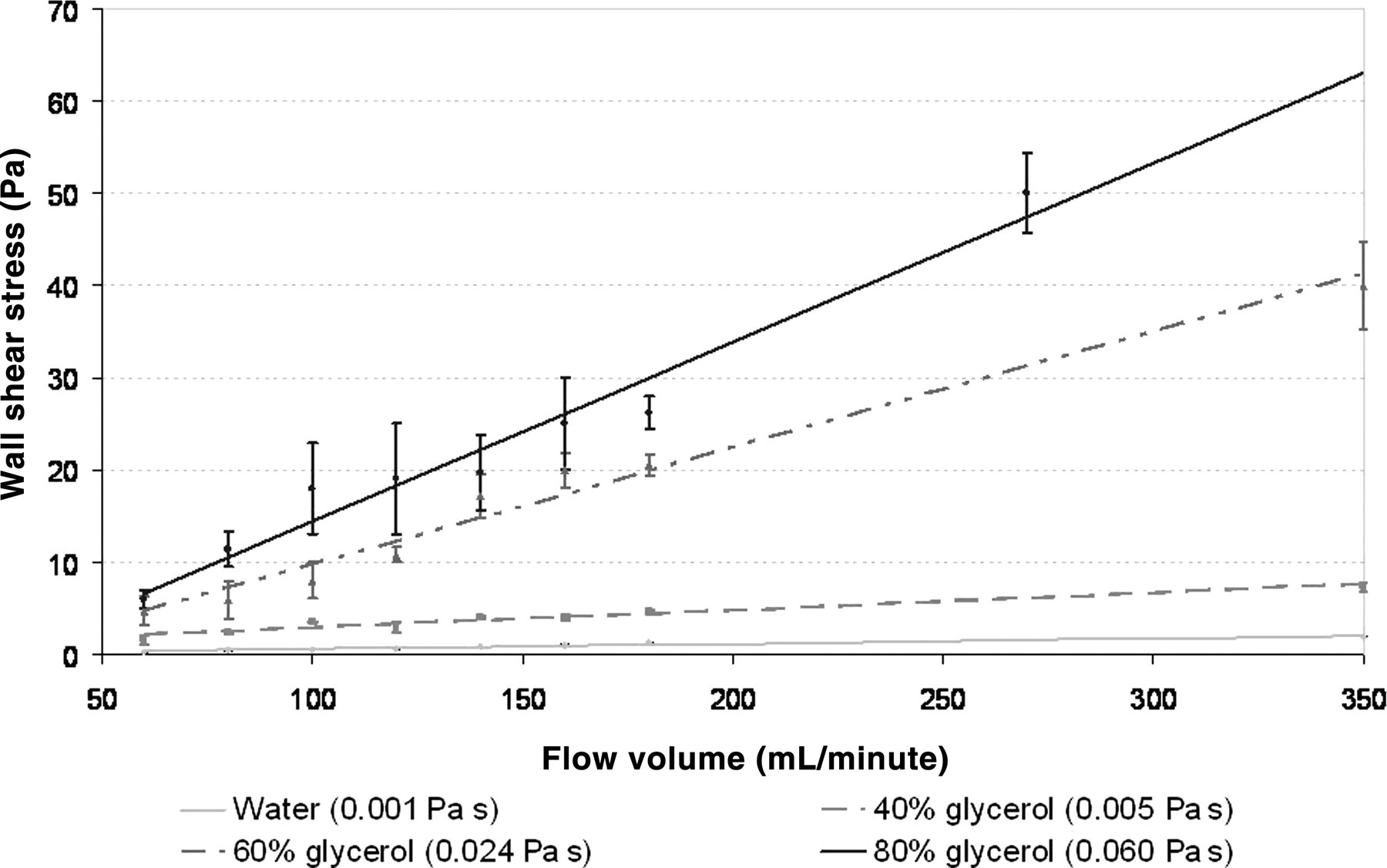
Electrostatic attachment
The microbubble UCA was found to remain attached to the agar via electrostatic forces up to a shear stress of 0.66 Pa. The IVUS images in Figure 8 demonstrate the echo observed from the attached bubbles at varying WSS. The echo from the agar surface with bubbles attached, up to a WSS of 0.66 Pa, was brighter than the echo from a plain agar surface. However, beyond 0.80 Pa WSS the echo from the surface to which the agent was attached was indistinguishable in intensity from the echo from plain agar.
Acoustic stability of the UCA over 24 hours shown as the mean backscatter against time. UCA, ultrasonic contrast agent
The RF analysis of the IVUS images also suggested that the UCA remained attached to the agar via electrostatic forces under very low WSS. These data are presented in Figure 9. This figure shows that the mean backscatter power from the contrast agent on the agar surface decreases rapidly as the WSS increases (P < 0.01). However, as there is still a greater mean backscatter from the attached microbubbles at 0.66 Pa, than from a plain agar strip, this confirms that the UCA remained attached to the agar surface up to this WSS.
IVUS images of microbubble contrast agent (arrowed), attached via an electrostatic mechanism, at varying WSS: (a) 0 Pa; (b) 0.30 Pa; (c) 0.44 Pa; (d) 0.66 Pa; and (e) 0.80 Pa, compared with (f) a plain agar surface. IVUS, intravascular ultrasound; WSS, wall shear stress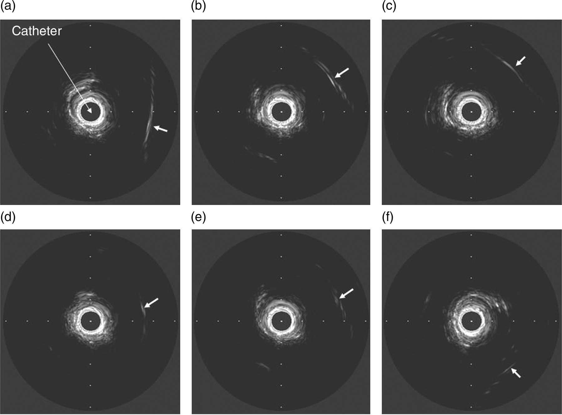
Streptavidin-biotin attachment
Under a streptavidin-biotin attachment mechanism, the UCA was found to remain attached to agar at 50 Pa WSS, the maximum attainable WSS within the flow chamber. Figure 10 shows IVUS images of the microbubble attached via a streptavidin-biotin link at varying WSS. The echo from the attached microbubbles at 0 and 50 Pa WSS was found to be brighter than the echo from a plain and streptavidin coated agar surface with no microbubbles, suggesting that the UCA remained attached for up to 50 Pa WSS via a streptavidin-biotin bond.
Mean backscatter power at varying WSS for microbubbles attached via electrostatic forces (error bars represent 1 standard deviation of four data-sets). WSS, wall shear stress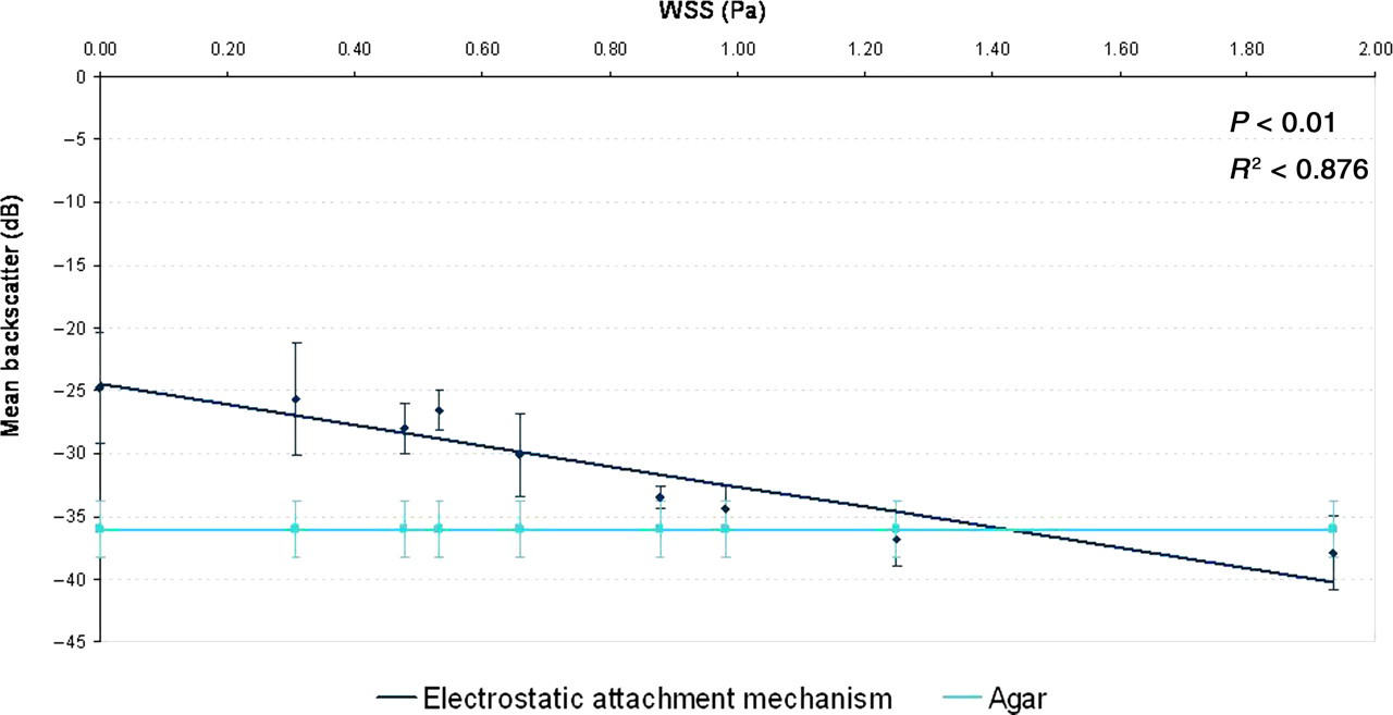
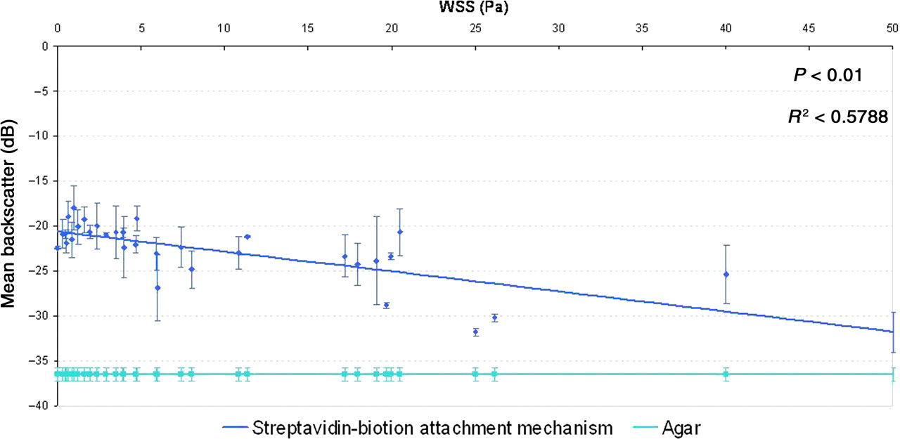
Analysis of RF data confirmed that the mean backscatter power from the microbubbles attached to the agar surface, via the streptavidin-biotin bond, was greater than the mean backscatter power from a plain agar surface (P < 0.01) (Figure 11). At 0 Pa WSS the mean backscatter power from the attached microbubbles was found to be −22.57 ± 3.71 dB, which is 13.97 ± 4.46 dB greater than the mean backscatter power from agar. At a WSS of 50 Pa the mean backscatter power from the attached microbubbles was 3.13 ± 1.23 dB greater than that from agar, confirming that the microbubbles remain attached to the agar under even under very high WSS (Figure 12).
IVUS images of the microbubbles (arrowed) attached to agar via a streptavidin-biotin link at (a) 0 Pa (no flow) and (b) 50 Pa WSS compared with (c) a plain agar surface and (d) a streptavidin-coated surface. IVUS, intravascular ultrasound; WSS, wall shear stress Mean backscatter power from microbubbles attached to an agar surface via a streptavidin-biotin link at varying WSS (error bars represent 1 standard deviation of four data-sets). WSS, wall shear stress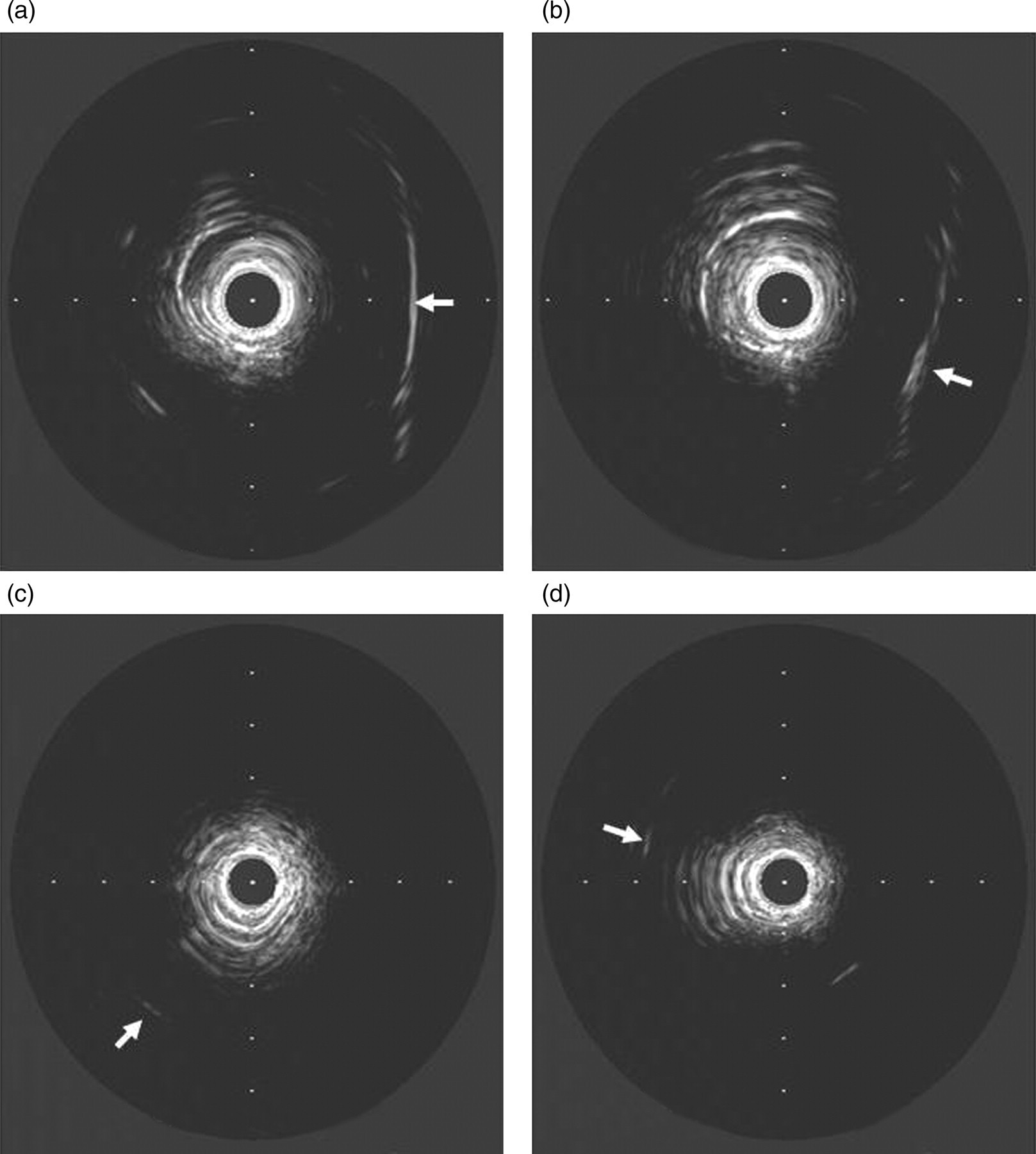
Discussion and summary
A novel flow chamber has been developed, which enabled investigation of detachment of an UCA exposed to known WSSs up to a maximum of 50 Pa. LDA was used to measure the flow velocity close to the wall to estimate the WSS at the position of the observation region of the flow chamber. The chamber was calibrated by assessing volume flow rates between 60 and 350 mL/minute for four different viscosity fluids produced from glycerol solutions. The flow chamber was shown to be suitable for assessment of UCA attachment and detachment under normal physiological WSS expected in human and small animal coronary arteries. A streptavidin-biotin attachment mechanism was investigated for a novel, lipid-based, microbubble UCA and compared with attachment of the agent via an electrostatic attachment mechanism.
Initial investigation of the attachment of the agent under varying WSS involved assessment of the maximum WSS withstood by bubbles attached by electrostatic attachment. The mean backscatter data presented in Figure 8 show that microbubbles remained attached to the agar up to a WSS of 0.66 Pa. This suggests that above this WSS the microbubbles have either been washed off the agar or destroyed. The IVUS scanner and transducer combination operates with an MI of 0.32 ± 0.02, which is below the accepted destruction threshold. 43 Conversely, microbubbles attached via a streptavidin-biotin mechanism appeared to remain adhered to the surface beyond this WSS.
Although we have described attachment in the absence of a streptavidin-biotin bond as electrostatic in origin, it is also possible that the attachment is due to a chemical bond between the microbubbles and the agar surface. 44–46 However, since WSS in human coronary arteries can reach 3.5 Pa, 36 this attachment, either electrostatic or chemical in origin, would not be suitable for targeting to coronary atherosclerotic plaques under physiological shear stresses. An additional consideration is that electrostatic charges may not exist on the plaque. The agent was found to detach rapidly from the agar surface as the WSS was increased, and at relatively low shear stresses of 0.66 Pa the difference in backscatter from the attached bubbles compared with the plain agar surface was approximately 3.80 ± 1.76 dB, compared with an initial difference of 12.68 ± 1.98 dB. This suggests that some microbubbles remained attached to the surface at this WSS. However, as the WSS was increased the mean backscatter power from the agar surface became comparable with that of plain agar, suggesting that all of the bubbles had detached.
In addition to an electrostatic targeting mechanism, a streptavidin-biotin link was assessed and shown to withstand a WSS of up to 50 Pa, making the streptavidin-biotin bond ∼75 times stronger than electrostatic attachment in the in vitro environment under flow. The rupture force for a streptavidin biotin bond has been reported to vary from 250 to 400 pN. 47,48 The strength of this bond, therefore, suggests that this attachment mechanism would be suitable for the attachment of antibodies to target this agent to areas of high shear stress, such as in narrowing of blood vessels due to the presence of atherosclerotic plaque in the coronary arteries and also in initial preclinical trials of the agent in healthy small animals where a WSS of 40 Pa has been recorded. 37
The streptavidin-biotin attachment mechanism is commonly used for conjugation of targeting antibodies or ligands to microbubble contrast agents; 30,34,49 however, due to toxicity resulting from retention in the kidneys, 50 streptavidin is not a suitable attachment mechanism for clinical studies. Future work will involve developing a more suitable method of conjugating antibodies to the microbubble.
Our investigations were carried out at room temperature (22°C) due to the nature of the set-up and the environment in which the calibration was carried out. However, due to the biological nature of the materials used in this investigation and intended clinical applications of these microbubbles, it would be valuable to collect results at body temperature (37°C) in future. Additionally, our investigations were carried out under laminar flow to assess accurately a constant WSS under which the agent would remain attached. Arterial blood flow is pulsatile and the agent would be subjected to varying degrees of WSS when targeted to atherosclerotic plaques.
The flow chamber was designed to investigate the strength of a streptavidin-biotin bond under WSSs expected in vivo to determine the feasibility of using this mechanism to attach antibodies targeted to atherosclerotic plaques to an UCA. Currently an investigation into the attachment of antibodies to the agent is being carried out; further to this the strength of attachment to a cellular surface will be investigated in vitro.
Conclusions
A novel flow chamber has been developed to test the adhesion of UCAs to a substrate under normal physiological WSS in the coronary arteries of humans and small animals. This flow chamber is suitable for investigating adhesion of UCAs up to 50 Pa WSS. The agent tested here was shown to remain attached to agar via a streptavidin-biotin link up to 50 Pa WSS, which is ∼75 times stronger than an electrostatic attachment mechanism. In addition, even at a high WSS of 50 Pa, the mean backscatter power from the agent was 3 dB higher than the backscatter from a plain agar surface alone thus enabling differentiation between the two surfaces.
A control investigation into the attachment of the agent to agar in the absence of the streptavidin-biotin bond confirmed that the attachment of the agent is due to the streptavidin-biotin bond and not other forces. Our investigations have confirmed that the streptavidin-biotin bond is able to withstand expected WSS within the human coronary arteries (3.5 Pa) and in mice (40 Pa) making the agent suitable for future in vivo applications. We conclude that targeted, lipid-based, microbubble UCA could feasibly attach under physiological flow conditions and would be suitable for further studies.
Footnotes
ACKNOWLEDGEMENTS
The author would like to acknowledge the EPSRC for studentship funds and the British Heart Foundation (grant number: PG/07/107/23895). The work presented here has also been submitted as part of a university thesis entitled ‘Attachment Mechanisms of a Novel, Targeted, Lipid-Based, Ultrasound Contrast Agent’.