Abstract

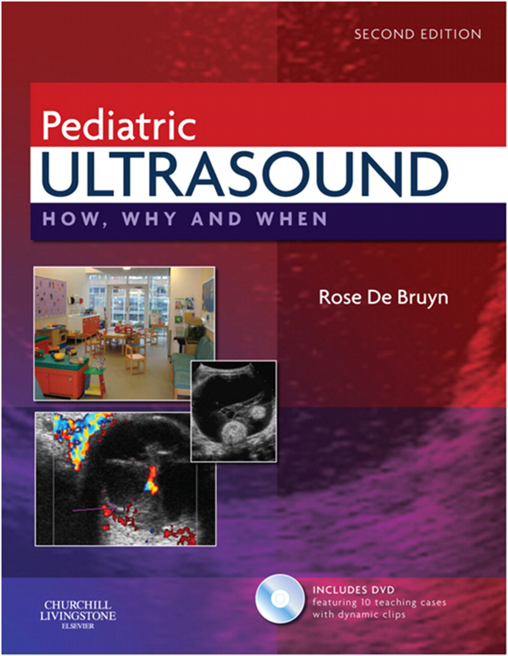
This paediatric textbook aims to provide those undertaking ultrasound examinations in children, with guidelines, normative charts and the framework necessary to obtain the maximum amount of information from ultrasound and help make a diagnosis. It certainly achieves and actually exceeds this remit and would be a useful acquisition for any sonographer or radiologist with an interest in paediatric ultrasound.
The opening chapter entitled ‘General issues of methods and equipment’ provides good practical advice and tips on ways to optimize the examination of children, which is particularly helpful for those starting out in paediatric ultrasound. This is followed by a chapter dealing with the prenatal sonographic diagnosis of congenital anomalies, with the focus on conditions that present for postnatal ultrasonography and leads very nicely to the paediatric ultrasound chapters.
This book is well laid out, easy to navigate and has a comprehensive list of chapters dividing paediatric ultrasound into anatomical areas. There are numerous good quality images throughout, providing clear examples of normal and abnormal appearances in children, which helps to make it very readable. One of the real strengths is the use of diagrams to assist in the explanations of embryology, anatomy and technique. Many of the chapters contain a section on embryological development, which along with diagrams giving explanations of congenital anomalies are extremely useful when performing ultrasound examinations in children. This is particularly well demonstrated in the chapters on the renal tract, the abdomen and bowel, and the head, neck and spine. Within the musculoskeletal chapter, diagrams of hip ultrasound technique and the corresponding ultrasound images are used to good effect.
Throughout the book there are comprehensive tables detailing normal measurements in children, imaging protocols and characterization of conditions. There are also many lists of useful tips and features or causes of conditions, making this book invaluable for reference. The pertinent points from each chapter are nicely summarized in a box at the end of the chapter.
An interesting chapter describing ocular pathologies and diagnostic ultrasound technique is provided by Ken Nischal from the Department of Ophthalmology at Great Ormond Street Hospital. Also from Great Ormond Street, Derek Roebuck gives an excellent account of cutting-edge paediatric interventional ultrasound in the final chapter.
A DVD accompanies this text and contains case studies and some video clips. While the case studies provide a good revision tool, rather like an e-learning quiz, the video clips are nice to see but do not add a great deal.
In conclusion, this book is packed with information, and is delivered in a clear and interesting format. It achieves what it sets out to do and highlights the strengths and weaknesses of ultrasound in many conditions. I would highly recommend it as an asset to any department in which paediatric ultrasound is performed.