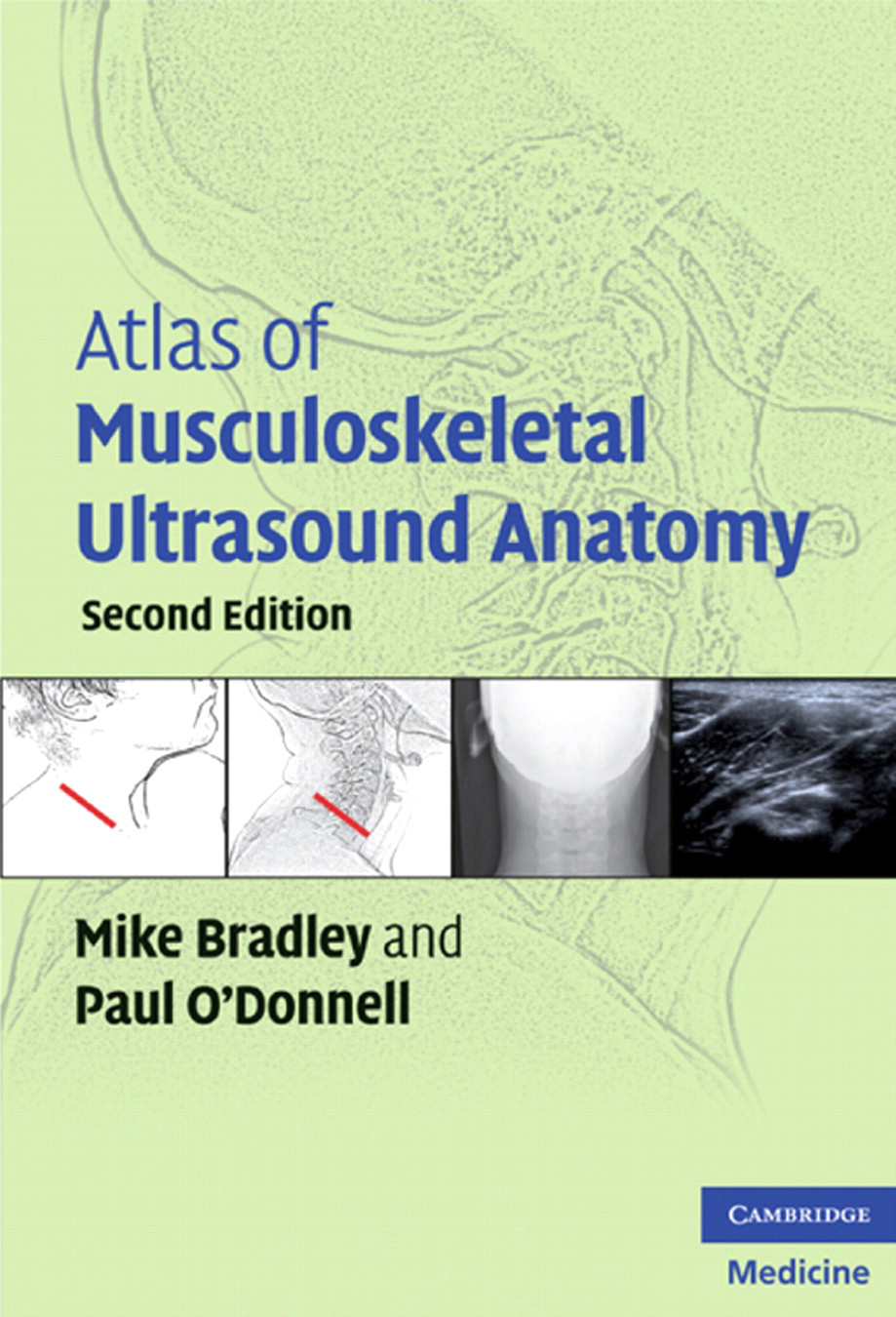
Abstract

The Atlas of Musculoskeletal Ultrasound Anatomy is a paperback atlas of normal ultrasound anatomy for imaging of the musculoskeletal system. The text aims to provide a quick reference of normal ultrasound appearances. This is supplemented with descriptions of important anatomical landmarks, as well as a guide to optimal ultrasound technique and transducer positioning. The second edition brings improved quality of ultrasound images due to state-of-the-art advances in transducer design and spatial resolution. The text is aimed at a variety of practitioners requiring knowledge of musculoskeletal anatomy including radiologists, sonographers, rheumatologists, physiotherapists, anaesthetists and orthopaedic surgeons.
The authors Dr Mike Bradley and Dr Paul O'Donnell have acquired remarkable expertise and are renowned in their field, having made substantial contributions to the literature. They have made every effort to provide high-quality ultrasound images with high-end equipment.
The book begins with a brief description of the principles and pitfalls of ultrasound and an overview of echogenicity of different tissues encountered during musculoskeletal scanning. The text is divided into chapters according to anatomical site, i.e. upper and lower extremities, abdomen and pelvis, and the indexing is clear and well organized. Each section provides a comprehensive description of muscle origins and insertions as well as a brief written summary of bony and neurovascular anatomy.
To assist in understanding, the ultrasound scans are correlated with surface anatomy and radiographic illustrations. Transducer position and orientation is depicted in each of these images to provide improved visuospatial understanding. The ultrasound images are of high quality and make use of panoramic or narrow fields-of-view to give the most appropriate demonstration of relevant anatomy. The images are clearly annotated to show important structures and their relations. In certain chapters, commonly occurring pathological conditions are described, for example, medial head of gastrocnemius semimembranosis bursa (Baker's cyst), and guidance is offered on transducer positioning.
The atlas gives an accurate overview of ultrasound anatomy, thus providing help to those who are already familiar with musculoskeletal ultrasound, as well as a starting point for beginners, irrespective of whether they are radiologists, orthopaedic surgeons, rheumatologists or allied health professionals. The text serves as a quick reference guide rather than a comprehensive text, to be employed as an adjunct to musculoskeletal ultrasound sessions. This would be useful for basic diagnostic imaging, but could be improved by featuring corresponding pathological anatomy, although this would require a larger, more in-depth volume and serve as more than just an anatomical guide.
Given the dynamic nature of ultrasound, it is often difficult to appreciate the anatomy from a static image. However, the atlas attempts to overcome this with a comprehensive review of the muscular anatomy in each section. The illustrations of surface anatomy and radiographic drawings are well chosen, but are of limited quality. They do, however, enhance understanding of transducer positioning and add to the value of this book.
Totaling 222 pages, this atlas achieves its goal to be a helpful pocket reference, rather than a textbook. There are few other such texts available for musculoskeletal imaging, with larger volumes serving as much more comprehensive works carrying a much greater price. The book is suitable for the intended readership, both for the experienced and inexperienced practitioner, as a basis for ultrasound review of the musculoskeletal system.