
Abstract
A departmental survey of personal methodology in the measurement of bile duct diameter was carried out by means of a representative diagram of a typical bile duct image configuration. The results revealed considerable differences in measurement practice, and factors that may explain the patterns of interobserver variation, among 20 respondents in this task, were postulated. There was considerable scope for error, by a factor of three times between the smallest and largest diameter selected noted within current practice variation. This reflects similar disparity found in the literature. Within the setting of the development of current therapeutic strategies, there may be a need for confirmation and/or re-establishment of contemporary normal ranges for biliary tree diameter.
A study of reasonable magnitude would be required to clarify the many and complex common and special causes of variation in ultrasonic evaluation outlined towards enhancing the utility of this useful and hitherto important ultrasonic sign.
A departmental survey was prompted by personal observation of heterogeneity in the measurement of bile duct dimensions measured on ultrasonic bile duct images in order to define the existence and extent of any variation.
If diversity sufficient to provide scope for clinically significant discrepancy was suggested or confirmed, then a search for reasons behind this might prove useful.
Contentious unresolved issues in the literature, some of which are noted to periodically re-emerge, may indicate that if personal practice variation has emerged it may well continue if divergence in the evidence base remains unresolved.
Material and methods – survey
A departmental survey of practice was undertaken to record which site operators would choose to measure (and where they would place their callipers in the process) the common bile duct (CBD) by means of a representative drawing (Figure 1) of a typical scan image. Eight possible site options were given, based on common positions quoted in the literature and used in practice, all the while acknowledging measurement is usually of the common hepatic duct in reality.

Diagrammatic part of questionnaire of the bile duct measurement options on representative section of the index scan. Respondents were also requested to identify their wall calliper placement sites – see options (a)–(e) in the text
The five options for calliper placement were listed as follows:
Outer wall to outer wall;
Outer wall to inner wall;
Inner wall to outer wall;
Inner wall to inner wall;
Inner mucosal line to inner mucosal line.
Respondents were asked not to discuss their questionnaire with others before completing it.
An investigation of bile duct mensuration practice using the same survey criteria within departmental textbook resources, a likely source of any practice variation, was also carried out.
Results
Two respondents did not specify their method of calliper placement as denoted by (–) in Table 1.
The survey response results (Table 1) showed surprising variability in choice of common duct measurement site.
Survey results – C, D and E all measured as the same diameter
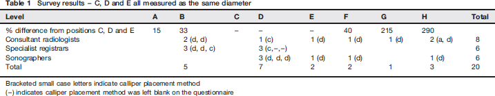
Bracketed small case letters indicate calliper placement method
(–) indicates calliper placement method was left blank on the questionnaire
Trends of note included:
Regarding variation among sonographers, half were centred on one site, the rest as a tail scattered distally;
Specialist registrars showed least variation – unified, single-site ultrasound training was thought to account for this;
Consultants exhibited most variation across the options;
• The modes were options D for the site chosen to measure diameter, and calliper placement method (d), i.e. inner to inner wall measurements.
Within 10 abdominal ultrasound textbook resources1–10 available in the department later compared in similar fashion to the questionnaire, variation was also evident as follows:
Position specified: one indicated A, 1 one C, 4 two D,9,10 one G, 2 and five were site unspecified.3,5–8
Calliper placement positions were unstated in five, and the other five described option (d).1,2,7–9
Three main categories for the upper limit of normal were noted included six defining 6 mm,2,5–8,10 three specifying 5 mm1,3,9 and one indicating 1 mm/decade of life. 4 Additionally, grey areas were used in three instances to be added to the ‘normal’ range limit above as ‘buffers’. Only beyond these added-on quantities was ‘abnormal’ status confirmed. These were given as 1,13,3 and 1 mm5 – in the last instance as an allowance for a post-cholecystectomy examination.
Discussion
Suspicions that the measurement point of the common duct was subject to variation intradepartmentally were confirmed by this survey. Low numbers preclude statistical analysis, but all respondents measure CBD diameter regularly in practice.
Variation in practice was also reflected in available departmental literature. Of note, textbooks were between 10 and 27 years old, many quoting sources well beyond that timescale; individuals may have adapted their own discriminating/coping methods in a response to the exigent lack of consensus therein, and also in wider literature review. The consultants and to a lesser extent the sonographers were trained in a variety of different centres. Also, there was an approximately 30-year spread of variation in the time periods spent developing experience in ultrasound scanning thereafter in these non-training graded staff. Prior discussion with colleagues may also have occurred. All of these factors may be relevant to consider as reasons behind the interobserver variation patterns.
More questions could have been added to the survey, such as an opinion on whether there is dilatation on choosing a particular option given a selection of dimensions or what would be considered their upper limit of normal. Low respondent numbers available to survey here could not have usefully informed these and more complex issues, including how any clinical and laboratory factors would merit consideration within any individual's final determination on duct size.
Contemporary treatment strategies are aimed at the earlier less complicated stages in the presentation of symptomatic disease; the introduction of laparoscopic cholecystectomy (LC) and endoscopic methods of treatment have greatly altered the patient experience of the treatment of symptomatic gallstones. 11
However, major problem areas remain: principally missed duct stones, CBD injury, which, although a recognized complication, will attract inordinate medicolegal scrutiny, 12 trocar-induced bowel damage and lost stones. The first two events if suspected, and uncertainty about anatomical detail, would certainly prompt common duct imaging with selective intraoperative cholangiography (IOC) and/or exploration. Surgeons might employ routine use of IOC for preventive reasons.
Common bile duct exploration (CBDE) is carried out either laparoscopically, which is less available in reality due to its technically demanding nature, or with an open conversion. There are slight variations in different world regions, as for example can be seen on ‘Imaging Pathways’, an informative Australian government website. 13
A risk stratification strategy for operative and other treatment options is now universally applied to the various imaging strategies available, towards producing the safest and most efficient process. This takes into account the risk of duct stones being present, followed by an assessment of the operative risk involved in the various treatment options for the individual patient; examples being age and co-morbidities like extreme obesity, gallbladder infection and pancreatitis.
Magnetic resonance cholangio-pancreatography (MRCP, Figure 2) is the imaging gold standard for evaluating the biliary tree today. Yet still duct mensuration is afforded much prominence when ultrasound is requested deployed earliest for biliary tree evaluation, as it is universally more accessible and introduces diagnostic anatomical evaluation soonest in the patient's journey.

MRCP image. The biliary tree was not dilated; this following a recent episode of dilatation with likely stone passage, and opiate administration for pain relief. Normal variant of pancreas divisum present.
Ultrasound is utilized as a prerisk assessment strategy, principally to establish a surgical causation and level of obstruction if present, in addition to being of partial use for detecting the presence of ductal stones often through the implication of observed duct dilatation rather than direct visualization of calculi. This can occasionally be taken a stage further by alluding to the presence of other pathology, usually for example by modifying the traditional tenet of Courvoisier's sign, where an enlarged gallbladder in the presence of jaundice implies a non-gallstone aetiology for duct obstruction. For ‘jaundice’ the ultrasonic surrogate will be ‘dilated bile ducts’, directing further evaluation when a dilated gallbladder is present. However, the exclusion value of ultrasound as the sole modality for any or all of the above is significantly limited.
Higher specificity and positive and negative predictive values for the component assays within liver function testing have more recently placed gamma glutamyl transferase (γGT) at levels above 90 units/L as being of considerable utility in predicting the presence of CBD stones in patients with diseased gallbladders. A negative predictive value of 97–98% has been calculated in two separate studies.14,15
The natural history of bile duct stone disease is important. Collins et al. 16 found a ‘natural history’ of 3.4% choledocholithiasis in nearly 1000 patients for elective cholecystectomy who all had an operative cholangiogram via a duct catheter retained for six weeks. However, they noted the incidence reduced spontaneously to 2.5% after re-examination at six weeks. Hence, 22 patients underwent endoscopic retrograde cholangio-pancreatography (ERCP), all therapeutic in intent, but notably with two failing due to inaccessibility of the biliary tree. This exemplified the difficulty of the task of balancing the benefits and risks, and implications for failures, of operative cholangiography and therapeutic ERCP. This ‘residual’ 2.5% group, where there is potential for long-term damage, emerges typically unpredictably despite passage through rigorous imaging preselection and therapeutic processes.
Enhancements in technology of the clinical and imaging environments, through endoscopy and via computing speed, respectively, have led to diversity in the surgical and other therapeutic and diagnostic regimens, applied to ‘stone’ principally, over time. Minimally invasive and endoscopic treatment modalities contain their own powerful diagnostic elements towards evaluation for duct stones, including intraoperative cholangiography (IOC) and choledocho-pancreatography (‘diagnostic’ ERCP).
MRCP came of age around the millennium with the development of heavily T2-weighted spin echo-based images. Various modifications to technique and sequences have kept sensitivity and specificity exceeding 90%.17–26 Stones and their mimics at either end of the biliary tree posed problems before recent enhancements targeted at these weaknesses. Now subtle anomalous pancreaticobiliary junctions and sub-3 mm stones are being reported. This consistency in accuracy has established this technique as the gold standard for common duct evaluation.
However, MRCP is not therapeutic whereas ERCP has therapeutic power exceeding 90%. 27 The success rate for stone extraction with ERCP reached 93% in the best series 20 years ago, which compared favourably with a 5% surgical failure rate.27,28 Treatment success rates for ERCP currently achieve 94% on average (see Table 2). 29
Summarized estimates derived by Brown et al. 29

Four (non-mutually exclusive) treatment options exist for intra-ductal stones at cholecystectomy:
Leave/delay for six weeks for a significant proportion to pass spontaneously;
LC-CBDE;
Open CBDE;
Postoperative ERCP.
Patients at low surgical mortality/morbidity risk with ductal stones are best managed by open cholecystectomy, with ERCP preferable for patients at high surgical risk and for those identified after LC.13,27 In the USA, a recent cost-effectiveness study by Brown et al. 29 recommends LC and routine IOC in elective cholecystectomy.
Problems defining a normal range for CBD diameter
In mechanistic modelling terms, we should consider the law of Laplace. Pascal's principle requires that the pressure is everywhere the same inside a distensible cylinder at equilibrium. However, there are great differences in wall tension at different parts of the cylinder. The variation is described by Laplace's Law. 30
The larger the conduit radius, the larger the wall tension required to withstand a given internal fluid pressure. Put another way, less pressure is required to distend a biological cylindrical conduit the wider it is. Blowing up a balloon illustrates the variables involved. Consider how difficult it is to start off with.
The biliary tree is of varying calibre and easier to distend at different sites through its course, with extrahepatic dilatation occurring first.6,30
Functionally, as for the urinary tract, dilatation does not always equate to obstruction.
The presence of the intact/functioning gallbladder is said to act as a secondary escape valve release for duct distension, requiring higher pressures to cause duct dilatation, and also hindering rapid ‘deflation’ when any obstruction is relieved.
‘Stress’ induced by cholecystectomy, especially with regard to a possible ‘sitemap’ for duct dilatation is an unresolved variable. Mueller et al. 31 (level G, [e] measurement method), among others, 32 argued that duct dilatation occurring post-cholecystectomy was a myth. Yet there is widespread ‘allowance’ for this situation in many quoted normal ranges.
Feng et al. 33 studied pre- and postoperative mean bile duct diameters in 234 cholecystectomies. Their figures of 5.9 and 6.1 mm, respectively, were statistically different. However their limit of normal, ‘6 mm or less’ includes these two results within the normal range. If ‘6’ is used one assumes deliberately this implies that 6.4 mm is the upper limit of normal applying the mathematical axiom of significant figures. Rounding to a single millimetre increases the ‘tolerance’, a common cause variation factor, by significant amounts, and much more significantly with smaller duct diameters. The authors further discuss an absence of consensus on CBD dimensions quoting source papers with 4, 6, 7 and 8 mm therein.
Majeed et al. 34 quote 5 mm, and Yang et al. 15 consider 11 mm as the upper limit for CBD diameter if the gallbladder is non-contracting. Terhaar et al. 35 specify <10 mm to complete the integer set 4–11. Horrow 36 measured at three places: D, G and at the superior level of pancreas. Perhaps practitioners should consider providing multiple measurements should we fail to confirm a single site which summarizes best.
The primary escape valve setting the pressure level is the Sphincter of Oddi. Variables here have long been exploited. Radiological examinations are described where morphine and fatty meal primed duct distension by closing the sphincter mechanism and emptying the gallbladder, respectively, simultaneously. 37 Duct dilatation was demonstrated in one ultrasonic study of opioid addiction. 38
Sphincter of Oddi tone, while relevant, is a less than predictable variable and is too problematic to assess with imaging. 39
Terhaar et al. 35 studied 42 ERCP-proven cases, in the process outlining the differential diagnosis in post-cholecystectomy syndrome. Following the logic of Mueller et al., 31 the ducts must not change and hence the differential is the same as for duct dilatation ab initio. Rutherford and Dewbury 40 list the causes of dilated extrahepatic ducts.
Anatomical variation and any demographic (obesity; extremely doubtful 32 ) or temporal change (debatable32,41) suspects for variability may require separate stratification. Perret et al. 42 concentrated on factoring in age but usefully informed how other confounding variables can be handled. Any statistical analysis therein could be even more problematic.41,43
Topal et al. 44 discuss variations in clinical methods leading to inaccuracies. Variation is embedded in small studies which are never the same, and are often reliant on the history of the institution from which they arise. Lindsell 45 stated in relation to intra- and interobserver variability that ‘no strategy should be based on the published work of others’, exhorting individuals to first undertake a study of their own accuracy. However, individual strategies risk inviting much more operator variation and consistently achieving the same (interobserver) results would be preferable. Given their undoubted power, larger studies, usually epidemiological or meta-analytical, should prevail, providing there is methodological purity.
An epidemiological study by Flum et al. 46 of 1.5 million cholecystectomies under US Medicare administration revealed that 75% were carried out laparoscopically and intraoperative cholangiography (IOC) was used in 40%.
There was a bile duct injury rate of 0.5% (detected within 1 year of operation), the presence of which tripled the death rate for cholecystectomy. Operator infrequency and increasing age also raised the mortality risk. They thus advocated routine employment of IOC and made no mention of other imaging besides. This implies that any imaging other than IOC, and which will include transabdominal ultrasound, may be superfluous at best, or even detrimental given still-acknowledged drawbacks, 47 to optimum operative practice.
However, Brown et al. 29 evaluated economically while applying meta-analytical methods in symptomatic cholelithiasis. For elective cholecystectomy, a 10% risk of stones averaged to a 64-year-old female patient presenting symptomatically. The main results summarized (and also see Table 2) indicate the following:
LC alone strategy – the cost per hospital day averted doubled (from $700 to $1400) as risk of CBD stone increased from 2 to 3%;
For a less than 1% risk of common duct stone being present, LC alone was cheapest and produced the shortest length of stay;
A negative serum γGT and non-dilated ducts on ultrasound (methodology not defined) equates to an intra-ductal stone risk of 2.1%. This practical cut-off suggests routine combined use of LC-IOC as a cheaper option, but entails a slightly longer length of stay.
Hence, even if ultrasound is less accurate, current resource limitations innately mandate that its ‘screening’ effectiveness both be retained and enhanced.13,29
Conclusion
A survey of personal methodology in the measurement of bile duct diameter on a typical image configuration utilizing various literature-derived reference points and commonly observed variations was carried out within this department, and results are tabulated herein.
Considerable differences in practice were revealed by this survey. Reasons behind the patterns of variations among 20 respondents in this task, also reflecting similar variation within the international literature, were discussed above. Considerable scope for error (to ×3 overestimation) was noted within current practice.
Ultrasound seems to be losing its veracity. Whether through a lack of precision or care, consistency in defining normal ranges is not being perceived within the literature for what should be easily defined and obtainable discriminatory data-sets.
If a ‘reliable’ measurement point or points exist(s) and is to be found, observing the panoramic bile duct delineation in Figure 2 indicates that MRCP studies would be the best complement to combine with our more ubiquitously available ultrasonic modality in any search.
Clarifying whether there is a need for a new normal range seems to require further study.
Meantime, pending a wider consensus, prudence dictates consideration of agreeing a unified intradepartmental strategy for CBD measurement. Additionally, before reaching a discriminatory conclusion in biliary tree evaluation, diligent account of all the individual clinical circumstances, including past and recent history, previous and recent biochemistry and imaging findings, if available, for each patient should be inclusive within standard procedure.
Acknowledgement
I am grateful to Mike Devlin of the Department of Medical Illustration, The Royal Infirmary of Edinburgh for providing the diagram in Figure 1, and to my medical and sonographer colleagues who responded to the survey questionnaire.
declarations
The author has no conflicts of interest to declare.