
Abstract
We present a case of a 43-year-old female with headaches, progressive paresthesias of the upper extremities, and vertigo. Ultrasonography could not visualize the left internal carotid artery (ICA). Magnetic resonance imaging (MRI) showed complete fusion of the C5, C6, and C7 levels, representing Klippel-Feil deformity. Angiography showed a unique abnormality of the aortic arch with complete absence of the left ICA. An embryologic defect associated with this type of abnormality is proposed, with defects of development of the third aortic arch and the aortic sac secondarily.
Variations in the arch and carotid artery anatomy are not uncommon. Usually such variations include the origin of the various vessels from the arch or origin of the vetebral arteries. Among the rare variations is aplasia of the internal carotid artery. It can be associated with fusion of the vertebra in the cervical region such as the case we are presenting here. This represents a unique finding in which a possible association between an abnormal aortic arch and Klippel-Feil deformity is suggested.
Case Report
The patient was a 43-year-old woman who presented complaining of headaches, vertigo, and paresthesias of the upper extremities. A diagnosis of transient ischemic attacks was made. Magnetic resonance imaging (MRI) of the brain was performed, which showed normal brain parenchyma but an absence of signal from the left ICA, suggesting occlusion of the distal portion of the internal carotid artery (ICA). Further evaluation led to carotid and vertebral ultrasonography, which showed absence of the left ICA. Magnetic resonance angiography (MRA) of the aortic arch and carotid arteries showed a string sign within the left ICA from the left carotid bifurcation ending at the base of the skull (Figure 1). MRI of the cervical spine showed congenital fusion of C5, C6, and C7, with posterior broad-based disk herniation at C3–C4 and C4–C5. The computed tomographic angiogram showed that the carotid artery in the foramen lacerum was not present on the left side. Moreover, the carotid canal in the brain, although patent, was extremely small compared with the one on the right and there was no flow of contrast (Figure 2).
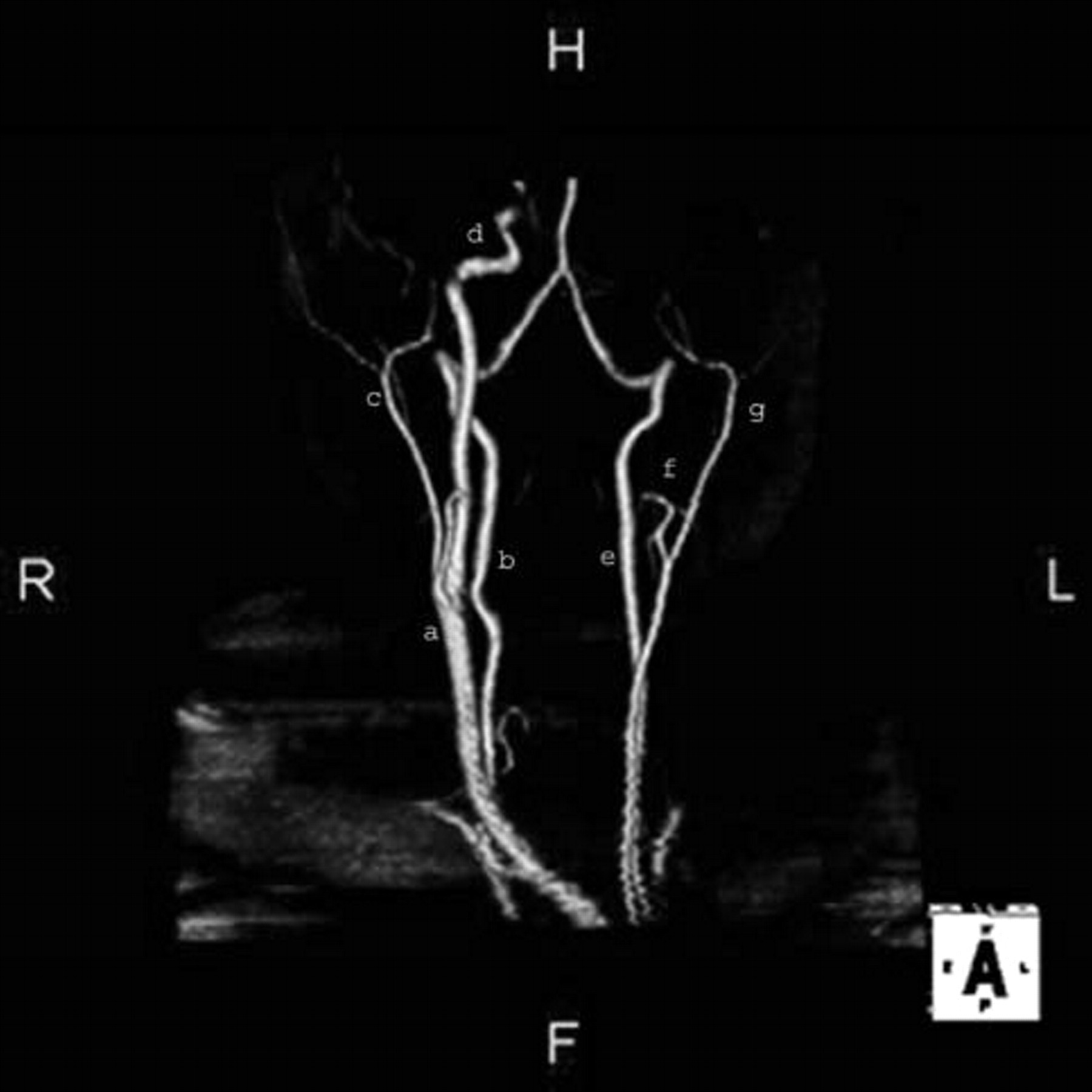
Magnetic resonance angiogram with three-dimensional reconstruction aortic arch and branches: (a) right common carotid artery; (b) right vertebral artery arising from the carotid artery; (c) right external carotid artery (ECA); (d) right internal carotid artery (ICA); (e) left vertebral artery, which arises from the aortic arch and forms the basilar artery; (f) small branch from the left ECA, which is thought to be the left ICA with dissection; (g) left ECA, exactly symmetric with the opposite side.

(a) Right carotid canal; (b) left hypoplastic carotid canal. Note the difference in size and volume of the left carotid canal (hypoplastic) compared with the one on the right.
Angiography demonstrated the right subclavian artery originating from the arch, absence of the brachiocephalic trunk, and the right subclavian artery, right common carotid artery (CCA), left subclavian artery, and left vertebral artery separately originating from the arch of the aorta. The right vertebral artery was originating from the proximal part of the right CCA. Intracerebrally, the right ICA was patent, as well as the anterior cerebral and middle cerebral arteries, with good communication between the right and left anterior cerebral arteries. There was no CCA on the left but a small external carotid artery (ECA) originating from the aortic arch (Figure 3).

Angiogram showing aplasia of the internal carotid artery.
Discussion
This case represents a young woman with agenesis or hypoplasia of the left ICA. The incidence of ICA agenesis is not exactly known; Lavaurs and colleagues reported two (0.33%) cases of ICA agenesis among 600 routine cerebral examinations. 1 Over the last 6 years, 732 carotid angiograms were performed at the Medical University of Ohio, with the current case being the only one with this abnormality, giving an incidence of 0.14%.
Two theories exist about the development of the carotid arteries. One theory suggests that the CCA and the proximal part of the ICA arise from the third aortic arch. 2 The other theory is that only the proximal part of the ICA arises from the third aortic arch, with the CCA developing from the aortic sac and the ECA arising from the first and second aortic arches. In both theories, agenesis of the ICA is always accompanied by absence of the carotid canal. Carotid artery development in the fourth embryonic week is necessary for the formation of the carotid canal in the fifth to sixth weeks of fetal life, 3 whereas the presence of a hypoplastic and small carotid canal (see Figure 2), as we propose in our current case, might reflect an obliteration of the ICA at a later stage of carotid canal development. The defective development of the third pharyngeal arch may induce the anomalies of the carotid artery system, as shown in experiments involving the genetic disruption of Hoxa3, which results in bilateral defects of the CCA. 4
Although variations in the number and arrangement of the great vessels of the aortic arch are not rare, the origin of the subclavian artery as the first branch of an aortic arch is extremely uncommon. 1 The embryologic origin of the subclavian artery may be explained by persistence of the fifth branchial arch with regression of the ipsilateral fourth branchial arch plus a segment of the dorsal aorta between the third and fifth arches during development. With cephalic migration of the seventh intersegmental artery, the future subclavian artery would arise from the fifth, not the fourth, arch. The vertebral arteries can arise from the aortic arch, the CCA, or the ICA. The vertebral artery arises from the postcostal longitudinal anastomosis between the C1 and C7 intercostal arteries and the cervical intercostal obliteration zone. 6
The major congenital abnormalities of the ICA can be classified as agenesis, aplasia, and hypoplasia, and they can be unilateral or bilateral. A 3:1 left-side predominance in agenesis of the ICA has been reported. 7 The term agenesis refers to a total absence of the organ, without any primordium observed. Aplasia is the term for the lack of development of the organ, although some primordium may be seen—in the case of the ICA, a remnant vessel, or some indirect sign of it, such as the presence of an ipsilateral carotid canal. In hypoplasia, the organ is present but has suffered incomplete embryonic development. Absence of the ICA is referred to as agenesis or aplasia. In this case, we presume aplasia of the ICA owing to the existence of the carotid canal in the base of the skull. 8,9
Although variations in the number and arrangement of the great vessels of the aortic arch are not rare, the origin of the subclavian artery as the first branch of an aortic arch is extremely uncommon. 1 The most frequent vertebral artery variant (2.4–5.8%) is the left vertebral artery arising directly from the aortic arch between the left CCA and the left subclavian artery. 5
Diagnosis of an aplasia of the ICA can be suspected by color flow Doppler ultrasonography. Nonvisualization of the ICA (whether a patent or an occluded vessel), the significantly reduced lumen diameter of the CCA, the absence of any sign of atherosclerosis in adjacent vessels, or carotid dissection should raise the suspicion of an aplastic ICA. 10 Although small-diameter carotid vessels may be seen in Takayasu's arteritis or other inflammatory processes involving the supra-aortic arteries, the inflammatory diseases often affect several arteries, and inflammatory changes can be proven by laboratory tests. 11 The bilateral presence of large vertebral arteries on MRA and angiography provides an indirect indication of long-standing collateral flow owing to either chronic vascular occlusion or a congenital anomaly.
ICA agenesis may be associated with intracranial aneurysms (25–34%), subarachnoid hemorrhage, cerebral hypoplasia, hemangioma, or anomalous vascular anastomosis. 12 The other rare reported pathologic abnormalities associated with ICA agenesis are corpus callosum agenesis, neurofibromatosis, meningocele, coarctation of the aorta, and cardiac anomalies. 1
Klippel-Feil syndrome refers to a segmentation defect of the cervical spine, which is often associated with anomaly of various other organs, such as the brain, kidney, and heart. It has also been associated with vascular anomalies, such as persistent trigeminal artery, 13 aortic coarctation, 14 and thoracic bifurcation of the CCA, 15 as well as with embryologic subclavian steal syndrome. 16 In 1986, Bavinck and Weaver presented the hypothesis of a subclavian artery supply disruption sequence as a potential explanation of the pathogenesis of the Klippel-Feil anomaly. According to their hypothesis, localized disruption of the vascular supply at specific points of the aortic arch complex during embryogenesis leads to disruption of normal organ development. 17
The association between Klippel-Feil anomaly and aortic arch abnormalities, such as the one we present in this case report, has been reported. The embryologic mechanism of this association is not entirely clear, although one may propose that it is a defect of the third aortic arch development. 15 This may point to a segmental developmental disorder leading to both the fusion of cervical vertebral bodies and the absence of the left ICA.