




Abstract
A patient with occlusion of multiple central veins from both dialysis and nondialysis catheters required permanent access for hemodialysis. Magnetic resonance imaging showed a patent left innominate vein. He underwent creation of a left axillary artery to internal mammary vein transposition fistula using the basilic vein from his right arm. The fistula has required one revision for outflow stenosis and one for aneurysmal degeneration. It continues to function well 3 years after placement. The internal mammary vein is an option for outflow when permanent hemodialysis access has failed in the presence of a patent innominate vein with occluded or severely stenotic ipsilateral subclavian and jugular veins.
Central venous catheters are frequently used for hemodialysis access. Up to 70% of patients requiring dialysis initiate their treatments with central venous catheters. 1 Despite concerted efforts, a quarter of some dialysis populations are catheter dependent. 2 Some patients with chronic renal failure present with a precipitous need for hemodialysis, whereas others develop problems with their existing vascular access and require temporary central venous catheters during repair of their access. Previous cannulation of the internal jugular or subclavian veins can produce a high incidence of central vein stenosis or occlusion. Occlusion of the subclavian, internal jugular, or innominate veins and/or the superior vena cava presents a substantial problem for placement of vascular access. The use of the internal mammary vein is described and can be used for suitable vascular access outflow in patients with multiple central venous occlusions.
Case Report
A 48-year-old white male who developed end-stage renal disease from chronic glomerulonephritis initiated hemodialysis through a right subclavian dialysis catheter in 1980. A left radiocephalic fistula was created at the same time. In May 1980, the patient received a living related renal allograft, which was subsequently lost to rejection. When he required a return to hemodialysis, his left forearm loop access was nonfunctional and a left subclavian catheter was placed for hemoaccess. A right forearm loop polytetrafluoroethylene (PTFE) access was placed, and a splenectomy was done in preparation for a second transplant. The patient received a second renal allograft in February 1984. The allograft functioned well until 2003 when he developed post-transplantation diabetes mellitus and a histoplasmosis infection. He was treated with amphotericin B and ultimately lost the function of his allograft. A right internal jugular dialysis catheter was placed for temporary hemoaccess. Permanent hemoaccess presented a challenging problem. Venous mapping showed a left basilic vein measuring 4.4 to 6.1 mm in diameter and a right basilic vein averaging 5.1 to 6.6 mm in diameter. Magnetic resonance imaging (MRI) of his chest, abdomen, and pelvis showed occlusion of both subclavian veins and the left internal jugular vein. The right internal jugular vein was stenosed at the base of the neck, and the left external jugular vein was severely stenosed at its insertion into the innominate vein (Figure 1). Both femoral veins were also found to be occluded (Figure 2).

Magnetic resonance venogram showing multiple central vein occlusions and stenosis.
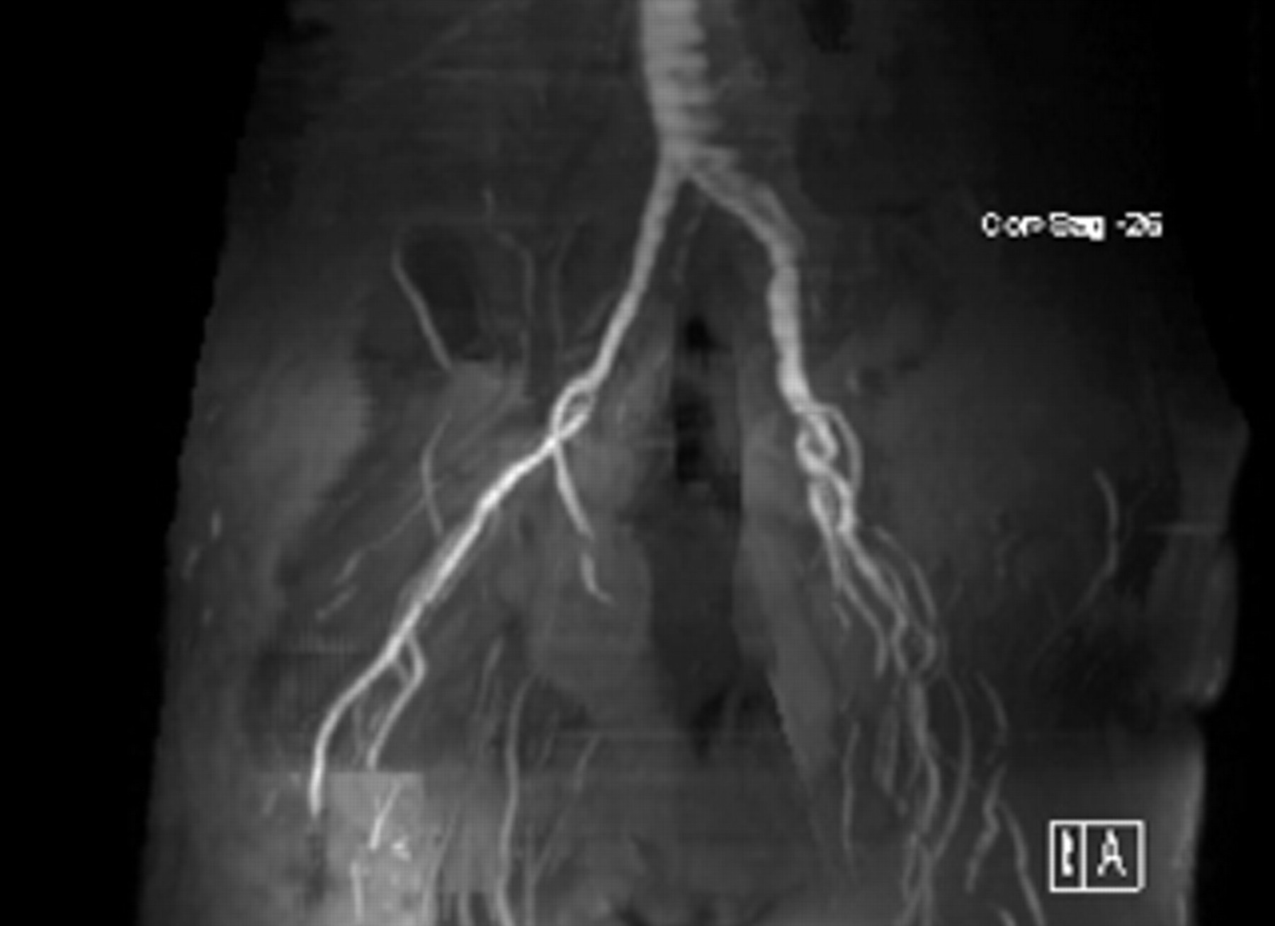
Bilateral femoral vein occlusions.
A peritoneal catheter was placed. This required repair of multiple ventral incisional hernias and significant adhesiolysis. Nonfunction of this catheter required its removal. Review of the MRI of the chest suggested that the left internal mammary vein could be used for central hemoaccess outflow. The medial portion of the left fifth rib was resected and the internal mammary vein was exposed. The basilic vein was removed from the right arm, and a left axillary artery to left internal mammary, basilic vein transposition fistula was created. A fistulogram at 1 month revealed a reasonably sized internal mammary vein (Figure 3). Three months later, the access failed. Thrombectomy and operative fistulography were done. The internal mammary vein was found to be stenotic at the basilic–internal mammary vein anastomosis. Outflow angioplasty was performed. Following revision, the fistula failed 1 week later. At this point, a decision was made to lengthen the fistula to improve the overall surface area available for cannulation and to permit the outflow anastomosis to be made to the more central segment of the internal mammary vein. Portions of the left basilic and brachial veins were used to lengthen and increase the diameter of the existing fistula. The outflow anastomosis was easily constructed after removal of the medial portion of the left second rib. The patient did require resection of a small aneurysmally degenerated segment of the vein. The fistula continues to function well during more than 3 years of follow-up (Figures 4 and 5).

Oblique view of the initial axillary artery to internal mammary vein, basilic vein transposition fistula showing a large central internal mammary vein.

Oblique view of the left axillary artery to internal mammary vein composite brachial and basilic vein transposition fistula.

Photograph depicting the left axillary artery to internal mammary vein composite brachial and basilic vein transposition fistula.
Discussion
The mammary vessels have long been used for free transfer grafts for breast and head and neck reconstruction. 3,4 Up to 20% of internal mammary vessels may be inadequate for reconstruction surgery in some series. 5 The internal mammary veins are one of five groups of veins that comprise the collateral venous network of the thorax. 6 This vein is usually suboptimal for use in vascular anastomosis below the level of the third rib because of its small diameter and multiple branches. 7 Dissection of the internal mammary vessels bilaterally in cadavers has shown vein diameters ranging from 0.64 to 4.45 mm at the level of the fourth rib. 8 The internal mammary vessels give direct perforators to the breast, with size ranging from 0.5 to 1.3 mm at the level of the second and third intercostal spaces. 9 Four different patterns of venous anatomy have been described. The most common was a single vein coursing medial and parallel to the artery until the fourth intercostal space, where it divided into medial and lateral branches with mean diameters of 2.7 and 1.8 mm, respectively. The medial and lateral veins are connected by intercommunicating branches. 10 This pattern is found to be different in the Asian population, with single veins found predominantly ranging in size from 0.8 to 4.8 mm. 11 The right internal mammary veins have been found to be larger than the left. 12 Use of a Doppler ultrasonic velocity detector can reliably identify the internal mammary veins and give a good estimate of their diameter. 13
The complications of central dwelling dialysis catheters have been described by multiple authors. Catheter-related venous stenosis has been reported to be nearly 40% in some central veins, compromising future access sites. 14 The extent of central venous obliteration from vascular access catheters has prompted innovative approaches to the creation of permanent vascular accesses for hemodialysis. With multiple subclavian and internal jugular vein stenoses or occlusions, the options are limited. The use of a PTFE graft from the axillary artery to renal vein has been described. 15 Bypassing the occluded axillary or subclavian vein by using the internal jugular vein may be applicable in some cases. 16,17 The use of the right atrium for fistula outflow has also been successful, although it should be used only for the patients in whom all access sites are exhausted owing to the magnitude of the procedure. 18,19
Use of the internal mammary vein for hemoaccess fistula outflow can be another viable alternative when there is extensive stenosis or occlusion of major central veins, particularly in the presence of a patent innominate vein with occluded or severely stenotic ipsilateral subclavian and jugular veins.