




Abstract
Cocaine-associated thrombosis has been reported in the literature with reports of vascular injuries to cardiac, pulmonary, intestinal, placental, and musculoskeletal vessels; however, injury of the pedal vessels is rare. We report on a 31-year-old man who presented 2 months following a cocaine binge with limb-threatening ischemia without an otherwise identifiable embolic source. Angiography confirmed extensive occlusive disease of the tibioperoneal vessels. The patient improved following therapy with heparin and a prostacyclin analogue. Cocaine-induced thrombosis should be considered in patients presenting with acute arterial insufficiency in the lower limb without any other identifiable cause.
Cocaine abuse is widespread and can lead to catastrophic drug-induced occlusion of the coronary and cerebrovascular vessels. Although there are numerous reports of other vascular injuries to pulmonary, intestinal, placental, and musculoskeletal vessels, occlusion of the pedal vessels is rare. We report on a young man who presented to our service 2 months after a cocaine binge with limb-threatening ischemia without an otherwise identifiable embolic source.
History
A 31-year-old male triathelete presented to the accident and emergency department having noticed discoloration of the fourth toe in his left foot and worsening intermittent claudication for the preceding 8 weeks, proceeding to pain in bed at night. He had been a smoker for 10 years but had discontinued 2 years previously. He had been running 10 km three times per week and swimming 1,500 m per week prior to this event. He initially denied any previous medical history or trauma to the leg; however, he did disclose on further questioning that following a family bereavement he went on a week-long binge in Europe of intranasal cocaine of unknown quantity, with his symptoms beginning on his return.
On admission, his left foot was cold and pale. He had an ulcer on the lateral aspect of his left foot and on the third toe of the same foot that was necrotic in appearance (Figure 1). Femoral and popliteal pulses were palpable bilaterally, but the dorsalis pedis and posterior tibial pulses were not palpable on the left side and the ankle-brachial indices (ABIs) were significantly reduced when compared with the other side (0.23 left and 1.08 right).
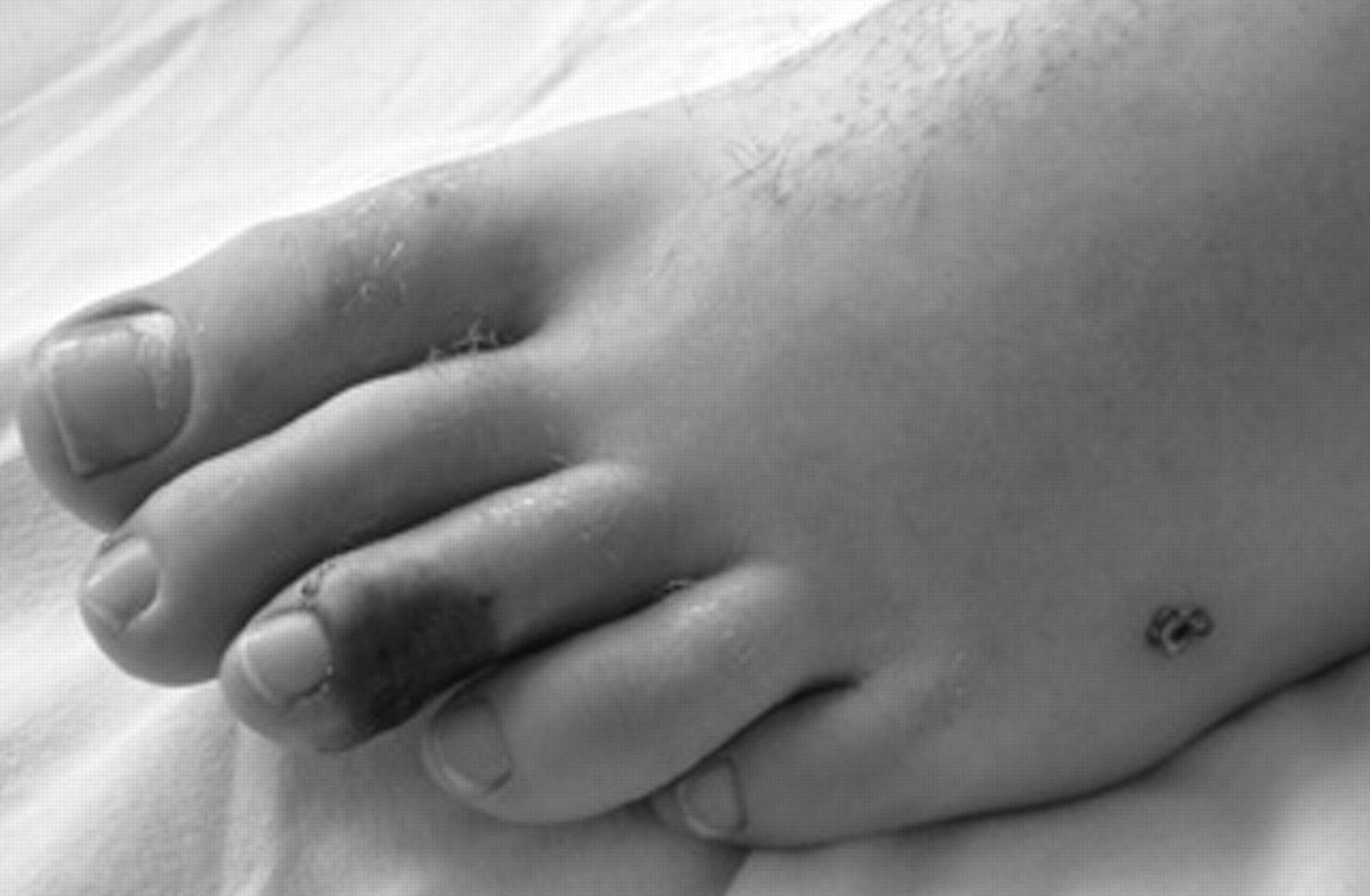
Photograph of the left foot on admission showing necrotic skin on the third toe and a necrotic ulcer on the lateral aspect of the foot.
Blood tests carried out included a complete blood count, liver function tests, urea, creatinine and electrolytes, lipid levels,

Angiogram images showing severe narrowing of the tibioperoneal trunk, with the left side being more severely affected than the right.
Intravenous heparin therapy was immediately commenced (the APTT was 70–90 seconds). Wound swabs were taken from the ulcerated lesions. Appropriate antibiotic therapy was undertaken in conjunction with the microbiology department. After 4 days, intravenous heparin therapy was continued without improvement; the patient then volunteered the additional information of his cocaine binge. Following case reports of successful treatment of cannabis- or cocaine-induced occlusive disease, he was commenced on an intravenous infusion of the prostacyclin analogue iloprost (1 ng/kg/min). 1 This was continued for 21 days and led to improvement in his symptoms and ABIs (0.61 left). He was discharged home, and when reviewed at 3 months, his ABIs were 0.71, he had recommenced training, and he was running 5 km three times per week.
Discussion
Cocaine is known to cause vasoconstriction and thrombosis of the arteries in the myocardium, central nervous system, muscles, kidney, uterus, and bowel. 2 Among the toxic vascular effects of cocaine, ischemic myocardial events have been experimentally confirmed in laboratory animals and healthy volunteers. Impurities and adulterants found in cocaine may exert a detrimental effect on the vasculature. Arsenic, which is a frequent contaminant of recreational drugs, is suspected to be an important trigger of thromboangiitis obliterans. 3 Coronary thrombus has been found in one-third of patients with cocaine-induced myocardial ischemia, with long-term cocaine use leading to premature atherosclerosis. 4 Various vascular injuries, such as vasculitis, superficial thrombophlebitis, subarachnoid and intracranial hemorrhage, pulmonary artery hypertension, pulmonary hemorrhage, skeletal muscle ischemia with rhabdomyolysis, and intestinal ischemia, have been reported following cocaine use. 5
Cocaine is a very lipophilic drug that readily crosses the blood-brain barrier, and it has been shown to potently induce cerebral artery vasoconstriction and cause chronic cerebral hypoperfusion in both animal models and human clinical studies. 6 It is a vasoactive drug with a multifactorial mechanism of action on the vascular system. 1 It causes the accumulation of noradrenaline at receptors by blockade of the transport system of the nerve terminal membrane. Catecholamine uptake is blocked in both the peripheral and central nervous systems. The result is increased sympathetic stimulation and increased presence of noradrenaline at the postsynaptic α-receptors. The subsequent vasospasm is of short duration. Although it has a relatively short half-life of approximately 1 hour, the major cocaine metabolite benzoylecgonine is still eliminated up to 10 days after administration in long-term cocaine users. 6 Cocaine alters prostaglandin production in rabbit endothelium, causing vasospasm, platelet activation, and thrombus formation. 1 The principle of prostacyclin treatment is restoration of normal endothelium and relief of vasospasm.
Thrombosis in the peripheral arterial system, associated with cocaine use, to the best of our knowledge, has been described only in seven cases. 1,7–9 In our case, the patient, although otherwise a healthy athlete, had a smoking history of 10 years but had been abstemious for 2 years prior to this event, which raised the possibility of his leg symptoms being due to Buergers disease; however, the temporal association of symptoms with the intake of a large amount of cocaine made the latter a more likely suspect. Popliteal artery entrapment in a young healthy athlete would also have been considered as a differential diagnosis; however, duplex ultrasonography and angiography showed no abnormality. Why his symptoms were unilateral is unknown; perhaps cardiac thrombi associated with the acute cocaine intake may have traveled distally and caused an acute occlusion.
Intermittent claudication in a previously healthy young man without any other precipitating factor is unusual. The initial onset of symptoms following a significant cocaine binge without another identifiable source would lead to the assumption that his foot ischemia and ulceration were cocaine related. The lack of response to 4 days of heparin therapy followed by a good clinical response to iloprost infusion would support further evaluation of its use in the treatment of peripheral ischemic complications associated with cocaine use.