




Abstract
Primary aortic tumors are rare and include a variety of histologic types, including aortic angiosarcomas, with less than 100 cases being reported since the first description of these tumors in 1873. The diagnosis of aortic angiosarcoma is usually confirmed by postoperative histopathologic examination or postmortem study. We present a case of primary aortic angiosarcoma presenting with intermittent claudication and radiologic findings of aortoiliac atherosclerotic disease treated initially with stenting; lower extremity embolic skin metastases developed during follow-up that prompted resection of the aortic bifurcation and restoration of the arterial continuity with a bypass. Despite postresection recurrence, a 3-year survival was achieved with aggressive multidisciplinary management.
Primary aortic tumors are extremely rare and include a heterogeneous group of aggressive malignancies. In 1873, Brodowski described the first case of primary aortic angiosarcoma, 1 and since then, less than 100 additional cases have been described. The diagnosis of aortic angiosarcoma is typically confirmed by postoperative pathologic examination or postmortem study. The prognosis remains dismal, probably because of the aggressiveness of this malignancy. 2 We present a case of primary aortic angiosarcoma with embolic lower extremity cutaneous metastases that was managed with an aggressive multidisciplinary approach followed by long-term survival and review the relevant literature.
Case Report
A 54-year-old Caucasian female presented to her primary care physician with the chief complaint of bilateral intermittent calf claudication. Her symptoms had started 2 months previously and were consistent with typical intermittent vasculogenic claudication at two blocks and were alleviated by rest. She denied any history of rest pain or buttock or thigh claudication. The patient denied any history of back disease, weight loss, or anorexia. Her past medical, surgical, family, and social history was unremarkable and noncontributory; the patient had no risk factors for vascular disease. She had never been a smoker. On physical examination, the patient was noted to have palpable upper extremity pulses and no carotid bruits. Lower extremity evaluation revealed bilateral palpable femoral and popliteal pulses. Pedal pulses were absent, but biphasic posterior tibial and dorsalis pedis Doppler signals were present, and the ankle-brachial pressure index was 0.7 bilaterally. Routine laboratory tests were within normal limits. A chest radiograph was negative for a mass.
A decision was made to obtain a digital subtraction aortoiliac angiogram (DSA) with bilateral lower extremity runoff. The DSA revealed an unusual large plaque in the infrarenal aorta. It was located posterolaterally approximately 4 cm below the takeoff of the renal arteries and extended to both iliac arteries. The superficial femoral artery, popliteal artery, and tibial trunk were all patent, with no significant hemodynamic stenosis. All three tibial vessels were continuous to the ankle. Based on the findings of the DSA, a decision was made to perform angioplasty and stenting of the aortic bifurcation and iliac arteries. Angiogram confirmed the presence of severe stenosis of the aortic bifurcation (Figure 1), which prompted us to proceed and employ two balloon-expandable stents (10 × 58 mm Omnilink, Guidant Co., Indianapolis, IN; now Abbott Laboratories, Abbott Park, IL) using the kissing stent technique. The procedure was performed with no complications and had good results, improving the patient's symptoms from two to five blocks.
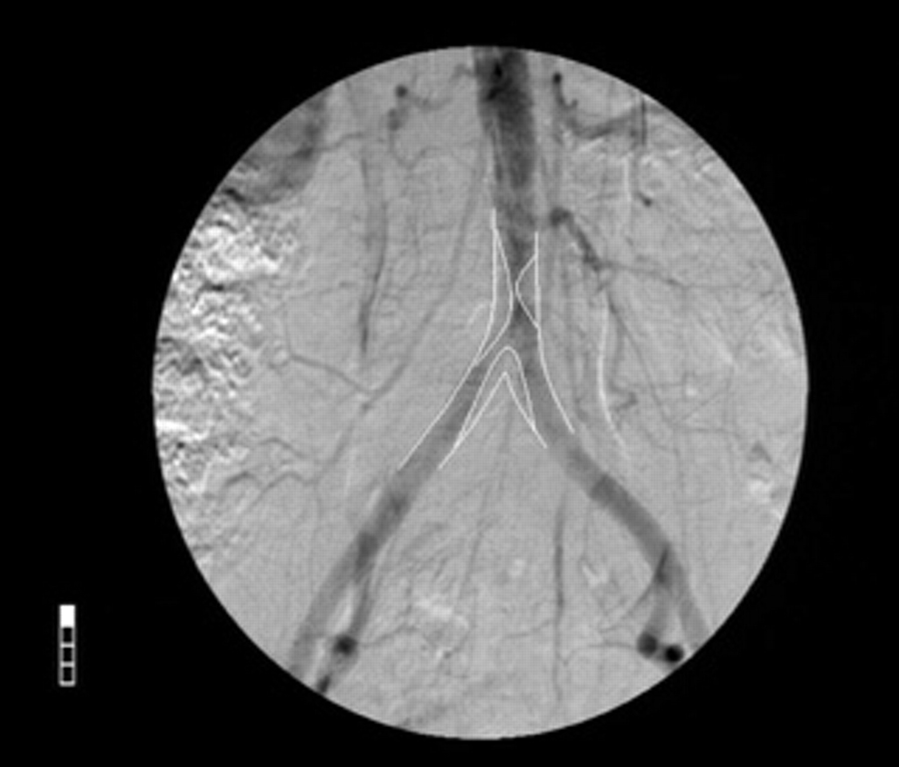
Digital subtraction angiogram of the aorta and iliac arteries showing severe stenosis of the aortic bifurcation that subsequently underwent successful stenting.
A month later, the patient presented with skin lesions in both legs. The lesions had the form of painless dark spots at the upper thigh, gluteal area, and leg that measured up to 1 cm in diameter (Figure 2). Biopsies of these lesions were taken, and the pathology results were consistent with a metastatic angiosarcoma, with anastomotic vascular channels lined by atypical endothelial cells of epithelioid type. Based on the recent history of aortic intervention, we strongly suspected that the primary tumor was the abdominal aorta.
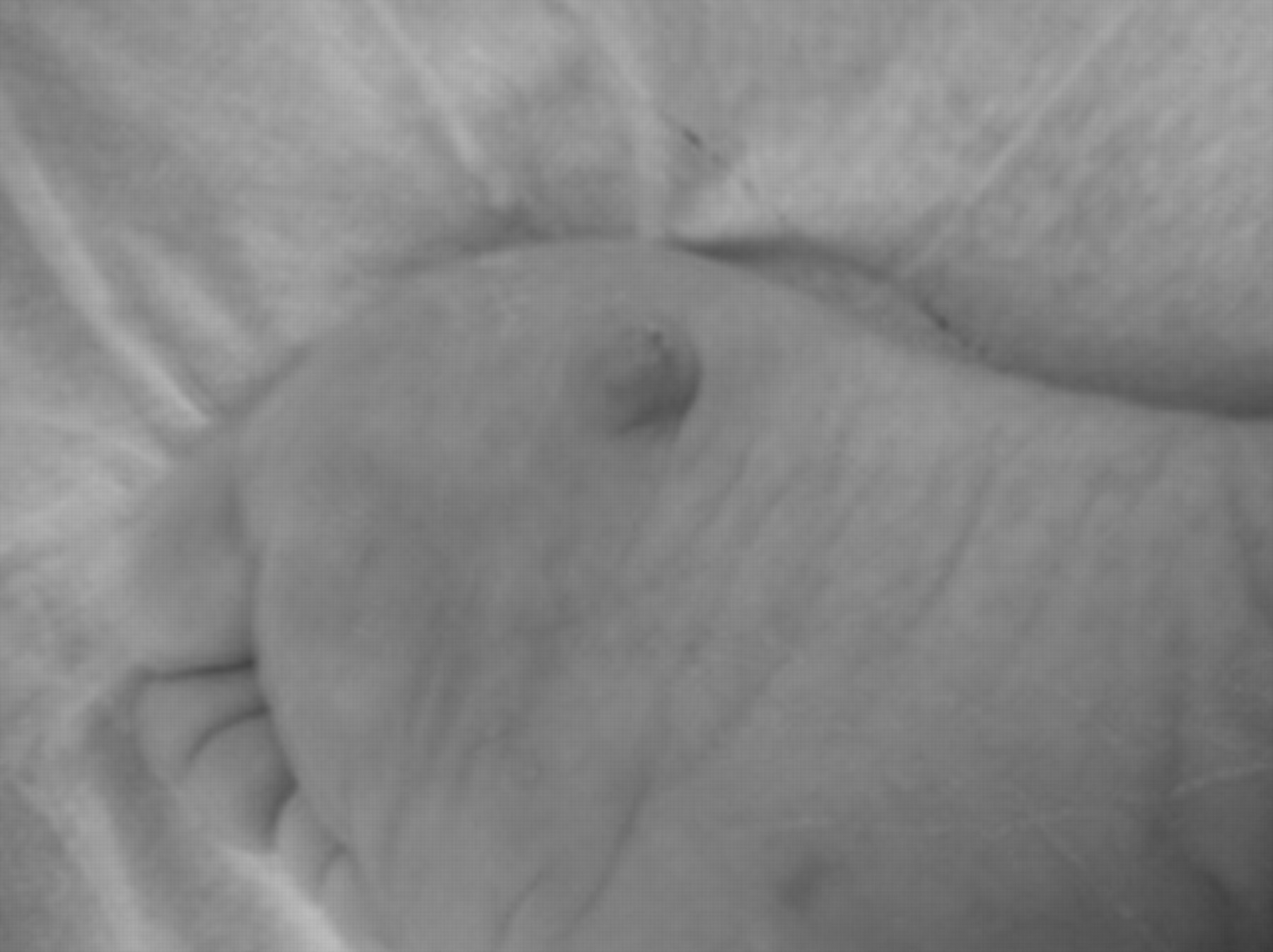
Skin nodule located in the plantar aspect of the right foot. This was painless, and the presence of focal necrosis was noted.
The patient was then referred to a medical oncologist and underwent a six-session course of chemotherapy with doxorubicin and ifosfamide, which resulted in partial remission; the skin lesions necrosed, but new ones appeared, and this was the indication for resection of the supposedly embolizing tumor. Preoperative chemotherapy was given to control the systemic disease and potentially to reduce the size of the tumor and make surgical removal easier. Additionally, abdominal aortic thrombosis was revealed on a computed tomographic aortoiliac angiogram, which was performed to explore a recent absence of femoral pulses (Figure 3). The patient was scheduled for elective excision of the aortic tumor and aortobi-iliac bypass. The procedure was performed through a midline transperitoneal approach. We mobilized the duodenum, opened the posterior peritoneum, and carefully dissected the aorta, from the left renal vein cranially to the bifurcation of the left and right common iliac arteries caudally; vessel loops were placed around each vessel. The inferior mesenteric artery was identified and noted to be occluded, and for this reason, a decision not to reimplant was made. We proceeded with anticoagulation (5,000 units of heparin administered as a bolus injection, intravenously), and vascular clamps were placed on the distal common iliac vessels and infrarenal aorta proximally to the lesion. A longitudinal aortotomy was made in the distal aorta and extended into the left and right common iliac arteries. An intraluminal soft tissue mass 4 cm long and 0.5 cm thick was noted approximately 2 cm distal to the orifice of the inferior mesenteric artery, which was very irregular in appearance. It had a grape-like appearance composed of numerous tiny spherical pieces of tissue that was extremely friable, producing severe stenosis of the aorta. The tumor extended distally into the left and right common iliac arteries. The distal aorta and proximal common iliac arteries containing the entire tumor mass were excised in toto. A 16 × 8 mm bifurcated graft (expanded polytetrafluoroethylene, Gore-Tex, W.L. Gore & Associates, Flagstaff, AZ) was used to restore anatomic continuity and anastomosed in an end-to-end fashion using 3-0 polypropylene sutures for the proximal and 4-0 polypropylene sutures for the distal anastomosis.
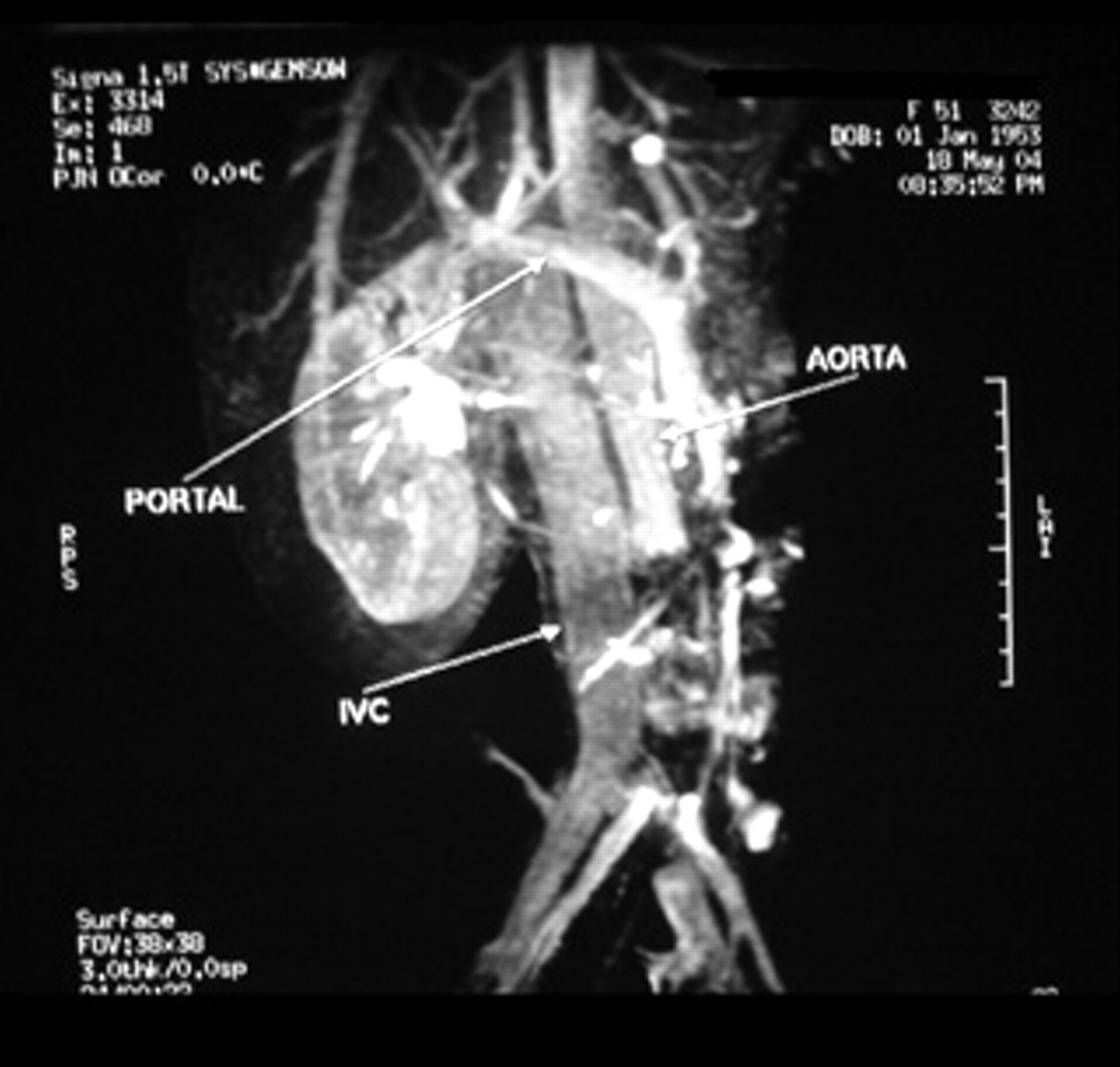
Computed tomographic aortoiliac angiogram showing abdominal aortic thrombosis.
Histology showed a high-grade malignant neoplasm with atypical polygonal endothelial cells with prominent nucleoli vacuolated or eosinophilic cytoplasm and increased mitotic activity. Immunohistochemistry was performed and revealed positivity of tumor cells for CD3H, CD31 and von Willebrand factor. Such findings were consistent with a high-grade epithelioid angiosarcoma (Figure 4). The postoperative course was uneventful, and the patient was discharged on postoperative day 5 with palpable posterior tibial pulses bilaterally and a significant improvement in her claudication. The patient was referred back to her oncologist and underwent a repeat complete course of chemotherapy (six sessions) using the same agents, with no response.

Histology showed infiltration of the aortic wall by a high-grade malignant neoplasm with atypical polygonal endothelial cells with prominent nucleoli vacuolated or eosinophilic cytoplasm and increased mitotic activity. Tumor cells formed small vascular spaces (hematoxylin-eosin stain; ×20 original magnification).
Seven months later, the patient returned with the chief complaint of increasing pain in the right shin. Physical examination and radiologic findings revealed a metastatic tumor of the right tibia. She was taken to the operating room, where resection and reconstruction with the Ilizarov method were undertaken, with a good outcome. Four months later, she presented with recurrent skin lesions of the right leg that necessitated a below-knee amputation performed 1 year later. The postoperative course was complicated by uterine bleeding owing to a uterine malignancy diagnosed on dilation and curettage histopathology specimens, followed by a radical hysterectomy with bilateral salpingo-oophorectomy; histopathology confirmed the presence of uterine metastatic disease. Fifteen months after the resection of the aortic tumor, metastatic lesions appeared in the right femur and left shoulder. The left shoulder disease was managed conservatively, whereas the right femur lesion was managed with a right total hip arthroplasty; this had a good result, and the patient was ambulatory with a leg prosthesis up to 3 years after the resection of the primary tumor, when she presented with acute bilateral lower extremity ischemia owing to thrombosis of the aortoiliac graft. She died of systemic complications of the ischemic event.
Discussion
We report on a patient with aortic angiosarcoma presenting with skin metastases following aortoiliac angioplasty for intermittent claudication. Multidisciplinary management with wide surgical resection, preoperative and postoperative chemotherapy, and aggressive treatment of metastatic disease were associated with long-term survival. Review of the literature identified 11 cases of aortic angiosarcoma with skin metastases. In 5 of the 11 cases, skin lesions were the first symptom (Table 1). 3–11 The mean age of these patients was 65.4 years (range 51–75 years). Men were affected more often than women (9 vs 2).
Presentation and Treatment Modalities
SFA = superficial femoral artery; NK = not known.
Primary aortic tumors are rare, with only 142 reported up to 2005. 12,13 Primary aortic angiosarcoma is even rarer, and since its first description in 1971, 11 only 24 cases had been described by 2005. 12 Three types of arterial tumors have been described in the literature: (1) intraluminal (polyploid), (2) intimal, and (3) adventitial. 14,15 Intraluminal and intimal types are associated with tumor embolization; this can involve not only the lower limb and skin 2,12 but also the viscera. 16 Arterial occlusion and tumor emboli to larger vessels have been described in the intraluminal type; in this type, the tumor has a focal attachment to the arterial surface, but its majority extends into the lumen as a polypoid mass, prone to break away. Less common are the adventitial tumors, which grow quickly and involve periaortic tissue and adjacent organs. 12,14 Wright and colleagues classified aortic neoplasm as intimal (obstructive or nonobstructive) or mural, originating from the media or adventitia of the aortic vessel wall. 17 Our case is an intimal nonobstructive tumor; this type of tumor grows along the intimal surface of the vessel with smaller intraluminal projections. Aortic sarcomas probably derive from intimal cells that show myofibroblastic differentiation. 18 The presence of a Dacron prosthesis has also been implicated. 19
Our patient presented with symptoms of stable intermittent claudication; this kind of symptom has been described previously in association with these tumors. 2 The most striking feature in our case was the temporal association between angioplasty and skin embolism, through the internal and external iliac arteries. Our case confirmed the crucial role of the skin histopathologist in the decision-making process, which was also shown by others. 9 In the era of preferential endovascular management of most types of severe vascular disease necessitating intervention, peripheral embolization would be the expected scenario as opposed to endarterectomy and direct diagnosis of the histology specimen. 20 In most patients, metastases coexist at the time of the initial diagnosis. 2 Malignant emboli may affect the skin, bone, pleura, and intra-abdominal organs, including the bowel, kidneys, spleen, and liver. 12,14
A variety of surgical procedures, including endarterectomy of the tumor, aortoiliac bypass, and resection with placement of an interposition graft, have been described. 2 Although there is no evidence that wide resection is associated with longer survival, it is more rational from the oncology standpoint. Chemotherapy with doxorubicin and ifosfamide has been proposed for the treatment of sarcomas, showing promising results in clinical trials. 21–23 In our patient, we employed aggressive preoperative and postoperative chemotherapy, which resulted in complete remission for some time and may have been associated with the long-term survival demonstrated. However, because of its rarity, the best treatment for aortic angiosarcoma is yet to be defined, and neither surgery nor adjuvant therapy provides a cure in the vast majority of patients.
The prognosis is usually dismal, and most patients die within the first year of diagnosis, with a median survival of 9 months being calculated from the series in Table 2; however, prognosis can be improved in selected patients with multidisciplinary management. 24 Almost all patients reported in the literature died from their tumor, apart from one patient who died of bronchopneumonia, 10 another patient who died of acute mesenteric ischemia, 8 and a third patient whose exact cause of death was not reported. 7 The prognosis depends on the histologic type, and long-term survival of several years in patients with the intimal type has been reported. 20,25 Our patient had this type of tumor.
Cause of Death in Patients with Angiosarcoma
In conclusion, despite the reported poor survival in patients with aortic angiosarcomas, our case suggests that long-term survival can be achieved with aggressive multidisciplinary management. Further research for better chemotherapy agents is justified.