




Abstract
Despite intense investigation over several decades to understand the mechanisms of vein graft failure, few therapeutic modalities have emerged. Emphasis using standard reductionist approaches has been focused on cataloging the components involved in the early events following vein graft implantation, but limited insight has been gained in understanding the dynamic interaction of these components. We propose that the application of systems theory offers the opportunity for significant advances in this area. Focused on modeling the dynamic relationships that define living organisms, systems biology provides the necessary tools to further our understanding of the complex series of overlapping biologic events on surgical implantation of the vein graft. Through the use of ordinary differential equation and agent-based modeling techniques, we present our ongoing efforts to define the nonlinear interactions between hemodynamics and vascular adaptation.
Analysis of the decision points leading to this costly miscalculation reveals several areas in which decisions were made based on limited or imprecise information. Specifically, two problems become apparent: (1) the current preclinical screening methods for identifying promising therapies that should move forward into clinical testing are inadequate and (2) tools to assist in the evaluation of early phase I/II clinical results, identifying those therapies most likely to be successful, are lacking. Underlying these limitations has been the traditional reductionist approach to these problems, with a focus on a specific intervention leading in a linear fashion to a final outcome, independent of the intervening pathways. Systems biology, with an emphasis on understanding the intervening components and providing predictive models to anticipate these results, offers an alternative approach.
Vein Graft Response to Injury
As outlined by Conte and colleagues, surgical vein grafting elicits a complex series of overlapping biologic events in the vein wall that begins immediately on implantation. 6 Repair of the damaged vein graft is initiated by the local synthesis of a wide variety of cytokines and growth factors. Chemokines induce the recruitment of neutrophils and mononuclear cells, further amplifying these pathways. Activated by these chemoattractants and mitogens, smooth muscle cells begin to dedifferentiate from a contractile to a synthetic phenotype and replicate within the media. By tracing the gradient of chemokines, liberated smooth muscle cells migrate from the media to intima, where continued proliferation and abundant matrix deposition are stimulated. Unchecked, these events lead to progressive thickening of the intima, narrowing of the lumen, reductions in graft flow, and intraluminal thrombosis.
Work in our laboratory has sought to provide insight into the complexity of these interconnected processes through transcriptional profiling. Using a rabbit vein graft model and a novel rabbit-specific Agilent microarray platform (Santa Clara, CA),
7
we examined the temporal variation in gene expression within the vein graft wall at 2 hours and 1, 7, and 28 days following implantation (Figure 1). At a false discovery rate of 0.10, 16% of genes were differentially expressed across all time points, with between 6 and 8% of the genes statistically different compared with normal vein at any single time point. Gene ontology analysis of these 497 differentially expressed genes (see Figure 1A) demonstrates early downregulation of intracellular maintenance activities, which recover to near baseline levels in the 1- to 7-day time frame. Coincident with this recovery is an increase in cell metabolism and matrix synthesis, which is initially noted at 1 day, with further expansion through 28 days. This coordinated shift in biologic processes within the graft is further illustrated by a principal component analysis (see Figure 1B), in which graft implantation induces a reproducible, patterned response of injury and repair. The difficulties inherent in potential interventions to redirect this response to a more favorable phenotype stem from the significant interconnectedness of the components that dictate these events (see Figure 1C). Given this complexity, the vulnerability of a single “bullet” approach, such as was attempted in the E2F decoy trials, is readily apparent.
A, Transcriptional analysis of rabbit vein grafts at 0.08, 1, 7, and 28 days following implantation demonstrates an early downregulation and then recovery of gene expression. B, Principal component analysis of these grafts illustrates the highly reproducible pattern that characterizes these changes. C, Analysis of the molecular pathways for these genes demonstrates the substantial interconnectedness among these elements.
Systems Biology and Vascular Remodeling
Despite intensive investigation over the last two decades using standard reductionist approaches, no clinically effective therapies have emerged to reduce vein graft stenosis and improve long-term outcomes. We propose that one of the primary flaws for limited progress in this field is not insufficient knowledge of the individual components of the system but the lack of a functional understanding of the interdependence and interaction of these components. This focus on understanding how properties emerge from the nonlinear interaction of multiple components is the hallmark of systems theory. In a general sense, systems biology encompasses the science of discovery, modeling, understanding, and ultimately engineering the dynamic relationships that define living organisms.8,9 Although a wide range of investigative approaches may be encompassed by this definition, Hood and colleagues extracted their thoughts of the essential features of contemporary systems biology (Table 1).8,10 Quantitative measurement of the dynamic behavior of interdependent components stands as the cornerstone of this approach.
Essential Components of Contemporary Systems Biology

Although multiscale mathematical models have been applied extensively to the area of cancer research, similar applications to the vascular system have been lacking.11,12 Estimates of vascular adaptation have been predominantly obtained through the use of empirical relationships, with no basis in the underlying physiology of the system.13,14 Among the few advances in the field has been the work of Serini and colleagues, who developed and experimentally validated a mathematical model of microvascular growth and reorganization based on the distribution of local chemoattractants as the fundamental mechanism for cell-to-cell communication. 15 More recently, Bailey and colleagues and Thorne and colleagues used an agent-based modeling (ABM) approach to examine the dynamics of leukocyte trafficking in the microvasculature.16,17 Although their model derivation is novel for its integration of a wide range of mediators and physiologic processes, the use of experimental data to support their fundamental assumptions or validate their predictions is limited.
Hemodynamics: Modulator of Vein Graft Adaptation
Fundamental to the systems biology approach is an understanding of the critical link between a system and its environment. Perturbations in the environment influence the structure and function of a system (Figure 2A). These changes then reshape the local environment, creating an inherent feedback loop between the system and the environment. This complex interaction may lead to a relative homeostasis, where changes in the environment and system converge to a stable phenotype (Figure 2B). Conversely, the absence of an inhibitory feedback loop between the system and the environment may result in a dynamic instability, with the environment inducing a progressive modification of the system that leads to critical failure. Early vein graft remodeling provides a classic example of this interdependence between a system and its surrounding environment.
A, Fundamental to systems biology is the concept that there is a dynamic interaction between a system and its surrounding environment. B, The nonlinearities of these interactions can lead to a local convergence and relative homeostasis of the system or induce progressive modifications of both the system and the environment that progress to critical failure.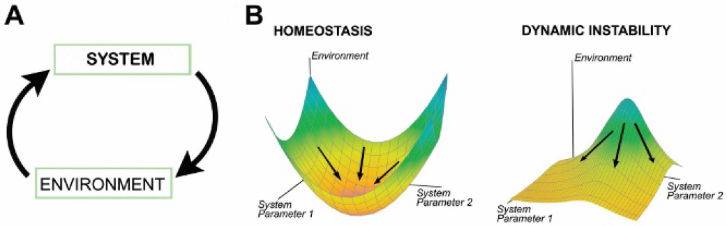
The acute transposition of a vein segment from a low-pressure/low-flow environment to the high-pressure/high-flow arterial system leads to significant structural changes within the wall. 18 This adaptation encompasses two distinct processes: intimal hyperplasia and wall (inward/outward) remodeling. 19 Intimal hyperplasia is characterized by migration of smooth muscle cells into the intima with proliferation and deposition of extracellular matrix, resulting in narrowing of the lumen. In contrast, remodeling is characterized by preservation (or loss) of lumen area through reorganization of the cellular and extracellular components within the media. Following vein graft implantation, both forms of adaptation are initiated, and the balance between these two processes dictates the thickness of the wall and the degree of luminal narrowing. 20
Vein grafts in the arterial system are exposed to four unique force vectors, tensile forces in the circumferential, radial, and longitudinal axes and surface shearing forces directed along the axis of flow. Considerable effort has been committed to understanding the impact of these various hemodynamic forces on the structure of the remodeling vein graft.21,22 Although confounded by an inability to clearly separate these variables, the bulk of the evidence suggests that medial thickening is correlated with circumferential tensile forces and intimal thickening is correlated with fluid shearing forces. Less well studied is the impact that these forces have on inward/outward remodeling, although an increase in cross-sectional area with increasing flows has been suggested.23,24
From a mechanistic perspective, the effect of physical forces on the biology of the vascular wall has been an intense area of research. Founded initially on the work of Gimbrone and his identification of the shear response element, a direct link between hemodynamics and transcriptional regulation within vascular cells has been firmly established.25,26 In addition, the fluid shear also has a significant influence on the kinetics of inflammatory cell binding to the endothelial surface and transmigration into the wall.27,28 A schematic representation of this response, illustrating the dynamic interplay between physical forces, cellular inflammatory elements, and an underlying gene regulatory network, is provided in Figure 3. The result is a highly integrated system in which local perturbations feed back to the other elements, leading to an updated but stable set point for the network or a condition with dynamic instability, characterized by early failure of the system. Detailed examination of our model system demonstrates such a critical recursive loop between the local hemodynamics and the regional biologic response of the vascular wall. Initial shear stress not only directs the primary set point for the gene network but also modulates inflammatory cell infiltration, both of which influence the cell- and matrix-based remodeling response and define the local modifications in conduit geometry. These morphologic changes induce perturbations in local shear and wall tension, resulting in new set points for the biologic response parameters.
The dynamic interplay between physical forces, cellular inflammatory elements, and an underlying gene regulatory network that comprise early vein graft remodeling. A critical recursive loop between the local hemodynamics and the regional biologic response of the vascular wall becomes apparent as a principal regulator of these events.
A frequent component of systems biology modeling, and a critical component of our model, is the integration of a multiscale approach. Implicit in this approach is the assumption that the tracking of repetitive, microscopic events can be used to predict the macroscopic function of a system. In the current system, molecular-level genomic information provides cellular-level predictions of proliferation and matrix syntheses that yield tissue-level estimations of remodeling, ultimately determining continued patency or failure of the vein graft.
Dynamic Modeling of Hemodynamically Driven Vein Graft Adaptation
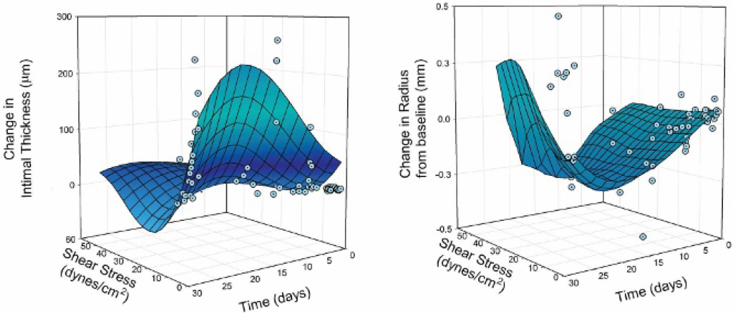
Based on animal experimentation by our group29,30 and others,21,24,31 the standard paradigm of vein graft remodeling has been established, in which reductions in wall shear stress induce an accelerated intimai hyperplastic response with narrowing of the lumen. Although attractive in its simplicity and likely valid within a uniform flow field, this relationship fails to explain the substantial heterogeneity in lesion development observed in a complex, three-dimensional geometry. Recent work in our laboratory redefines these concepts, demonstrating that the dynamics of the complex flow field are critical in understanding the remodeling process. Using a rabbit vein graft, focal stenosis model, we identified a biphasic relationship between shear stress and the resulting hyperplastic response. In a uniform, laminar flow field, where low to moderate shears are encountered, the standard inverse correlation between shear and intimai thickening is observed. But in areas of segmental narrowing, there is a critical shear threshold at approximately 10 dynes/cm2, above which increasing shear stress leads to marked augmentation in the rate of intimai thickening (unpublished data, 2008). The result is rapid, segmental narrowing of lumen, secondary to destabilization of the physiologic homeostasis between outward remodeling and wall thickening. In an effort to further explore the nonlinear behavior of these events, several modeling approaches have been explored.
Ordinary Differential Equations
Ordinary differential equation (ODE) modeling consists of establishing a series of mathematical relationships that describe the sequential change in the components of the system over time. Derived from the observed and hypothesized interactions among the various components, these expressions provide the general framework for the structure of the system. Integration of experimental data to estimate the unknown parameters transforms these relationships into a tool useful for predictive modeling. These equations describe the collective behavior of the system and thus rely on a large number of individual elements to maintain accuracy. When the number of elements becomes small, stochastic variability in the system can dominate, leading to substantial reductions in the accuracy of this approach. Because of this reliance on bulk average properties, detailed understanding of the spatial heterogeneity that may occur within a system is lacking and is a recognized weakness of the ODE approach.
We recently published a mathematical model of shear-regulated intimai hyperplasia based on experimental data obtained from the rabbit vein graft construct.29,30,32 Extending the work of previous investigators,24,31,33 an inverse correlation between intimai thickness and shear was assumed via the following expression:
Ordinary differential equation modeling of vein graft adaptation illustrates the dynamic interaction between intimal hyperplasia and shear stress as a function of time. Similar nonlinearities are observed in shear-dependent outward remodeling.
Agent-Based Modeling
ABM is a mathematical approach for analysis of dynamic systems in which elements evolve through a number of discrete time steps governed by a set of rules based on the states of the neighboring cells.34,35 Rather than attempting to define the behavior of a complex system at the level of the continuum, via a system of coupled differential equations, ABM breaks the complex system into discrete components (or elements) that are governed by a set of simple rules. The complex behavior of the system emerges then from the local interaction of the elements. This type of modeling is “bottom up,” inasmuch as all measured parameters and outcomes from the model are generated by the actions of the agents. Such approaches have been applied to the development of three-dimensional scaffolds for tissue engineering applications with novel results.36,37
We applied these concepts to develop an ABM approach to vascular remodeling. Capitalizing on the inherent circumferential symmetry of the vein graft, a polar coordinate system is employed (Figure 5). Also benefited by the longitudinal (axial) symmetry of the vein graft, reduction to a two-dimensional geometry is accomplished. The graft wall is divided into a grid with each element measuring 10 × 10 μm, consistent with the approximate size of a smooth muscle cell (SMC). All elements will be populated with either cell or matrix in a random fashion. Changes in the model configuration occur using a two-step process: the first step determines the change in the number and composition of the element within the grid, and the second step resets the outer radius of the wall based on the extent of inward/outward remodeling and locally redistributes the elements to within this redefined grid. At the start of the simulation (t 5 0), no macrophages will be present in the tissue. On initiating the model, the rate and location of monocyte entry into first (luminal) row of cells will be dependent on the local shear stress. On entry into the wall, these elements exhibit a biologic effect on SMC proliferation and apoptosis. Macrophage migration is simulated by a random walk approach, as recently described by Perez and Prendergast.
38
Each macrophage is allowed to move through the three-dimensional matrix, without a preferred direction of motion (anisotropic).
Agent-based model of vascular adaptation. Changes in the wall structure are initiated in step 1 by the stochastic probability of cell proliferation/apoptosis, matrix synthesis/degradation, and inward/outward remodeling. Step 2 induces inward/outward remodeling of the wall and commences monocyte influx/efflux and a random walk of existing monocytes.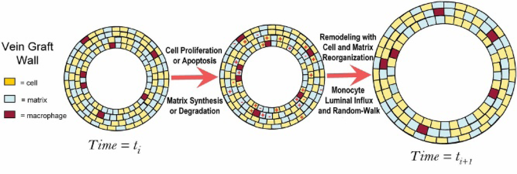
Application to Human Vein Graft Failure
Despite evolving evidence in animal models, the application of these concepts to the understanding of human vein graft failure remains poorly defined. Much of our current understanding of hemodynamics and its impact on human vein graft adaptation comes from the work of Fillinger and colleagues, who identified shear stress as a primary driving force for outward dilation within the first several months following implantation. 39 Further support has been added by Owens and colleagues, who confirmed this observation and suggested that system inflammation functions as an important modifier of shear-induced outward remodeling.40,41 Unfortunately, examination of these nondiseased segments provides only limited value in understanding the active lesion remodeling that characterizes human vein graft stenosis.
Parallel to the basic science and computational efforts detailed in this proposal, our laboratory has been active in the development of a translational research program. Critical to these efforts is imaging of in vivo human vein grafts, with particular emphasis on evolving vein graft stenotic lesions and their response to the hemodynamic environment. Figure 6 illustrates our ongoing clinical research in which computed tomography was used to obtain accurate geometries along the length of the vein graft. Coupled with the hemodynamic information obtained from Doppler spectral analysis, dynamic shear stress profiles are calculated and mapped to the three-dimensional graft geometry. Sequential examination provides a powerful tool to understand the influence of physical forces on lesion development. We are actively developing these technologies for integration into the predictive models outlined above.
Computed tomographic scan reconstruction of a human vein graft and estimation of wall shear stress along the length of the graft. Graphs demonstrated wall shear stress at 7 and 17 weeks. Note that the region of elevated wall shear stress in the distal graft at 7 weeks has demonstrated significant improvement following outward remodeling in this segment.
We propose that advanced understanding of vascular remodeling will be facilitated by the integration of these systems-based approaches into translational research. With further development, the remodeling events in the complex geometry of the human vein graft can be directly integrated into the model, and the primary focus of our future research efforts will be validation of these results in humans and development of a package for use by clinicians and clinical researchers.