




Abstract
Venous and arterial identity is predetermined in the embryo, with embryonic vessels expressing Eph-B4 differentiating into veins and vessels expressing ephrin-B2 differentiating into arteries. The specialized membrane organelles lipid rafts and caveolae serve as localized domains for proteins to interact with one another and play a role in signal transduction and vesicular trafficking. Several tyrosine kinase membrane receptors, including Eph-B1, have been colocalized to caveolae. These data suggest that caveolae and Eph receptors may have coordinated roles in determining vessel identity, not only during embryogenesis but perhaps also during adult vascular remodeling and angiogenesis.
Preferential Expression of Arterial and Venous Markers in Mouse Models
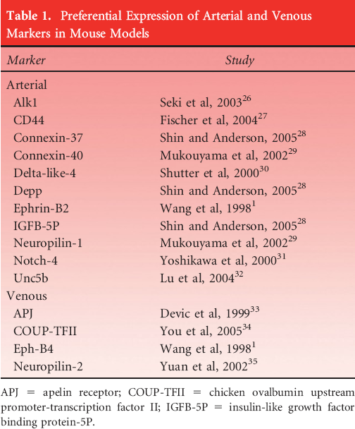
APJ = apelin receptor; COUP-TFII = chicken ovalbumin upstream promoter-transcription factor II; IGFB-5P = insulin-like growth factor binding protein-5P.
Vascular markers have recently generated interest owing to their dynamic involvement in both with the vasculogenesis of an embryo and with remodeling later in adult life, as seen during angiogenesis. Markers previously thought to be quiescent in the adult may, in fact, play a significant role in vascular remodeling. This is of particular significance when looking at the role of these proteins in vein graft remodeling or for potential therapeutic application such as in therapeutic angiogenesis. In particular, markers of interest include the Eph family of tyrosine kinases and their respective ligands, the ephrins. Other proteins and structures of interest are caveolins, the major component of the cell membrane organelle caveolae. These unique membrane structures play an as yet incompletely elucidated role in both vasculogenesis and angiogenesis. However, their special role in membrane trafficking and organization leads us to believe that they are more than silent bystanders to endothelial proliferation, migration, and tube formation.
Lipid Rafts
Lipid rafts were first hypothesized toward the end of the last century. 4 These specialized domains of membrane are very specifically enriched with lipids such as glyco-sphingolipids and cholesterol and certain types of lipidated proteins; lipid rafts have the unique biochemical property of being insoluble to most detergents, especially to the nonionic detergent Triton X-100 at 4°C. 4 Lipid rafts are distinct areas in the plasma membrane with order and structure among an otherwise sea of random lipids. Many functions have been proposed for these membrane domains, including involvement in cell signaling, endocytosis, vesicular trafficking, and cytoskeletal function.5,6 However, their role in these various cellular functions is still under debate.
Lipid rafts are thought to serve as localized domains for proteins to interact with one another. For example, many proteins have a glycosylphosphatidylinositol (GPI) “anchor” added to their C-terminus; this posttranslational modification selectively targets proteins to the apical side of the plasma membrane and particularly to lipid rafts. Thus, the GPI anchor and lipid raft are thought to form a site where proteins can organize and serve as a specific domain, that is, to serve as a site of communication or signaling. The related membrane organelle, the caveolae, shares similar properties to the lipid raft but with some specific alterations (Figure 1).
Caveolae and lipid rafts. The area delineated by black bars shows an increased concentration of glycosylphosphatidylinositol (GPI). A, This demonstrates the general morphology of a typical caveolae. Caveolin proteins hold the structure in its characteristic shape. The caveolae is also distinguished by its high concentration of cholesterol. B, Here a lipid raft is shown simply as a collection of proteins linked to GPI. The raft shows no three-dimensional structure other than being an enriched and linked area of proteins and lipids.
Caveolae
Caveolae, Greek for small cave, are specialized membrane organelles characterized by their flask-like shape. These small (50–100 nm) membrane invaginations are virtually indistinguishable from other lipid rafts and also share the property of being insoluble to typical detergent preparations. Caveolae and lipid rafts are cholesterol-enriched domains of the cell membrane separate and distinguishable from other membrane domains. Their function has been implicated in various processes, including cholesterol transport and signal transduction. 7 Various proteins have been localized to caveolae, including caveolins, endothelial nitric oxide synthase (eNOS), and several types of tyrosine kinases, 7 including the protein Eph-B1. 8 Localization of these proteins specifically to the caveolar domain implies that they may serve an important role in signal transduction. Although these specialized areas of membrane have been compared to clathrin-coated pits, it is clear that they serve quite different functions. Clathrin-coated pits serve as the main entrance for endocytosis of products to be degraded in lysosomal pathways. Caveolae on the other hand, appear to be involved in endocytosis of macro-molecules not destined for degradation. This property has made caveolae a target for several types of pathogens including SV40 virus and bacteria.9,10 It has been proposed that pathogenic organisms may use the caveolar endocytosis pathway as a means for bypassing otherwise lethal lysosomes associated with clathrin-coated pit endocytosis. Given that caveolae could not have evolved to facilitate pathogen entry into cells, it is thought that they may be a cellular adaptation aiding transcytosis and controlling regulation of linked proteins. These are some of the properties that make caveolae an intriguing area of study in vascular physiology. For example, the protein caveolin-3 has recently been shown to be an arterial endothelial marker linking the family of caveolins to vascular identity. 11
Caveolins are a family of proteins that are essential to the formation of caveolar organelles in endothelial cells.12,13 Three proteins of this family have been described to date, including caveolin-1, caveolin-2 and caveolin-3. Caveolin-1 is critical to caveolae formation, and its absence inhibits caveolae production. 14 Although caveolins have been shown to be dispensable to embryogenesis, as homozygous caveolin-1-knockout mice are viable with only minor phenotypic abnormalities, the role of caveolins in angiogenesis is thought to be critical. 15 Various studies have shown that caveolin-1 and other proteins localized to caveolae are involved in cell signaling and fine modulation of such signals.12,16 An example of this was shown by the Sessa group in the study of caveolae involvement with the protein eNOS. 17 They reported that wild-type and caveolin-1-overexpressing mice both express the same amount of eNOS. Interestingly, however, they find that more eNOS was caveolae associated with the mice overexpressing caveolin-1 than in those not overexpressing it. In this same study, the authors also reported that caveolin-1 overexpression via transgenic mice reduces vascular permeability. This is thought to be due to decreased eNOS-dependent blood flow. Other studies have more specifically shown the interaction of eNOS with caveolin-1. Feron and colleagues showed using coimmunoprecipitation that nearly all the eNOS in endothelial cells is associated with caveolin-1. 18 They further suggested that the targeting of eNOS to caveolae in endothelial cells may represent an intrinsic feature of the signal transduction pathway associated with nitric oxide in these types of cells. These data, in conjunction with what we know about the expression of Eph-B1 and its localization to caveolae, open up further questions about the role of these membrane organelles in vasculogenesis and angiogenesis.
Ephrins/Eph
Eph receptors and their respective ephrin ligands represent a family of tyrosine kinases implicated in many varied functions, including embryonic development, axon guidance, and cell migration and adhesion. 3 The interaction of these proteins is critical for both embryonic vasculogenesis and later in adult life angiogenesis and vessel remodeling. The ephrin-B2 ligand is found exclusively on arteries. It is thought that this ligand determines the fate of a nascent artery and then persists as a marker into adulthood. The role of ephrin-B2 and other arterial markers is still under investigation. Disruption of the ephrin-B2 gene prevents venous remodeling in angiogenesis to branched structures. 19 However, since venous fate is thought to be predetermined genetically, the finding that both arterial and venous angiogenesis is disturbed in the presence of only ephrin-B2 inhibition shows that a critical interaction must occur between ephrin-B2 and Eph-B4, allowing for proper formation of each type of blood vessel.
Homozygous knockouts of ephrins are lethal during embryogenesis beyond midgestation. 20 Embryos died at embryonic day 10 and exhibited pale yolk sacs and growth retardation with minimal development of vasculature. Although the critical importance of these markers is clear in early life, their purpose in adult life has yet to be fully examined. Boundary formation between arterial and venous systems is thought to be regulated in part by the interactions of ephrin-B ligands with Eph-B receptors. Several groups have looked at this relationship between Eph receptors and ephrin ligands. Recent detailed analysis of mesenchymal tissue involved in angiogenesis revealed that selective lack of endothelial ephrin-B2, in spite of expression of ephrin-B2 in other tissues, is lethal. 21 These results suggest that specific vascular expression of this ligand is critical for both embryogenesis and vasculogenesis. 21 More work testing the importance of mesenchymal ephrin-B2 still remains to determine the role of this ligand in adult angiogenesis.
The ephrin-B2 ligand is found not only in larger vessels but persists even into the smallest microvessels and capillaries. 22 The interaction of ephrin-B2 with its cognate receptor Eph-B4, found predominantly on venous endothelium, determines vessel identity and regulates coordinated angiogenesis. 1 This process may also involve reverse signaling between the Eph-B4 tyrosine kinase and ephrin-B2 during the formation of new capillary networks, such that the ligand ephrin-B2 also generates a new signal within its cell. It is thought that reciprocal signaling may produce an inhibitor response and prevent fusion of small capillary systems into larger formed vessels. 23 These interactions are complex, and more investigation will be necessary to fully understand the roles of these markers in angiogenesis. 3
The interaction of ephrin-B ligands with Eph-B receptors is modified by other angiogenic factors such as vascular endothelial growth factor (VEGF), Ras GTPase activating protein (RasGAP), and possibly eNOS. 2 VEGF in particular has been shown to be a strong modulator of endothelial proliferation, and its role in Notch signaling has been shown to promote differentiation of endothelium into arterial vasculature. 24 Graded signaling by this growth factor affects downstream expression of both Eph-B4 and ephrin-B2 genes, implying a role for VEGF as an active participant in vascular differentiation. This also suggests a role for VEGF and its downstream pathway members as potential regulators of angiogenesis. Thus, targeting expression of Eph-B4 and ephrin-B2 and driving vessel identity may prove to be a clinically useful therapeutic strategy.
Summary
Figure 2 illustrates the theorized relationship between caveolae and the Eph family of tyrosine kinases. As previously mentioned, eNOS has been shown to colocalize within caveolae, although the exact relationship of this protein to caveolin-1 or caveolin-3 is not fully understood. One can postulate that if caveolae are critical to Eph receptor activity, an organism lacking caveolae would exhibit abnormal vasculogenesis; in fact, caveolin-1 knockout mice show vascular hyperpermeability and other defects. Recently, it had been demonstrated that caveolin-1 and Eph-B1 are linked to one another within caveolae.
8
As such, caveolins are likely to be important in the regulation of Eph receptors; this putative regulatory role suggests a further role for caveolins in the determination of vascular identity. Additional work investigating the role of caveolins as determinants of vascular identity needs to be completed, especially their role in the regulation of Eph receptor forward signaling and Ephrin ligand reverse signaling. In addition, the role of caveolin-3, found primarily in differentiated smooth muscle cells,
25
in vascular identity has yet to be fully elucidated.
This is a closer look at the caveolae with Eph receptors illustrated. The relationship of caveolins to Eph-B tyrosine kinases is still not fully understood. This shows the receptor in proximity to caveolin and thus linked to the caveolar space.