




Abstract
In the present report, we describe a two-stage technique of combined basilic and brachial vein transposition. Our patient had a brachial-basilic vein fistula created, but during the second stage for the transposition, a low basilic-brachial vein confluence was found. Instead of abandoning the procedure, the brachial vein was mobilized and transposed to primarily constitute a usable fistula, which subsequently was successfully used for hemodialysis. A detailed description of our technique is provided. Surgeons should be aware of this alternative procedure to maximize fistula creation rates.
In an effort to decrease the prevalence of hemodialysis catheters and prosthetic grafts in patients with end-stage renal disease (ESRD), the use of an autogenous transposed basilic-brachial fistula (TBBF) 1 has been strongly advocated by the most recent update of the Kidney Disease Outcomes Quality Initiative (KDOQI) guidelines. 2 Although the question whether this procedure should be performed in one 1 or two stages 3 remains unanswered, acceptable midterm patency rates have been reported. 4,5 As more surgeons become familiar with the indications, technique, limitations, and complications of this type of autogenous fistula, some have realized that transposition of the brachial vein is also feasible, either as a one-stage 6 or a two-stage procedure. 7 In the present report, we describe the technique of a two-stage, combined basilic-brachial vein transposition procedure suitable for patients with the low basilic-brachial vein confluence anatomic variation.
Technique
Both procedure stages are performed on an outpatient basis, under regional (interscalene brachial plexus) ± local anesthesia with intravenous sedation. The index upper extremity is placed on an arm board and circumferentially prepared to the shoulder and axilla.
During the first stage of this procedure, the basilic vein is identified and mobilized through a transverse antecubital fossa incision. Patient is anticoagulated with 3,000 units of unfractionated heparin, intravenously, and an end-to-side fistula is created with the brachial artery or in the case of a high bifurcation the radial or ulnar artery using 7-0 polypropylene suture (Prolene, Ethicon Inc, Somerville, NJ). Following a period of at least 6 weeks to allow the fistula to mature, we proceed to the second stage, which involves full mobilization of the basilic vein and its continuation-the brachial vein-through a longitudinal incision along the medial aspect of the arm so that the brachial vein is mobilized in its entire length by dividing all tributaries with 4-0 silk suture and/or surgical clips for the distal vein stumps (Figure 1). The medial antebrachial cutaneous nerve and its branches are carefully protected to preserve sensation of the medial forearm. After this, the patient is given 3,000 units of unfractionated heparin, intravenously. The anterior surface of the arterialized basilic-brachial vein is marked using a marker pen to prevent it from being twisted, and after a suitable period of time, the proximal part of the fistula near the anastomosis is controlled with a bulldog clamp followed by transection of the basilic vein and the brachial vein distally that is ligated. A bulldog clamp is placed on the distal brachial vein to allow dilatation of the basilic-brachial vein with saline to ensure that there are no stenotic areas or leaking points (Figure 2). We prefer to transpose the basilic-brachial vein instead of superficializing it to avoid obligatory injury of the medial antebrachial cutaneous nerve and trapping of the vein within the scar tissue and to improve accessibility of the vein during hemodialysis because the vein is tunneled laterally to its original location. Accordingly, a superficial anterolateral arm tunnel is made between the top and bottom of the incision with a sheath device (arteriovenous sheath tunneler, C. R. Bard, Inc., Murray Hill, NJ), with care taken not to make the tunnel too deep that it could make fistula cannulation difficult (Figure 3). The basilic-brachial vein is then placed inside the arm tunnel, with care taken not to twist the fistula, using its top markers. The vein is distended again with saline to ensure that it expands and is not twisted. Reanastomosis of the two ends of the basilic vein is subsequently performed in a standard end-to-end fashion with 7-0 polypropylene suture (Prolene) (Figure 4). The bulldog clamp is released from the proximal fistula part, and the anastomosis is tested for a major leak that would necessitate placement of repair sutures. Gelfoam soaked in thrombin solution (bovine thrombin, Johnson & Johnson/King Pharmaceutical, Bristol, TN) is used as a complement to achieve hemostasis from suture holes. At this point, the fistula is palpated and should have a strong thrill. The wound is then checked for hemostasis and closed with a continuous 4-0 absorbable suture for the subcutaneum and stainless steel staples for the skin (Figure 5). Wound suction or heparin reversal is not used routinely. In case of complications, the patient is observed in the short stay unit.
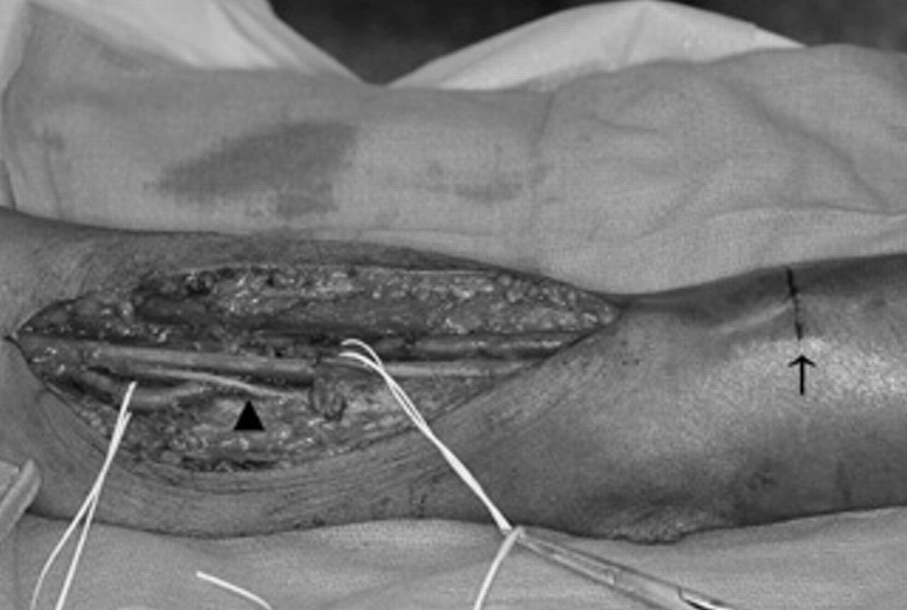
Through a longitudinal incision along the medial aspect of the upper arm, full-length mobilization of the short basilic vein segment and the brachial vein is performed by dividing all tributaries with 4-0 silk sutures and/or surgical clips. The scar of the first stage of the procedure antecubital fossa is marked and shown with an arrow and the medial antebrachial cutaneous nerve with an arrowhead. The brachial vein is surrounded with vessel loops.
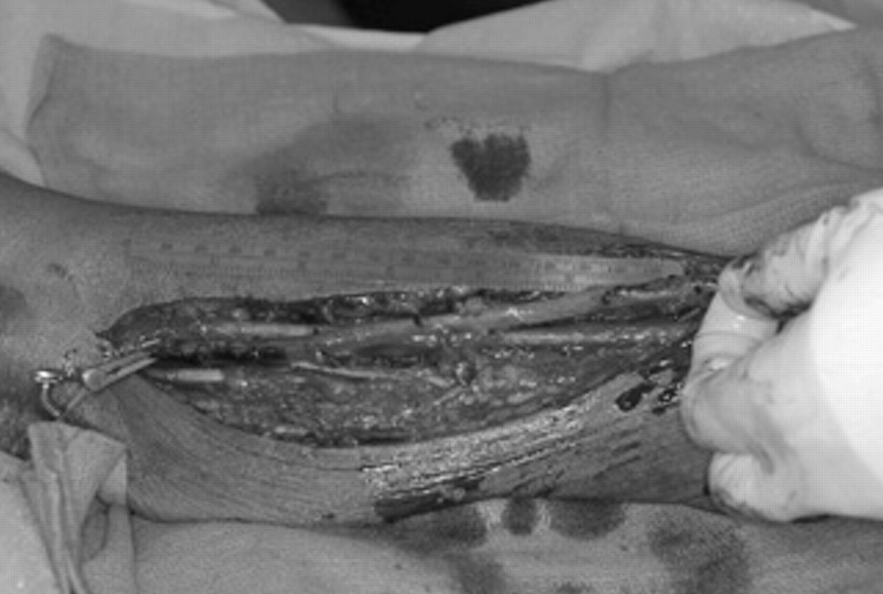
The basilic vein is transected distally, and in preparation for the transposition procedure, it is dilated with saline to ensure the absence of stenotic areas.
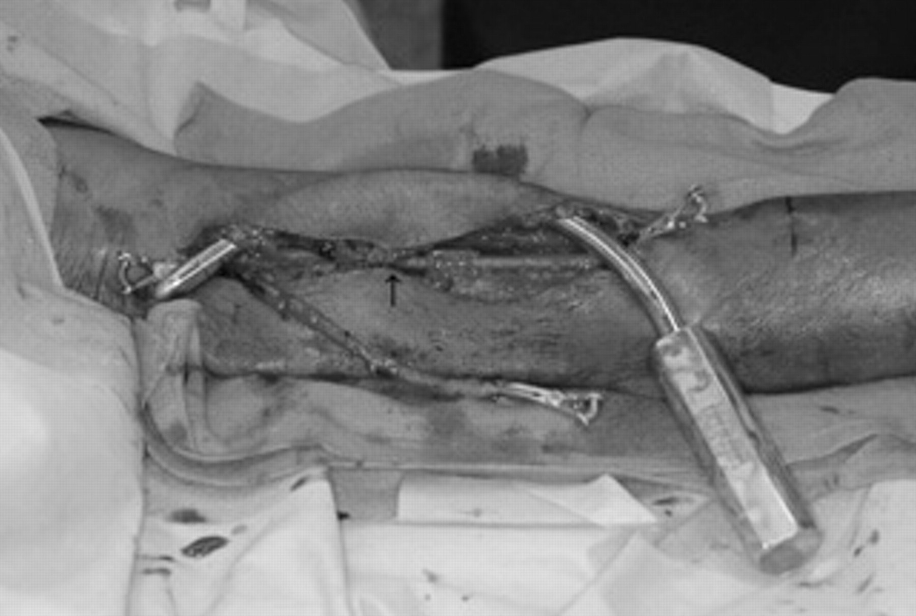
A superficial anterolateral arm tunnel is made between the top and bottom of the incision with a sheath device, a tunneler, in preparation for the brachial vein transposition. To facilitate creation of the tunnel, the wound is partially closed with a subcutaneous suture (arrow).

The brachial vein is placed inside the arm tunnel with care taken not to twist the fistula, using the marks made with a marker pen. Reanastomosis of the two ends of the basilic vein is performed (arrow), and a thrill is reestablished.
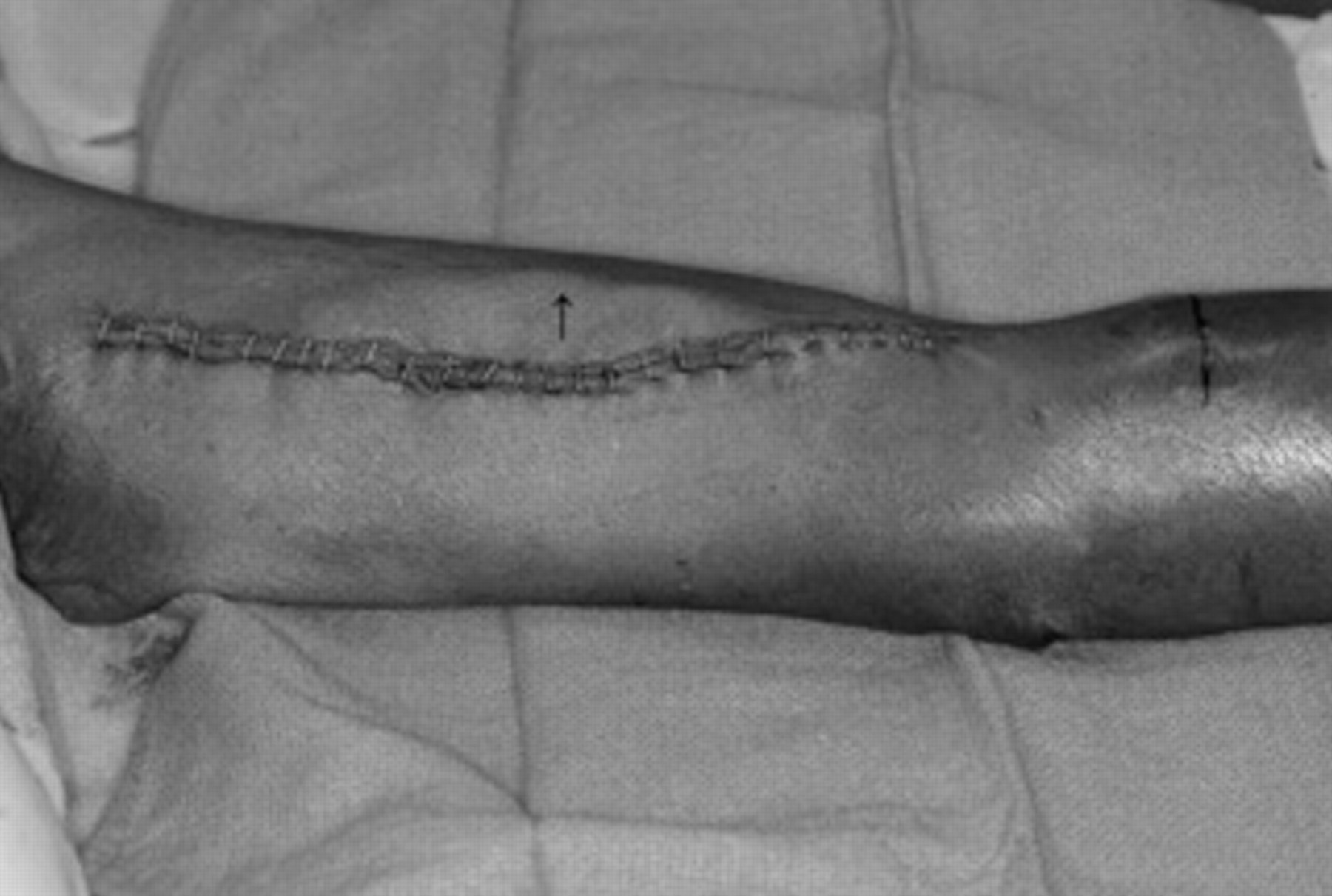
Wound closure with stainless steel staples for the skin is performed. The subcutaneous course of the transposed vein is easily visible (arrow).
Case Report
A 67-year-old female with stage IV chronic kidney disease (CKD) owing to renovascular disease and possible diabetic nephropathy was referred for vascular access creation in anticipation that hemodialysis would soon be needed. The patient had a brachial-basilic fistula created that, after an uncomplicated course, was allowed to mature for a period of 56 days, when the patient was brought back to the operating room for the transposition procedure. In the meantime, the patient had started hemodialysis through a right internal jugular vein tunneled cuffed catheter. On arm exploration, there was a low basilic-brachial vein confluence, so a combined basilic-brachial vein transposition was performed, as described above. The procedure was uneventful, and the patient was successfully dialyzed 7 weeks later and had the tunneled catheter removed.
Discussion
The KDOQI guidelines state that in patients with ESRD or CKD, in anticipation of hemodialysis not suitable for radiocephalic or brachiocephalic fistula, TBBF should be the preferable option because of its autogenous nature and based on results from recent studies that have shown a reduced reintervention rate compared with prosthetic grafts. 4,5 TBBF should be preferred in candidates with suitable anatomy. Extended use of upper arm deep veins, that is, the brachial veins, described for the first time in 1993, 8 was recently revived and reported to be successful in avoiding the use of prosthetic material, 6,7 but it remains unclear whether this procedure should be performed in one or two stages. In the current report, we describe a two-stage transposition procedure of a short basilic vein and its brachial vein continuation in a patient with anatomy that was unsuitable for traditional TBBF.
According to classic anatomic descriptions, the basilic vein begins in the ulnar part of the hand dorsal venous network, continues up along the posterior surface of the ulnar side of the forearm, and inclines forward to the anterior surface below the elbow, joins the median antecubital vein and then runs upward along the medial aspect of the upper arm, perforates the deep fascia somewhat below the middle of the arm, and runs along the medial side of the brachial artery up to the lower border of the Teres major muscle, where it joins the brachial vein to form the axillary vein. 9 In our case, we encountered an anatomic variation of a low basilic-brachial vein confluence and took advantage of the fact that the deep system-the brachial vein-did mature to allow transposition of the basilic-brachial vein complex and prevented use of a prosthetic graft. Consistent with previous reports, veins in this area are well known to have variable anatomy 8,10 ; a low basilic-brachial vein confluence is not uncommon, necessitating harvesting of its continuation as brachial vein to gain the necessary length so that the patient can have a usable fistula, 11 but experience with this technique remains largely anecdotal; other authors consider combined basilic-brachial vein transposition controversial because of its extensive nature in the ESRD patient population. 12 However, our patient had successful and uncomplicated construction of such a basilic-brachial fistula.
A major advantage of the two-stage technique, indicated for veins with a diameter less than 4 mm, 13 is that the arterialized vein has a thickened wall, which makes dissection during the second stage easier. A thin-walled vein can also be easily damaged, necessitating repair, and this might lead to complications such as postoperative bleeding and wound hematomas and long-term sequelae such as stenosis formation. 14 Additionally, a thin-walled vein is more prone to get twisted when placed inside the tunnel of a one-stage procedure, leading to early fistula thrombosis.
A major concern when harvesting a deep vein or using it as a fistula is venous hypertension; although this was not seen in two studies on brachial vein transposition, their authors suggested that clinicians should be vigilant in recognizing this potential problem. 6,7
A variable maturation rate of brachial vein transposition has been reported, 7,15,16 which might reflect different selection criteria in patients with variable venous anatomy. In our case, the fistula matured, with no need for any ancillary procedure. Regarding the long-term patency of this kind of fistula, a 76% 1-year primary patency has been reported 7 ; direct comparison between one- and two-stage procedures, alone or in combination with basilic vein transposition, deserves further investigation.
In conclusion, basilic-brachial vein transposition is feasible as a tertiary access option in selected patients. Surgeons should be aware of this procedure to offer their patients the highest quality of care.
Footnotes
Acknowledgment
Financial disclosure of authors and reviewers: None reported.