




Abstract
Cases of mural aortic arch thromboses are generally associated with diffuse atherosclerosis of the aortic arch and have primarily been detected in elderly patients. However, the presence of mural thrombi in the aortic arch in young patients without diffuse atherosclerosis has rarely been reported. We describe a case of a hypercoagulable young patient with arterial embolism in whom investigations revealed a mural pedunculated aortic arch thrombosis without clear diffuse atherosclerotic lesions.
The primary source of peripheral arterial embolism (PAE) is cardiac in > 85% of cases. Owing to newer sophisticated imaging techniques, noncardiac sources of PAE have been detected with increasing frequency. Among these noncardiac sources, the aorta has been reported in up to 5% of cases to be the origin of PAE. 1 Mural thrombus in the aorta is commonly observed in the setting of aneurysmal disease and significant atherosclerotic occlusive disease. 2 Although uncommon, symptomatic arterial embolism from either of these aortic disease conditions is recognized. 2 In contrast, mural thrombus with subsequent arterial emboli originating de novo from the nonaneurysmal, nonatheromatous aorta is exceedingly rare. 3 In this case report, we demonstrate the presence of a large, mural thrombus in a morphologically normal aorta as the source of PAE.
Case Report
A 42-year-old man of Pakistani origin presented with pain and pallor of sudden onset in his left upper limb. His past history included hypertension, hypercholesterolemia, sleep apnea, diabetes mellitus (type 2), and two previous deep venous thromboses owing to an underlying factor V Leiden mutation. He had no previous medical history of arrhythmia, ischemic heart disease, or stroke; was a smoker for 15 years; and had a body mass index of 28. Prior to his presentation, he was given conflicting advice regarding his requirement for ongoing anticoagulation and discontinued it. Physical examination revealed cooled and mottled fingers in the left hand. His left axillary pulse was palpable, but distally in the left arm, no pulses were present. The examination was otherwise unremarkable.
A successful left brachial embolectomy was performed with postoperative angiography, and the patient was anticoagulated. Investigations were performed to discover a potential source of the embolus. The resting electrocardiogram and Holter monitoring for 24 hours disclosed a normal sinus rhythm without any pathologic findings. The transthoracic echocardiographic examination showed no structural or functional cardiac abnormalities. Computed tomographic (CT) angiography of the thoracic aorta revealed a significant mural thrombus extending into his left subclavian artery from his descending aorta (Figure 1), and this was confirmed by transesophageal echocardiography (Figure 2). A full hypercoagulable workup (antiphospholipid antibodies, protein C, protein S, antithrombin, factor V Leiden, prothrombin G20210A mutation, and homocysteine) was repeated in consultation with our hematology service, and a factor V Leiden mutation was the only abnormality detected. The case was discussed with our cardiac surgery colleagues, and plans were made to proceed with a left thoracotomy and thrombus removal from the arch under circulatory arrest and cardiopulmonary bypass. Prior to that surgery being carried out, the patient suddenly embolized to his left lower limb and developed an acutely ischemic limb requiring popliteal artery embolectomy. Repeat CT angiography of his thorax revealed that the thrombus in his aorta was absent and had embolized to his leg. Thus, our plans for thoracotomy and thrombus extraction were cancelled, and his anticoagulation continued. His postoperative course was complicated by development of a delayed compartment syndrome 1 week following the popliteal embolectomy, which required fasciotomies. The patient was discharged with lifelong anticoagulation.
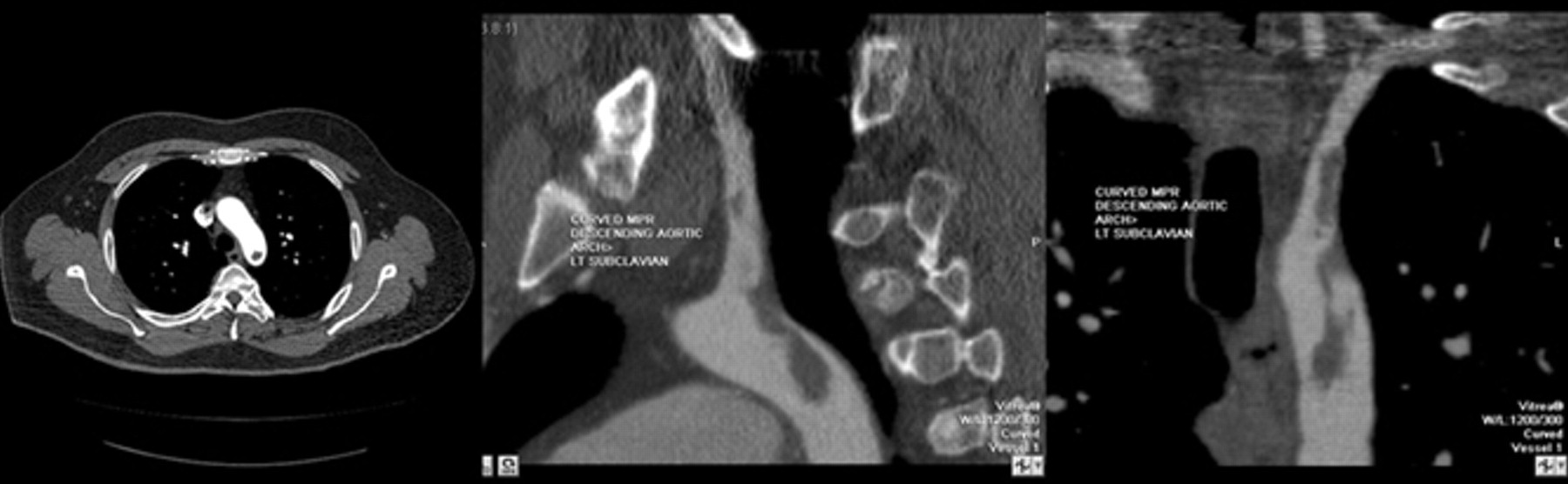
Axial, sagittal, and coronal computed tomographic thoracic angiographic images revealing a thrombus involving the proximal 3 cm of the left subclavian artery and extending 3 cm caudally into the thoracic aorta.

Transesophageal echocardiography revealed a pedunculated mobile mass arising from the descending aorta (arrow).
Discussion
Since the initial description by Weismann and Tobin in 1958, 4 aortic mural thrombus has been accepted as a definite clinical entity and a source of arterial thromboembolism. 5 The mural thrombus is usually located in the abdominal aorta, but, albeit infrequently, it can occur in the thoracic aorta. 6 Although most thoracic thrombi are seen with atherosclerotic disease, several other etiologies have been described, including aneurysms, dissections, trauma, malignancy, hypercoagulable states (polycythemia and antithrombin III deficiency), and systemic fungal infections. 7 Generally, thromboembolic events are associated with advanced age with the thrombus arising from complex and ulcerated atherosclerotic plaques. Interestingly, the region around the left subclavian artery seems to be one of the predisposed localizations for thrombus formation. 8 However, as this case report (in accordance with a few previously published studies), demonstrates, thrombus can also affect younger patients.
In the study by Laperche and colleagues, the mean age of the 23 patients with aortic thrombus was 45 ± 8.4 years; the youngest patient was 34 years old. 8 In their study, smoking was the leading risk factor (n = 16), followed by hypercholesterolemia (n = 11) and elevated fibrinogen levels (n = 10). In our patient, the risk factors of smoking, hypercholesterolemia, and factor V Leiden mutation were present. Also reported in this article was histopathologic examination in all patients undergoing surgery for thrombectomy, revealing microscopic features of atherosclerosis limited to the insertion site. Furthermore, Perler and colleagues previously reported thromboembolic events originating from a morphologically normal aorta in two young women. 9 Both patients were taking oral steroid medications, and both patients were heavy cigarette smokers. These cases and a review of the previous literature suggest that the development of aortic mural thrombi, at least in some patients, may not always result from diffuse and advanced atheromas but may constitute a separate and distinct clinical entity of a premature atherosclerosis. In idiopathic cases, it seems that the ductus arteriosus remnant can be a site of origination. 10 As the aorta of the patient described in this case report had normal dimensions and was free of any visible atherosclerotic intimal lesions, the additive prothrombotic effects of hypercoagulability (factor V Leiden mutation), cholesterol, and excessive smoking seem most likely to be the cause of the thrombus formation. Cigarette smoking induces global changes in both peripheral and central vascular function. 11 Some authors postulate that the most important effects of cigarette smoking in promoting atherosclerosis and thromboembolic complications may be endothelial disturbance and fibrin formation. 12
Aortic mural thrombus may be either asymptomatic or symptomatic. Owing to new and sophisticated imaging techniques in recent years, such as spiral CT scan, magnetic resonance imaging, and transesophageal echocardiography, noncardiac thrombi are an emerging clinical entity as incidental findings and in patients presenting with unexplained distal embolization or luminal occlusion. Among these noncardiac sources, the aorta has been reported in up to 5% of cases to be the origin of PAE. 1 However, although mural thrombus in the aneurysmal or atherosclerotic aorta with protruding atheromas may be the source of major arterial embolism, 2 emboli originating from a nonatheromatous and nonaneurysmal aorta, as described in our case, are a rare event. 3 These aortic lesions can cause either macroemboli or atheroembolization (microemboli).
When considering therapeutic options for thoracic aorta thrombi, it is necessary to view them as a heterogeneous group rather than a single entity, each having a different clinical course and prognosis depending on its nature and etiology. Karalis and colleagues reported a 73% incidence of embolic events among patients with pedunculated and highly mobile aortic thrombi, as opposed to only 12% when those thrombi were layered and immobile. 13 Several treatments have been used in different forms with variable success for management of aortic thrombus, including anticoagulant therapy alone, 8 thrombolysis, 14 and thromboaspiration and surgery. 15 Most of the literature addressing treatment options pertains to patients with peripheral or visceral macroembolic events. In such situations, systemic anticoagulation has been considered the mainstay of treatment. 6 However, the risks associated with anticoagulation and the risk of recurrent embolism cannot be accurately estimated because of the limited number of reported patients. Recently, there have been several reports regarding successful operative thrombectomy of lesions in the thoracic aorta. 6,16–18 We recommend surgical thrombectomy rather than anticoagulant regimen in young patients, in the presence of a large hypermobile thrombus (as in this case report), and in patients with recurrent embolic events if the surgical risks are acceptable. The optimal management of asymptomatic thoracic aorta mural thrombi has not been clearly established. 17 Endovascular stent grafting provides a new, minimally invasive therapeutic option in the treatment of symptomatic mobile thoracic aortic thrombus 19 ; however, its role in the setting of aortic thrombosis regarding the long-term outcome is not established yet.
As a consequence of the widespread availability of advanced diagnostic tests, thoracic aorta thrombi will continue to be diagnosed with increased frequency, particularly in asymptomatic patients. Embolic events arising from thrombi within a nonaneurysmal, nonatherosclerotic aorta are a rare but possibly emergent event. Clinicians have to be aware of the higher risk for developing thromboembolic complication in the presence of prothrombotic risk factors such as smoking and hypercoagulability, not only in the venous system but also in the arterial vascular system. Further studies and reports are needed to outline the natural history of these thrombi and outcomes of various therapeutic interventions to define the optimal treatment strategy.
Footnotes
Acknowledgment
Financial disclosure of authors and reviewers: None reported.