




Abstract
Secondary aortoduodenal fistula is an uncommon but potentially fatal complication that can occur after aortic reconstruction surgery and usually presents with upper gastrointestinal hemorrhage. Taking into account the accompanying multiple comorbidities of those patients, conventional open surgical repair carries with it significant mortality and morbidity rates. The purpose of this case report is to describe the successful combined endovascular and open surgical repair of an acutely ruptured aortoduodenal fistula in a 67-year-old male patient. Four months after the procedure, the patient remains well.
Aortoenteric fistula (AEF) is one of the most severe complications of abdominal aortic reconstruction surgery and usually presents with exsanguinating upper gastrointestinal hemorrhage. Left untreated, it has a 100% mortality rate. Conventional surgical repair is associated with a perioperative mortality rate of 21 to 59% 1–5 and frequent major complications. 6 Staged extra-anatomic bypass grafting, aortic graft excision, graft excision, and in situ bypass with an allograft, an antibiotic-impregnated prosthesis, a silver-coated prosthesis, or an autogenous vein are current surgical treatment options. 7 The current report presents an endovascular graft repair of a secondary AEF in an acutely bleeding patient.
Case Report
A 67-year-old man who had undergone repair of an infrarenal abdominal aortic aneurysm with a Dacron tube graft 5 years previously was admitted to the emergency department complaining of back pain over the previous month. Four months prior to admission, he was admitted elsewhere with a history of fever and pain in the left inguinal region. On that occasion, an abdominal ultrasound examination was negative, and the patient was treated conservatively with oral antibiotics.
The patient was referred to Urology with a working diagnosis of renal colic. On the following day, the patient suddenly had an episode of massive hematemesis and hypotension. An urgent gastroscopy revealed bright red blood spurting from the third part of the duodenum. An urgent computed tomographic (CT) scan with intravenous contrast and 5 mm slices revealed a para-aortic thrombus and thickened aortic wall below the origin of the renal arteries (Figure 1).

Computed tomographic scan demonstrating inflammatory lesions around the graft with perigraft fluid collection, soft tissue thickening, and enhancement of the periduodenal fat.
Resuscitation included intubation and transfusion of 7 units of packed cells and 5 units of fresh frozen plasma. In this setting, a decision was made to attempt endovascular repair of the aortoduodenal fistula by placing an endoluminal stent graft across the defect at the upper suture line between the aorta and the old Dacron graft. The patient was transferred to the operating room in a state of hypovolemic shock with systolic blood pressures ranging from 50 to 70 mm Hg, hematocrit 17%, and hemoglobin 5.1 g/dL. The endovascular repair was carried out under general anesthesia, after full preparation for open surgery should conversion have been required. A vertical groin incision was used to expose the left and the right common femoral arteries. The patient was fully anticoagulated with heparin at this time. A baseline angiogram was carried out to delineate the renal arteries. A Talent (Medtronic Hellas, Athens, Greece) main body AF 3016C155AX and contralateral iliac limb IW1416C90AX were successfully deployed.
The groin incisions were closed in a standard fashion. After the endovascular repair, a laparotomy was performed by general surgeons, during which the duodenal fistula was identified, its margins were resected, and the resultant defect was sutured in two layers. The patient was extubated the following day in the intensive care unit. A triple-antibiotic regimen of ciprofloxacin, clindamycin, and vancomycin was started. The patient had temperature spikes of 37 to 38°C. The hemoglobin and hematocrit remained stable in the postoperative period.
A follow-up spiral CT scan at 3 months showed the para-aortic thrombus mass to be resolving, with no evidence of endoleakage (Figure 2).
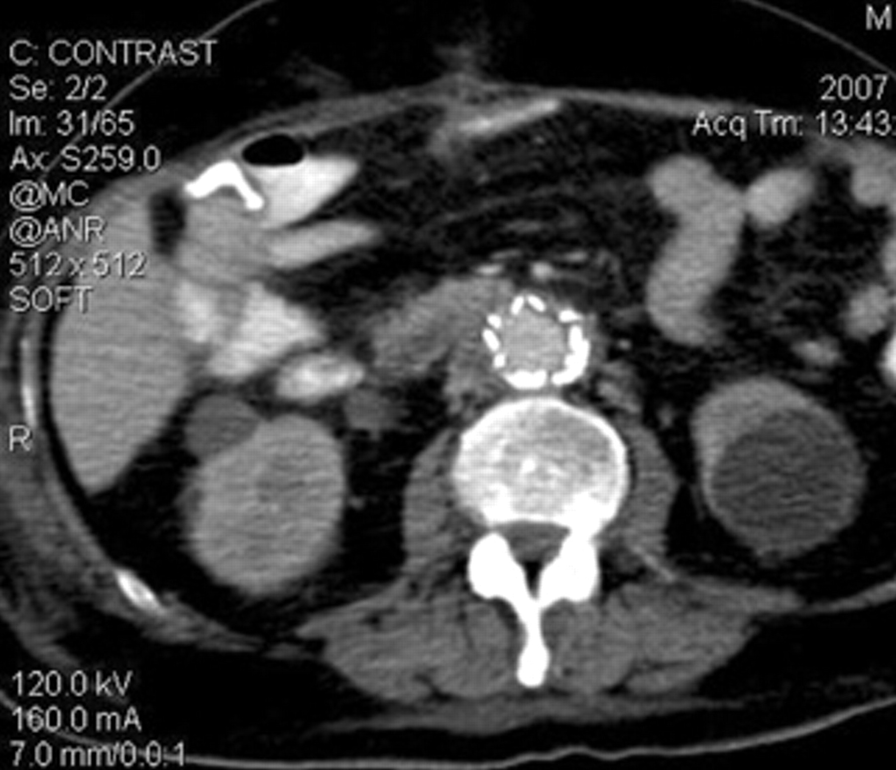
Computed tomographic scan showing the endovascular stent graft into the old Dacron graft.
Discussion: Review of the Literature
AEF can form between the aorta and any part of the gastrointestinal tract, although they most commonly occur in the third part of the duodenum.
Primary AEF in 80% of cases have an atherosclerotic or aneurysmal etiology but may also occur owing to other causes, such as gallstones, radiation injury, swallowed foreign bodies, and cystic medial necrosis. 8 Most commonly, secondary AEF are due to a communication between a prosthetic graft and the duodenum. 9 This complication is estimated at 0.5 to 2.5% following aortic prosthetic graft surgery. 10 The injury may occur within weeks or decades following surgery. 11 Mechanisms include mechanical erosion of the suture line, suture line disruption with pseudoaneurysm, and fistula formation. AEF after stent grafts from direct erosion was recently reported. 12
The cornerstone logic is the endovascular exclusion of the AEF. The technique may be the only treatment required in some patients, or it could serve as an adjunct to patient stabilization, prior to more definitive treatment (“a bridge to open surgery”). 6,13–16 However, this technique has a few limitations. It is an unsuitable treatment for patients who do not have a sufficient length of normal aorta below the renal arteries. The use of the stent graft relies on the presence of an aortic cuff between the suture line and the renal arteries. It is not possible to delineate the suture line on a CT scan. 7
Although endovascular repair seals the fistula, it does not physically disrupt it. In most cases, the fistula is between the proximal suture line and the duodenum. However, it has been reported that distal anastomosis or the body of the graft is responsible in 19% and 14% of cases, respectively. 11
Endovascular treatment may effectively control bleeding, but it cannot, however, deal with graft infection. Antibiotic coverage should be guided from the blood culture results, or, if not available, patients should be started on broad-spectrum antibiotics that should cover the most common organisms responsible: Proteus mirabilis, methicillin-resistant Staphylococcus aureus, Escherichia coli, S. aureus, and Staphylococcus epidermidis. 17–19 Antifungal treatment may also be required. The length of treatment required is not established but may range from a period of 6 months to indefinitely. This approach is supported by observations following open surgery. 17,20
Table 1 summarizes a series of published endovascular treatments for AEF. Of the 28 cases reviewed, 4 patients (14%) died of sepsis after the endovascular repair. Endovascular repair allows for hemodynamic stabilization, and when combined with broad-spectrum antibiotics and drainage or diversion when necessary, sepsis may be controlled for prolonged periods. Initially, it appears that it controls acute hemorrhage, but recurrent infection may remain a problem. The outcome over time should be evaluated. In summary, endovascular treatment in the management of AEF should be further evaluated. 21
Published Cases of Endovascular Management for Aortoenteric Fistulas
GI = gastrointestinal.
Footnotes
Acknowledgment
Financial disclosure of authors and reviewers: None reported.