




Abstract
We describe the successful treatment of a large scalp pseudoaneurysm using direct thrombin injection.
Keywords
There is an increasing drive toward minimally invasive treatment of pseudoaneurysms. In 1986, Cope and Zeit described direct thrombin injection into aneurysms to induce thrombosis. 1 Since then, there has been an increase in the application of this minimally invasive treatment, particularly for iatrogenic femoral pseudoaneurysms following femoral artery catheterization. 2 Other pseudoaneurysm sites that have been treated by either direct thrombin injection or endovascular thrombin injection include the common iliac artery, 1 common femoral artery, 1 peroneal artery, 1 accessory hepatic artery, 1 aorta, 3 pulmonary artery, 3 gastroduodenal artery, 3 left gastric artery, 3 and renal artery. 3
Generally, the treatment is quick, safe, and efficacious when compared with open surgery alternatives. However, complications have been reported. 4,5 We describe what we believe to be the first report of the use of thrombin to treat a scalp pseudoaneurysm.
Case Report
An 85-year-old male with a history of Parkinson disease, dementia, depression, and a left sphenoid wing meningioma fell in the nursing home where he was living. At the time of the fall, he was taking a number of medications, including clopidogrel. He suffered an open dislocation to a finger, a laceration to an elbow, and a laceration to the left side of the scalp. The finger injury was reduced and treated with antibiotics, and the elbow and scalp lesions were simply dressed. Following discharge, a large lump developed on the scalp at the site of the laceration that would bleed intermittently. Three weeks following the fall, he was referred to the vascular clinic. On examination, there was a large lump on the left side of the scalp (Figure 1). Of note was the expansile pulsation of this lump and evidence of recent bleeding. Figure 2 shows the computed tomographic scan with contrast confirming the presence of a pool of contrast within the lump. The pseudoaneurysm was thought to have arisen from a branch of the superficial temporal artery. The pseudoaneurysm nature was confirmed on a color duplex scan. Given the frail nature of the patient and his multiple medical problems, we opted to treat the pseudoaneurysm using direct thrombin injection.
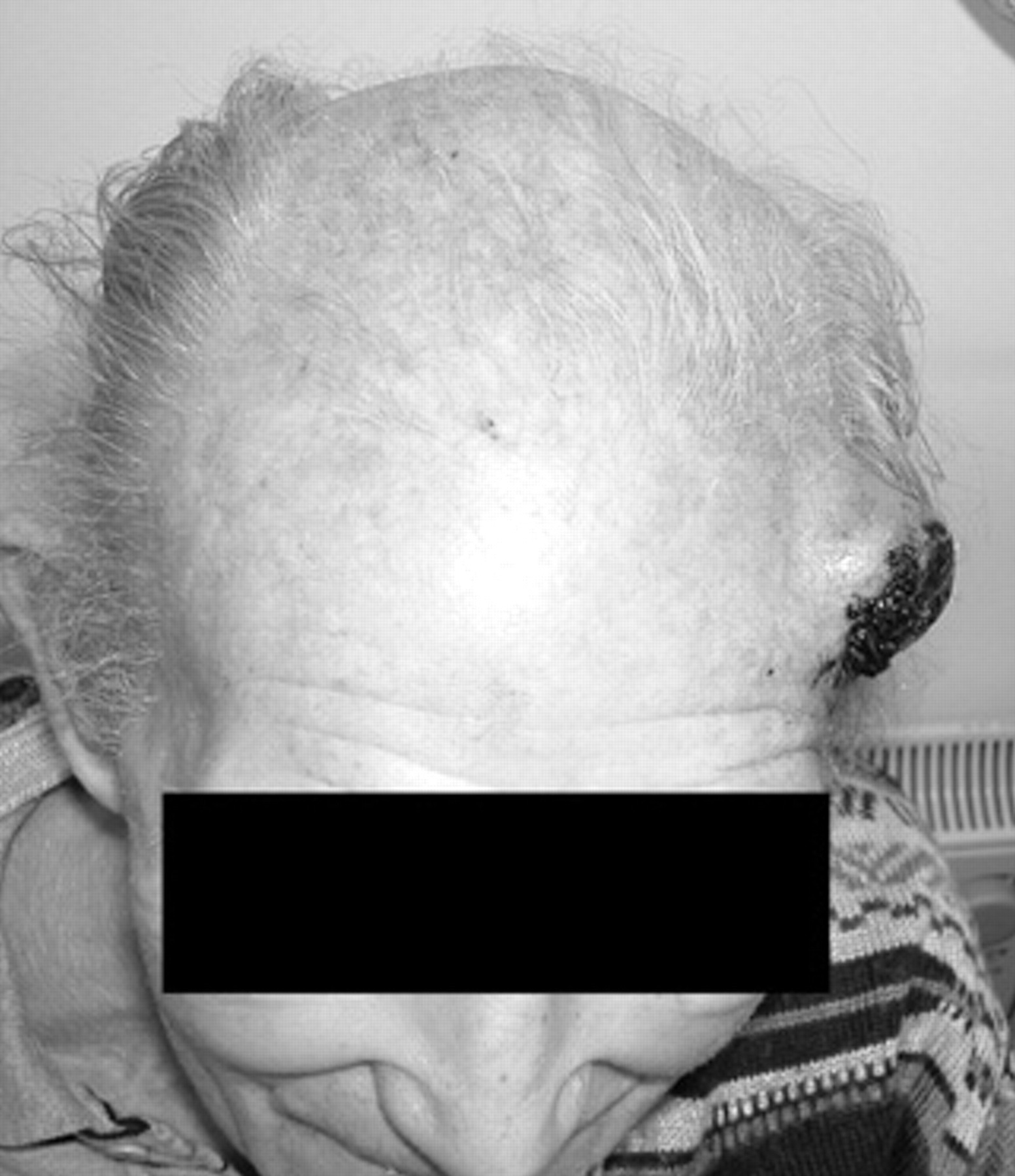
Scalp pseudoaneurysm.

Contrast-enhanced computed tomographic scan confirming the presence of the pseudoaneurysm (note also the left sphenoidal meningioma).
Under local anesthetic and ultrasound guidance, 0.2 mL of the thrombin solution component (350–500 IU/mL) of Tisseel (Baxter, Austria) was injected into the lumen of the false aneurysm. No proximal vessel compression was used to reduce flow. Immediate thrombosis of the false aneurysm was noted. The lesion was no longer pulsatile. Ten weeks later, the lesion had completely healed (Figure 3).

Completely resolved lesion at 10 weeks.
Discussion
There are many other minimally invasive techniques to deal with peripheral false aneurysms, including embolization coils, detachable balloons, stent grafts, and other thrombogenic materials (eg, Gelfoam, Upjohn, Kalamazoo, MI). In this case, the distance from common femoral arterial access, the small size of the affected vessel, and the overall procedural difficulty made other endovascular approaches less desirable, although not impossible. Open surgery was considered and would have been employed if the thrombin injection had failed.
Conclusion
We describe a quick and efficacious technique for the outpatient treatment of a large scalp pseudoaneurysm.
Footnotes
Acknowledgment
Financial disclosure of authors and reviewers: None reported.