




Abstract

Why dialysis access? Seemingly, we could stick to sexier topics of major aortic disease and limb salvage. For several reasons, dialysis access is an extremely important topic. Foremost among these reasons is that dialysis access is frequently neglected as an important part of surgical training in residency. In many residency programs, surgical care of this group of patients is often relegated to the junior residents, with senior trainees preferring to focus on “the big cases.” In fact, dialysis access cases do not count toward case counts for vascular surgery residents to document completion of training. Thus, many surgeons get most of their experience from conferences such as this one and on-the-job trial and error. Interventional nephrology, a rapidly growing group of physicians, is almost entirely trained by postresidency programs, including conferences and minifellowships. The need for ongoing education in this field is real.
The dialysis patient population continues to grow and the access choices continue to become more complicated. There is no foreseable solution to this problem. Increasing incidence of obesity and diabetes and longer life expectancy seem to be the main drivers behind the expansion in dialysis population, but the evidence is not completely clear. Unlike cororary disease, statins, antiplatelet drugs, and coated stents have not led to a decreased need for interventional treatments. The only cure for dialysis dependence is kidney transplantation, but numbers of organs remain extremely limited and wait times for transplantation continue to grow. Dialysis access for these patients remains crucial to survival as it provides their “lifeline,” connecting them to dialysis machines for life-sustaining renal replacement therapy.
Outcomes fall short of hopes and expectations in many areas of dialysis. The advanced morbidity and mortality of these patients limit their treatment options. Nearly every trial in clinical research has dialysis dependence listed in the exclusion criteria because of these associated outcomes. Recent trials of arteriovenous fistulae and grafts documented embarrassingly poor outcomes, with less than half of fistulae progressing to usable maturity and only a quarter of grafts remaining functional without additional procedures, with no clinically significant benefit of adjunctive medical therapies.
The growing population has tremendous socioeconomic consequences. The cost of dialysis patients is disproportionate to their numbers, consuming far more Medicare dollars than their share. This has led to US government initiatives with advancing criteria of “Fistula First” and the formation of the end-stage renal disease networks tasked with education and tracking of outcomes. Pay for performance seems imminent, and already some areas of the country have seen decrees to dialysis units to redirect referral patterns for access creation based on outcomes. In this era of economic troubles, improved outcomes and reduces cost are a necessity.
Dialysis access also provides some unique opportunities. Throughout the body, vascular interventions fail largely because of intimal hyperplasia. This is true for coronary stents, peripheral angioplasty, and bypasses. This failure mode is arguably the most aggressive in dialysis access, with rapid development of proliferative changes at sites of surgery or intervention. These areas are unique in that they are relatively superficial, making them easy to image with a variety of different modalities, and require frequent intervention, opening the possibility of obtaining tissue for further study. More focused research in dialysis access and dedication of more funding could result in advances that would be widely applicable to many areas of cardiovascular disease.
In the following pages, many areas of dialysis access are discussed in abstract form. Troublesome areas of access failure, central venous stenosis, and complications of access are all discussed, in addition to general outcomes. Careful planning paradigms and algorithms are presented because both routine patients and the most challenging groups require innovative approaches, put forward by leaders in this field.
Ultimately, we feel that conference attendees get the most benefit from focused, in-depth exploration and explanation of a single disease process and can most rapidly incorporate them into their practice and stimulate new concepts for research and clinical care to benefit patients. We hope that you enjoy and benefit from the following abstracts and welcome you to join us next year in Houston for our next installment of the Total Endovascular Series.
Transhepatic Catheters: Are They Really Worthwhile?
Ali Amin, MD, FACS, FACC, RVT
Reading Hospital and Medical Center, Reading, Pennsylvania
The prevalence of chronic kidney disease is substantially greater than the number of patients with end-stage renal disease. There are 350,000 patients on hemodialysis in the United States, and the population is projected to grow by 7% per year. Unlike other forms of end-stage organ failure, end-stage renal disease manifested by renal failure is unique in having three modalities of therapy: (1) hemodialysis, (2) peritoneal dialysis, and (3) renal transplantation. Each form of renal replacement therapy has its own risks and benefits. Kolff first employed hemodialysis in the late 1940s for the treatment of acute renal failure. Scribner developed vascular access in the early 1960s. This enabled the use of hemodialysis as a chronic therapy.
All three treatment modalities have evolved significantly over the past few decades. The selection of a particular treatment modality is dependent on the clinical setting and patient preference. These modalities should be viewed by the physician and the patient as alternative and complementary therapies. To perform hemodialysis on a repetitive basis, access to circulation is essential. In 2002, the National Kidney Foundation published the Disease Outcomes Quality Initiative (DOQI) guidelines. These were based on a systematic literature review. A uniform format for summarizing strength of evidence was developed based on an evaluation of study size, applicability, results, and methodological quality. The Kidney Disease Outcomes Quality Initiative (KDOQI) recommends placement of an arteriovenous fistula. The arteriovenous fistula is the gold standard for hemodialysis access. Synthetic arteriovenous grafts can be used when a native arteriovenous fistula cannot be placed. This modality has a higher rate of thrombosis and infection than a fistula. The third option is percutaneous dual-lumen catheters, which are preferentially placed in the internal jugular vein and tunneled under the skin.
Although dual-lumen tunneled catheters provide immediate and convenient access to the circulation and are adequate for hemodialysis, they have a high rate of infection and clotting. Therefore, every attempt is made for the patient to have a noncatheter hemodialysis access for hemodialysis.
Generally speaking, catheter malfunction is the most common noninfectious complication of central venous catheterization. Malfunction is defined as either (1) failure to achieve a blood flow rate of at least 300 mL/min on two consecutive occasions or (2) failure to achieve a blood flow rate of 200 mL/min on a single occasion. Infection is the most common complication of venous catheterization for venous access. This may occur in up to 40% of patients.
Recurrent vascular access failure remains a major cause of morbidity in patients receiving long-term hemodialysis. When standard access sites, including the femoral vein, occlude, unconventional access methods become necessary. These unusual sites may include placement of a catheter in the recanalized veins or small collateral veins, transthoracic cuff catheters, and a right atrial catheter.
Transhepatic catheter access is an alternative modality in patients with no other options for hemodialysis. These patients have often exhausted traditional sites from prior surgeries and catheter placements. The exhausted sites include internal jugular, external jugular, subclavian, femoral, inferior vena cava, and translumbar. Transhepatic central venous catheters can be placed safely with minimal complications. They have a low infection rate, which can be managed by catheter exchange over wire. However, they are associated with a higher thrombosis rate and lower primary patency.
In summary, transhepatic dialysis catheters should be used only in patients who have exhausted traditional and alternative accesses for hemodialysis.
Physical Examination and Patient Evaluation
Gerald A. Beathard, MD, PhD, FASN
Lifeline Vascular Access,
Austin, Texas
Thorough evaluation of the patient with end-stage renal disease prior to the placement of a dialysis vascular access will definitively enhance the chances of success. A careful review of the patient's medical history is important. There are comorbidities and mechanical factors that can materially influence the decisions that are necessary in placing an access.
Creation of an arteriovenous fistula (AVF) has significant cardiovascular effects, particularly increased blood flow and cardiac output. Patients with severe heart failure (New York Heart Association class IV) owing to intrinsic heart disease are at considerable risk for worsening cardiac function with the creation of an AVF. This risk is greater with the upper arm than with the forearm.
The total progression from recognition of the need for dialysis to successful dialysis with an AVF can take 4 to 6 months. This decreased life expectancy this time may be too much to make an AVF practical.
Diabetes is associated with peripheral artery disease, which can hinder the maturation of a distal AVF. A plain radiograph of the hand and forearm has been recommended to evaluate the diabetic patient. If no vascular calcification is seen, the construction of a distal fistula should be considered. Obesity and diabetes often go together. Veins tend to be deeper, which can be a definite problem; transposition should be expected. With increasing age comes an increasing incidence of peripheral vascular disease as well as other comorbidities that could potentially have an effect.
Mechanical factors such as peripherally inserted central catheter (PICC) lines are very destructive to peripheral veins. The leads for pacemaker and implantable cardioverter-defibrillator devices are inserted through the cephalic or subclavian veins and can lead to stenosis and obstruction of these major veins very quickly. Stenosis of the central veins occurs in up to 50% of patients following the use of a central venous catheter. A failed access takes with it venous anatomy that is no longer available for the construction of a new vascular access. This can be especially problematic if the failures have been multiple and bilateral. Vascular surgery or trauma can result in damage to vessels that can affect the creation of an access.
Three evaluations of the arterial system should be performed: pulse examination, differential blood pressure measurement, and the Allen test. Special care should be directed toward detecting venous problems. The detection of regionalized edema such as one arm or the breast should be cause for concern. The two arms should be compared to determine if their size is comparable. With central venous obstruction, collateral veins begin to form. Often these are superficial enough to be apparent on physical examination. It is usual for a patient with chronic renal failure to arrive at the point of initiation of dialysis without having had some type of central venous catheterization. Evidence of these prior episodes of cannulation should be sought and documented.
Vascular mapping including the arterial and venous systems is mandatory. This may be done using ultrasonography, angiography or both.
Access in Children
Mary L. Brandt, MD
Baylor College of Medicine, Houston, Texas
Access for hemodialysis in children is a surgical challenge because of the size of the vessels and the special physiology of the patients. Primary arteriovenous fistulae, with a lower rate of secondary failure and complications, are preferable for long-term hemodialysis access in children. The decision to determine which patients are “too small” for this surgical approach varies from institution to institution, based on the experience of the surgeon and the availability of microsurgical techniques. Ultimately, the decision is a balance between the risk of primary failure (and subsequent loss of that site for a future fistula) and the complications of central venous access, which may also prevent creation of effective future access. Placement of an arteriovenous graft should be considered only when all options for an adequate primary arteriovenous fistule have been exhausted. The use of a central venous catheter should be reserved as a “bridge” to a more permanent access or reserved for children so small that the risk of primary failure of an arteriovenous fistula is unacceptably high. In all cases, access must be planned before the procedure, with the long-term need for dialysis in mind. Whether the surgeon creating the access is a vascular surgeon, pediatric surgeon, or transplant surgeon, it is imperative that the surgeon who is providing the access be an active participant in the decision-making process, be educated about the special needs of children in renal failure, and be aware of the issues surrounding the choice of access for each patient.
Anesthesia for Dialysis Access: What Is Best?
Guy L. Comeaux, MD,* and Eric K. Peden, MD†
† Methodist DeBakey Heart & Vascular Center, Houston, Texas
Since the advent of surgery, there has been a quest for safe, reliable anesthesia. Access surgery is quite diverse, and no single method of anesthesia suits all cases and patients. Renal failure automatically escalates these patients on the American Society of Anesthesiologists’ risk scale to a level 4 of a possible 5. Poorly controlled comorbidities such as severe hypertension, an increasingly elderly population, and frequent noncompliance make any anesthetic option seem quite risky. The options for anesthesia include local anesthetics only, sedation plus local anesthetics (monitored anesthesia care [MAC]), regional anesthetics, total intravascular anesthesia, and general anesthesia. High levels of inactivity, known cardiac disease, or several risk factors for coronary disease would suggest that nearly all of these patients should have an extensive cardiac workup preoperatively, which is simply impractical.
Our approach at The Methodist Access Center (TMAC) has been to do MAC for endovascular access procedures and regional anesthesia for open procedures. General anesthesia is reserved for very few patients. We have adopted an ultrasound-guided approach for regional blocks that has been previously described. The block is performed at the supraclavicular level. The ultrasound probe is positioned over the subclavian artery above the clavicle and the area is prepared. A small amount of lidocaine is used at the needle entry site. Insulated 22-gauge needles, 50 to 90 mm in length, are used. The needle is inserted at the lateral end of the probe in the plane of the ultrasound beam. The needle can be seen approaching the brachial plexus and puncturing the investing sheath. Location is confirmed with injection of small amounts of saline followed by a test dose of epinephrine-containing solution. Location can also be confirmed using a nerve stimulator. The local anesthetic is 0.5% ropivicaine combined with 2% lidocaine in equal amounts. Volumes used vary according to the patient's size, 25 to 40 mL. This combination affords rapid onset (lidocaine) and duration (ropivicaine). Clonidine, 100 |g, is added to the local anesthetics to improve the effectiveness of the block.
Complications are most commonly incomplete pain relief, which can generally be easily supplemented with additional sedation and local anesthetic, with rare conversions to general anesthesia. Pneumothorax, hematomas, intraneural injections, and intravascular injections have been exceedingly rare.
Patient acceptance of this regional anesthetic regimen has been excellent. Patients generally recognize the benefit of avoiding general anesthetic, and as long as they are sedated and pain is controlled, satisfaction levels are high. Use of regional anesthesia has led to lowered hemodynamic instability, reduced use of vasopressors, and a shorter turnaround time between cases. The anesthetic approach for dialysis patients continues to evolve. At our access center, regional anesthesia has facilitated steady growth in procedural volume while improving patient safety.
Access in the Morbidly Obese
Ingemar J. Davidson, MD, PhD, FACS,*
Matthew Mulloy, Ramesh Saxena, Namita Singh
The University of Texas Southwestern Medical Center at Dallas,
Dallas, Texas
Since 1995, the average body mass index (BMI) of hemodialysis (HD) patients has risen by 13%, mirroring the expanding obesity epidemic in the United States. Consequently, the number of obese dialysis patients (BMI > 30) has increased over the last decade. The impact of BMI on dialysis access outcomes has been difficult to elucidate. Some studies show inferior outcomes for dialysis access procedures; others do not reveal a BMI-associated outcome effect.
Any algorithm for creating dialysis access in the obese must include consideration of peritoneal dialysis (PD). Although the placement of PD catheters in obese individuals may be technically challenging, a recent study showed no significant risk of higher BMI on PD catheter survival. Thus, when planning a lifelong dialysis access strategy, PD should be considered the first modality in all suitable cases, regardless of weight, followed by appropriately planned HD.
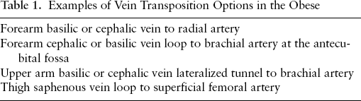
The successful placement of dialysis access must include proper preemptive planning, a detailed case-directed history, and a detailed physical examination. Vascular mapping with duplex Doppler ultrasonography performed by the surgeon, in conjunction with a dedicated ultrasound technologist, is the mainstay of preoperative planning. The only difference in the obese patient is the impact of excess subcutaneous fat. The burden of abundant fat is a double-edged sword. The same fat that makes veins run deep beneath the skin, causing fistulae constructed from these veins to be difficult to cannulate, also protects against iatrogenic damage prior to surgical access placement. Therefore, the normally superficial veins of the arm may be deeper, patent, and of adequate size for fistula creation. Fistula accessibility can be modified by performing “superficialization” procedures (Table 1) on veins that meet the criteria for use accept deeper than 5 to 6 mm from the skin surface. We prefer transposition vein surgeries as one stage rather than allowing the vein to “mature,” only to perform superficialization (or transposition) in a subsequent procedure. As part of a long-term strategy, we always consider a forearm loop polytetrafluoroethylene (PTFE) graft in cases of inadequate forearm veins but useable antecubital veins. Forearm PTFE loop graft placement, preceding upper arm vein transposition access surgery, will “build” upper arm veins (cephalic and basilic) for future use when the forearm access fails.
Examples of Vein Transposition Options in the Obese
The Infected Graft: Salvage or Sacrifice?
Ingemar J. Davidson, MD, PhD, FACS,*
Matthew Mulloy, Suzanne Wada
The University of Texas Southwestern Medical Center at Dallas,
Dallas, Texas
Dialysis access graft infections occur in up to 35% of patients over the lifetime of the graft. Early infections are associated with the surgical procedure, whereas needle cannulation induces later events. Sixty to 70% of infections are caused by gram-positive bacteria (Staphylococcus aureus, coagulase-negative Staphylococcus, and Enterococcus). Twenty to 30% involve gram-negative bacteria (Klebsiella, Pseudomonas, Enterobacter, and Escherichia coli). Empiric antibiotic combinations include vancomycin for gram-positive bacteria and ticarcillin-clavulanate or piperacillin-tazobactam for broad gram-negative coverage. The increasing severity and management of graft infections are exemplified in the scenarios below:
Minor surgical trauma. Erythema and warmth along the graft, which are sometimes noticed early after surgery and last for days, are not caused by infection. Antibiotics may not be prescribed.
Localized erythema. Localized erythema (< 1 cm) is associated with infection around needle puncture sites. Empiric antibiotic coverage is as suggested above. This site must not be used for cannulation, and progress is monitored closely. This scenario often precedes significant infections, as outlined below.
Small, fluctuant (1—2 cm), nonpulsatile mass, without skin breakdown, drainage, fever, or chills. Purulent material or hematoma cannot accurately be delineated by examination. Incision and drainage are indicated. Extensive dissection must be avoided, cultures taken, the content gently expressed, and the wound packed with gauze. Adjust antibiotics based on culture reports. Wound care can be managed expectantly, even if a small segment of graft was initially exposed.
Short-segment graft infection (2—4 cm). Localized segment of erythema around a cannulation site with a history of spontaneous (pulsatile) bleeding requires surgical revision dictated by the clinical history, access anatomy, and surgeon's experience. Ultrasonography is useful to assess the extent of the infection and to guide surgical strategy.
Long-segment graft infection (4—10 cm). A longer segment of graft involved in an infection is commonly associated with other local pathologic access processes, such as graft stenosis, perigraft scarred tissue, hematomas, or pseudoaneurysms from repeat cannulation trauma. Surgical strategies may include proximal and distal control with bypass grafting and excision of the infected segment.
Full-length graft infection. The most severe infection involves the entire graft, including the anastomoses. The most dangerous scenario is a pulsating and expanding hematoma or pseudoaneurysm. The patient is commonly septic, with fever and chills, or may just have positive blood cultures. Technically challenging cases occur when the anastomosis is high in the axilla or groin. Adequate proximal and distal arterial control includes exploration of the subclavian and iliac arteries, respectively. With more distal anastomotic sites, the surgeon can use a sterile, pressure-controlled tourniquet. At exploration, the authors leave a short graft remnant (1—3 cm) at the arterial anastomoses covered with tissue. The majority of wounds will heal. Should infection recur, that is, cutaneous fistula, delayed vascular repair with removal of the entire graft remnant and artery repair is performed, now in a stable patient with less tissue inflammation.
The Clotted Graft and Fistula: The Right Way to Do It
Mark G. Davies, MD, PhD, MBA
Methodist DeBakey Heart & Vascular Center, Houston, Texas
Occlusion of arteriovenous access sites is a common and often frustrating clinical event. Currently, array of pathways and venues provide declotting services for occluded arteriovenous access. The correct approach is still not clear. Endovascular and open surgical strategies are currently employed to manage patients with thrombosed vascular hemodialysis access. Population-based studies on the treatment of thrombosed autogenous arteriovenous fistulae have shown similar outcomes for surgical and endovascular intervention in terms of primary success. The long-term primary and secondary patencies are slightly better for surgical treatment. The results of endovascular and open thrombectomy on prosthetic arteriovenous grafts are also comparable. Autogenous arteriovenous fistula thrombosis has clear differences from arteriovenous graft thrombosis. Thrombus volume present in an arteriovenous clotted fistula can be quite variable. A fistula might thrombose with minimal or no thrombus. At other times, there is moderate-to-severe thrombus burden that accompanies arteriovenous fistula clotting. An unsuccessful percutaneous graft thrombectomy is more likely in forearm than in upper arm grafts and more likely if there is a lesion in the draining vein, the central vein, or the graft itself. Although percutaneous balloon angioplasty to correct an underlying stenosis might be all that is needed to declot an arteriovenous fistula with no thrombus, thrombectomy is required to successfully declot an arteriovenous fistula with moderate thrombus. Open or percutaneous thrombectomy is always required in arteriovenous grafts. Aggressive declot therapy adds on average an additional 12 months of functionality (defined as continued dialysis access), which in many cases triples functional longevity. Surgery achieves superior patency rates compared to repeated radiologic interventions and should be considered if reocclusion occurs within a month following endovascular intervention. The degree of commitment of physicians appears to be as important as the type of technique used, as is the ability to convert from endovascular to open intervention.
Performing the Perfect Access Angiogram
Bart L. Dolmatch, MD
UT Southwestern Medical Center,
Dallas, Texas
It is possible to talk of many things about the perfect arteriovenous (AV) access angiogram (fistulogram). However, all discussion begins with an understanding of the patient, the access, its problem, and the possible ways that AV access breaks and can be fixed. There are perfect fistulograms for looking at immature AV fistulae, AV access in a swollen extremity, aneurysmal AV access, distal hypoperfusion, and the clotted AV access. Regardless, however, the AV circuit must be evaluated as a complete circuit that starts at the heart and ends at the heart.
Although inflow arteries are not often imaged, it is easy to transduce pressures from a vascular access by extrinsically occluding the venous outflow. This gives information that can help diagnose a central arterial stenosis (subclavian, axillary, brachial arteries) and in some cases direct angiography to these central arteries.
In many cases, preprocedure ultrasonography can be used to identify vascular anatomy that is preferred for vascular access-or those that should be avoided (pseudoaneurysms, thrombosed segments, small caliber arteries or veins).
Imaging of AV fistulae may be accomplished with vascular access achieved into an artery or a vein. When the artery is used, arterial anatomy and flow in all distal arteries (especially important when evaluating steal and immature AV fistulae) are shown. When access is made into the vein, a decision must be made: either inject into the vein and use compression of outflow to permit reflux across the AV anastomosis or place a retrograde catheter that will cross the anastomosis into the artery. Neither venous puncture technique gives the “best” angiogram because attempts to reflux across proximal stenosis may be unsuccessful, whereas retrograde catheterization may leave a catheter across a stenosis and reduce imaging of the outflow vein. Furthermore, when venous access is made, there may be venospasm, and it can be difficult to discern spasm from fixed stenosis at the puncture site.
Direct arterial puncture in AV fistulae can be done for nearly all upper extremity AV circuits with a 3F (approximately. 20 gauge) micropuncture access with ultrasound-guided retrograde brachial artery puncture at the antecubital fossa. Injections of 3 cc contrast per second for 2 to 4 seconds is reliably achieved with a power injector, and the 3F catheter can be used for imaging during subsequent interventions from a second puncture in the vein.
AV grafts are more “standard,” typically requiring direct AV graft puncture with a high certainty of contrast reflux into the inflow artery during graft or venous outflow compression. However, ring-reinforced expanded polytetrafluoroethylene (PTFE) grafts may be difficult to compress, and, on occasion, an occlusion balloon may be needed to adequately occlude venous outflow.
Complete AV access angiography includes two views (as close to “orthogonal” as possible) of all vascular segments in the extremity, as well as breath-held frontal imaging of the central veins. Magnification should be used whenever possible. “Collateral veins” without demonstrable stenosis should be evaluated with a second projection angiogram, and, on occasion, pressure measurements will be necessary to uncover a stenosed valve or other angiographically subtle stenoses.
Even the best fistulogram, however, has limitations. Some angiographic 50% diameter stenoses may not cause dysfunction, whereas 30% ones may. It is important to strive for the best angiographic study but to remember that angiography is mostly an anatomic rendering of a very dynamic AV circuit. The pump (heart), viscosity of blood, volume of flow, and absolute lengths and diameters of stenotic lesions all contribute to the adequacy of blood flow in an AV circuit.
Stents and Stent Grafts: Help Me or Hurt Me?
Bart L. Dolmatch, MD
UT Southwestern Medical Center,
Dallas, Texas
Stents and stent grafts, of course, can only help me. They bail me out of trouble, give superior cosmetic results to percutaneous transluminal angioplasty (PTA), and provide diversity to my interventions. When used in approved ways, they can even generate revenue! What they do for patients, however, is not so simple.
Bare metal stents, remarkably, have never been approved for arteriovenous (AV) access use, and only the Wallstent (Boston Scientific, Natick, MA) has received clearance from the Food and Drug Administration (FDA) for use in central veins. Nevertheless, metal stents have been widely adopted for treatment of AV access stenosis and PTA-related complications.
Initial enthusiasm for bare metal stents in AV access intervention waned once it was demonstrated that stent-related patency was no better than successful PTA. Updated clinical studies with nitinol self-expanding biliary stents and the Wallstent in AV access reiterated the limited patency following stenting. The National Kidney Foundation Kidney Disease Outcomes Quality Initiative (KDOQI) practice guidelines for AV access intervention espouses limited use of stents, reserving them for the small subset of interventions where PTA fails, causes rupture, or has frequent recurrent stenosis-when surgery is not feasible! Most stents will develop Instent restenosis, and there is no consensus as to the appropriate way to manage this problem. Repeated PTA, repeated stenting, revision surgery, and abandonment for a new access are possible solutions, at least in the short run. Regardless of the solution, Instent restenosis is a vexing problem that will likely require repeated interventional treatment.
Stent grafts (or covered stents), unlike bare metal stents, may have a brighter upside. Not only can they afford excellent technical results, like bare metal stents, but there is also a growing body of clinical evidence that covered stents may provide better patency than bare metal stents or PTA. Shemesh and colleagues reported superior patency when expanded polytetrafluoroethylene (ePTFE)-covered stents were compared to bare stents for treating cephalic arch stenoses. Gupta and colleagues reported a small series in which AV fistulae were salvaged with excellent patency using ePTFE-covered stents when PTA had essentially failed. Finally, Haskal reported 6-month AV graft circuit patency that was superior for an ePTFE-covered stent compared to PTA. Largely based on the clinical data reported by Haskal, the Flair covered stent (Bard Peripheral Vascular, Tempe, AZ) received FDA approval for use as a primary treatment for AV graft venous anastomotic stenosis at the end of 2008.
A lot remains to be learned about covered stents in AV access, and many controversies remain. The role of covered stents for treating aneurysms and pseudoaneurysms, venous stenoses in AV fistulae, and central venous stenoses remains unclear. However, early results with covered stents in AV access intervention are encouraging.
Access in the Elderly
Hosam F. El Sayed, MD
Methodist DeBakey Heart & Vascular Center, Houston, Texas
Once seen as a temporary means of rescuing individuals from uremic coma, dialysis has allowed millions of patients with end-stage renal disease (ESRD) to survive for many years. There has been a striking increase in the incidence of ESRD over the last 30 years, with an average annual increase in the number of new cases being 6 to 7% per year in the United States.
The growing proportion of elderly patients mainly accounts for the total increase in number. Since 2000, in the United States, the adjusted incident rate for patients aged 45 to 64 years has increased by 2.4%. In patients aged 75 years or older, the increase is five times greater (11%). There is an even more striking increase in octogenarians and nonagenarians where initiating dialysis is considered. In the United States, this group has nearly doubled in less than 10 years. A similar trend is also observed in Europe.
Elderly dialysis patients constitute a special group of patients with unique characteristics. Older dialysis patients are frailer, and other medical conditions are more likely to occur with advancing age; furthermore, as vascular disease and diabetes are frequent causes of ESRD in the elderly, this further increases the risk of cardiovascular death. Older patients are more likely to have malnutrition, walking disability, and cognitive impairment, all of which affect quality of life and mortality in that special group of ESRD patients.
Because of the characteristics of this group, several issues need to be addressed. The first issue is whether we should offer dialysis to those patients. It is now agreed that age alone should not be the reason to withhold renal replacement therapy in older patients. However, severe mental impairment, the burden of severe comorbidities, life expectancy, and quality of life should all be considered in the decision making between starting dialysis versus conservative therapy.
The second issue is what the best form of renal replacement therapy is for this group of patients. Only a few elderly patients are on peritoneal dialysis (PD) compared to younger patients or to elderly patients on hemodialysis (HD). Although well-designed controlled trials are lacking, many reports agree that both dialysis methods are comparable in terms of patient and method outcome. HD required the elderly patient to be admitted to a daycare service, whereas PD can be provided in the patient's home. Also, HD is associated with problems related to the creation and maintenance of dialysis venous access. On the other hand, older patients are much more vulnerable to problems associated with aging that may affect their level of independence, such as anxiety, depression, dementia, visual impairment, and cognitive impairment, all of which interfere with self-performing PD; therefore, nephrologists may be reluctant to recommend PD for their elderly patients owing to their physical and mental disabilities.
A third issue is the type of vascular access if HD is used. In older uremic patients, the arteries are profoundly altered by atherosclerosis and the accelerated process of aging typical of renal failure. Veins are often thin and small caliber, with lumens that are altered owing to previous blood sampling, which can limit the option of using a native arteriovenous fistula (AVF). These factors pose challenges in the creation of the vascular access. Latos and colleagues found that only 14% of patients over 65 years of age on dialysis had an AVF, whereas 52% had a prosthetic arteriovenous graft and 34% a central catheter. Furthermore, if AVF is planned, there is an ongoing debate as to whether a forearm radiocephalic or a proximal arm AVF should be created as a first choice.
Elderly uremic patients pose unique and complex problems related to dialysis that continue to be challenging in the different levels of their care.
Scope of the Problem and Manifestations
Hosam F. El-Sayed, MD
Methodist DeBakey Heart & Vascular Center, Houston, Texas
The number of patients requiring hemodialysis and the duration needed for dialysis are continuously growing. Central venous stenosis (CVS) has emerged as a significant clinical problem in those patients. It is well known that CVS potentially compromises the patency and the flow in vascular accesses. Also, CVS can cause venous hypertension, which may lead to incapacitating symptoms in patients who may require ligation of the access for symptomatic relief. Consequently, this problem has been and continues to be a major impediment to dialysis access management.
The incidence of CVS in hemodialysis patients has been reported in the literature to be 11 to 40%. Although CVS in those patients can occur secondary to the high-flow state inducted by the creation of an arteriovenous shunt, with resultant regions of increased turbulence, the main cause remains central venous vascular access and instrumentation, particularly when the subclavian vein is accessed. The most frequent causes now are central venous catheters or cardiac devices. Approximately 50% of patients on dialysis have a history of exposure to subclavian vein catheterization. A Belgian study detected significant subclavian vein stenosis or obstruction by venography in 19% of patients who had received dialysis through a subclavian catheter. The incidence of stenosis correlates with the duration of the central venous catheter use.
Pacemakers and implantable cardioverter-defibrillators are frequently and increasingly used in patients with end-stage renal disease treated with hemodialysis. A 10-year review of pacemaker insertions at one institution found a 71% incidence of significant subclavian vein stenosis. Another review of defibrillator lead placements found that 14 of 30 patients had > 50% subclavian stenosis. However, in the absence of an ipsilateral arteriovenous fistula or graft, such central vein stenoses are rarely symptomatic in comparison to patients on hemodialysis who have a high-flow ipsilateral arteriovenous access.
Although most thrombi were peripheral in venographic reviews of peripherally inserted central catheters (PICCs), 12% of these thrombotic events were central vein thrombi. Considering that the incidence of use of PICCs in hemodialysis is 38%, the overall incidence then has significant direct implications on all hemodialysis patients.
CVS can be totally asymptomatic. It can also manifest with hemodynamic clues of elevated central venous pressures on the hemodialysis machine or increased urea recirculation on dialysis (> 15%). If severe enough, CVS will present clinically with manifestations of venous hypertension. These include prolonged bleeding from the puncture sites following dialysis. In addition, significant arm swelling and pain as well as dilated veins around the shoulder region in an attempt to bypass the obstruction can occur. In some severe cases, patients can have neck swelling up to the full extent of CVS syndrome.
The management of CVS in dialysis patients is aimed at prolonging the functional use of the access and relieving the symptoms. The treatment can be simply ligating and abandoning the access, which, although it does not treat the stenosis itself, usually is enough to relieve the symptoms of venous hypertension in those patients. However, to maintain using the access and relieving the symptoms, endovascular intervention or open surgical repair is required to relieve the CVS. Multiple questions and concerns regarding both endovascular and open treatment modalities remain unanswered, which adds to the complexity of the problem.
Last-Ditch Catheters: Options and Outcomes
Marc H. Glickman, MD
Eastern Virginia Medical School, Norfolk, Virginia
The largest growing patient population on dialysis is the patient who is catheter dependent. Many of these patients have lost their ability to have either an upper extremity or lower extremity conventional access owing to either central vein occlusion or central vein stenosis. These anatomic disabilities have occurred owing to prior catheter placement.
In May 2008, the Food and Drug Administration approved a new device, called the HeRO catheter. This device has been approved for patients who are catheter dependent owing to central vein problems. This device has both an expanded polytetrafluoroethylene (PTFE) component, which is subdermal, as well as a nitinol-wrapped silicone outflow catheter, which is placed through either a stenosis or an occlusion of the central vein. Katzman and colleagues reported a high success rate and a low complication rate in patients who have had a HeRO device placed. We have reported our experience with over 50 HeRO catheters placed in both internal jugular veins and subclavian veins. We have a 98% success rate for access placement in these very difficult patients. Our experience demonstrates a very high success rate, a low bacteremic rate, and improved patency using clopidogrel as adjunctive pathology. We recently reported the placement of a HeRO catheter, placing the outflow component in the retroperitoneal suprarenal vena cava in a patient who was dependent only on transhepatic catheters owing to both superior vena cava and inferior vena cava occlusion.
The HeRO device offers patients a significant alternative to tunnel dialysis catheters. This procedure can be performed with low morbidity and a low complication rate and provide patency similar to that of an arteriovenous graft. We have opted to place a HeRO device prior to placing a leg graft in patients with central vein stenosis or occlusion.
The HeRO device, a new product, offers significant improvement in outcomes and should be considered in those patients who are catheter dependent.
Access Trials: What's New!
Marc H. Glickman, MD
Eastern Virginia Medical School, Norfolk, Virginia
New access studies today surround three concepts: new materials for prosthetic grafts, new pharmacologic and genetic approaches to reduce intimal hyperplasia and therefore improve graft patency, and endovascular approaches for fistula and graft maintenance.
Two new prosthetic grafts undergoing investigation this year include an early stick graft designed by W.L. Gore (Newark, DE) and a new graft material that promotes endothelial ingrowth, with heparin bonding developed by NanoVasc (Alameda, CA). The new early stick graft from Gore, called AccuSeal, is a layered graft of expanded polytetrafluoroethylene (PTFE) with the Propaten-heparin component in the inner portion. This graft will undergo a nonrandomized study in the second quarter of 2010. The NanoVasc graft will undergo the first human implantation in Europe again in the second quarter of 2010. It is hoped that these two studies will offer new products and improved performance for our patients with end-stage renal disease.
Reduction in intimal hyperplasia at the venous anastomosis is important for improved graft survival. Four studies have been under way for arteriovenous graft (AVG) improvement. These studies include the Pervasis study, which is an allograft of human aortic endothelial cells that produce substances that promote and reduce intimal hyperplasia. Proteon study is a pancreatic elastase that destroys the elastase in the distal vein; again, the purpose is to reduce the development of intimal hyperplasia. There is the Veg F study of genetic modulation of the distal anastomosis that reduced intimal hyperplasia by altering the genetics of the endothelium of the vein. Lastly, the use of a sirolimus wrap, Coll-R, has been studied at Washington University, and early data suggest both safety and improved primary patency of the AVG in a small cohort of patients.
There are two randomized studies assessing using covered stent grafts and comparing their performance to that of standard percutaneous transluminal angioplasty of venous outflow stenosis. These studies are being sponsored by Gore and Bard, respectively, concerning their covered stents. Data so far are not available, but progress is under way for recruitment in each of the studies.
Putting It All Together: Road Map for Success
Thomas S. Huber, MD, PhD
University of Florida College of Medicine,
Gainesville, Florida
The Kidney Disease Outcomes Quality Initiative (KDOQI) practice guidelines and the Fistula First Breakthrough Initiative (FFBI) have emphasized the construction of autogenous arteriovenous hemodialysis accesses (arteriovenous fistulae [AVF]). Mature AVF are generally accepted to be the optimal access given their better long-term patency, lower complication rates, and reduced cost. Unfortunately, not all patients are candidates for AVF, and the increased emphasis on AVFs has resulted in the adverse consequences of decreasing their overall maturation rate and increasing catheter use. The prevalence of AVF across the county has fallen short of the 2009 FFBI target (66%), and the incident rate of patients starting dialysis with an AVF was only 13% in the United States Renal Data System. These limitations of AVF have led to an increased emphasis on creating a safe, inexpensive access that ensures adequate dialysis (ie, “functional access”), regardless of type. In our practice, patients are evaluated using an algorithm designed to identify all upper extremity access options with an emphasis on the construction of AVF. Patients undergo both arterial and venous imaging in the noninvasive vascular laboratory. Invasive imaging with catheter- or computed tomography-based arteriography and/or venography is used selectively to confirm the initial plan. Criteria for a suitable outflow vein for an AVF include a diameter ≥ 3 mm, a length spanning the forearm/arm, and the absence of an ipsilateral central vein stenosis (or occlusion). Criteria for a suitable inflow artery include no hemodynamically significant proximal stenoses and adequate diameter (ie, brachial ≥ 3 mm, radial ≥ 2 mm). The hierarchy of access procedures includes the radiocephalic AVF > radiobasilic AVF > brachiocephalic AVF > brachiobasilic AVF > forearm/arm prosthetic (arteriovenous graft). During the prospective validation, our algorithm, a successful, mature AVF, developed in 70% of patients. The Hemodialysis Fistula Maturation Consortium, a multi-center trial funded by the National Institutes of Health and designed to identify the predictors of AVF maturation and the underlying mechanisms, may help further clarify the optimal permanent access. Maintaining hemodialysis access is a difficult problem that requires a lifelong plan and committed providers.
Recognizing and Treating Steal Syndrome
Thomas S. Huber, MD, PhD
University of Florida College of Medicine,
Gainesville, Florida
Hand ischemia is one of the most troublesome or worrisome adverse outcomes after an upper extremity arteriovenous hemodialysis access. However, it is somewhat debatable whether it represents a true “complication” or, more appropriately, an adverse, hemodynamic outcome within the standard of care. A variety of different terms and acronyms have been used to describe the phenomenon, but the most commonly used descriptive term is simply “steal syndrome.” The construction of an arteriovenous fistula establishes a low-resistance circuit through which the blood flow is preferentially directed based on the pressure gradients. This “physiologic steal” syndrome is tolerated in the majority of cases but becomes problematic (or patients develop hand ischemia) when the compensatory changes are inadequate to meet the metabolic needs of the tissue. A variety of clinical risk factors and noninvasive studies have been used as predictors, but, collectively, they are not sufficiently accurate to determine when hand ischemia is inevitable. The diagnosis can usually be made based on the history and physical examination, with additional imaging (noninvasive or invasive) reserved for equivocal cases. The differential diagnosis of postoperative hand pain or problems after an access is fairly limited, and foremost on this list is access-related hand ischemia. The treatment goals for patients with hand ischemia are to reverse the underlying process and to salvage the access. However, treating the hand ischemia is foremost, and simply ligating the access will reverse the symptoms, presuming that it is performed in a timely fashion. There are a variety of treatment options (eg, ligation, proximalization, banding, distal revascularization and interval ligation [DRIL]), and they should be viewed as complementary, but simple ligation and DRIL are the most common. The decision about the remedial treatment is contingent upon a variety of factors, including the underlying cause of the ischemia, the utility or potential utility of the access, future access options, patient comorbidities, and the available conduit. The DRIL procedure likely represents the best option for most patients and has withstood the proverbial “test of time” in terms of relief of symptoms and access salvage. In our own experience, the DRIL procedure relieved the precipitating symptoms in the majority of patients, with a success rate approaching 90%. This clinical improvement was supported by the hemodynamic changes as measured by the wrist/brachial indices and digital/brachial indices. Furthermore, the objectively documented, midterm patency rates for the DRIL bypass were also very good, and neither the patients’ hands nor accesses appeared to be adversely affected or compromised by the procedure. It is incumbent on all access surgeons to be familiar with every aspect of access-related hand ischemia, including the preoperative predictors, definitive diagnostic approach, and various treatment options.
Arteriovenous Fistulae: Options for Creating Functional Autogenous Access
William C. Jennings, MD, FACS
The University of Oklahoma College of Medicine,
Tulsa, Oklahoma
Arteriovenous fistulae (AVF) are the preferred vascular access for hemodialysis, with lower mortality and morbidity rates, along with lower costs, when compared to access by catheters or grafts. AVF vascular access in the United States has increased 60% in recent year owing to the efforts of the National Kidney Foundation Kidney Disease Outcomes Quality Initiative (KDOQI), the National Vascular Access Improvement Initiative “Fistula First,” and others. Our surgical goal has been to construct a functional and durable autogenous access for each of our patients.
In addition to technical skill, creating successful AVF requires surgical planning for adequate access flow volume, reasonable cannulation sites, outflow maturation/vein size, minimizing steal risk, and more. Working knowledge of the many AVF options maximizes the opportunity for success. Ultrasound (US) vessel mapping is critical in planning functional AVF and, we believe, yields the most useful information when performed by the operating surgeon.
Radiocephalic arteriovenous fistulae (RC-AVF) remain the recommended first consideration for initial access placement, although several authors have reported RC-AVF to have high failure rates owing to thrombosis or failure to mature. We found that careful patient selection will identify suitable candidates for RC-AVF and reported > 95% functional cumulative patency rate with a 2-year follow-up. Most patients in our practice require more proximal AVF constructions. US examination is a key element in identifying patients not suited for RC-AVF and defines the best site and vessels for other AVF options.
Our second vascular access choice, and most common access operation, has been a midarm AVF using the proximal radial artery (PRA) for inflow when feasible. We feel that the risk of steal syndrome is lower with PRA-AVF than with brachial artery inflow. Bidirectional flow AVF are often feasible, offering the potential for both forearm and upper arm outflow in addition to intervention access through the forearm segment and/or increased flow for marginal fistulae during the first critical weeks after surgery prior. When using the brachial artery for AVF inflow, we limit the anastomosis to ≤ 75% of the diameter of the brachial artery to minimize the risk of steal syndrome.
We have reported favorable outcomes with both basilic and brachial vein transposition AVF. Basilic veins are used for primary transpositions when the vein is ≥ 4 mm in diameter; otherwise, they are completed as staged procedures. Almost all brachial vein transpositions are created with staged operations. Other complex access operations are useful although rarely needed, such as femoral or saphenous vein transposition AVF. We find smaller saphenous veins to be less likely to mature and limit AVF use to those veins ≥ 6 mm in diameter.
If an AVF fails to mature within 4 to 6 weeks, we obtain a fistulogram with intervention as indicated. We do not view an interventional procedure as a surgical failure but, when needed, as an important part of successful AVF maturation and maintenance. Problems with established AVF, such as poor inflow, recirculation, prolonged postcannulation bleeding, or high venous pressures, require clinical and US examinations, guiding the interventionalist's fistulogram and angioplasty as indicated.
Our vascular access practice evaluates patients referred from local and regional physicians in over 50 dialysis units, in addition to patients from cities and centers outside the region and other states. Using the many AVF options available, we reported functional cumulative patency exceeding 90% with a 2-year follow-up. In patients in whom our AVF failed, the majority had a later successful AVF created, yielding a patient-specific autogenous access rate > 95%.
Coagulation and Access: What to Do?
Jeffrey H. Lawson, MD, PhD
Duke University,
Durham, North Carolina
Complications associated with vascular access grafts and fistulae are common. Some reports suggest that nearly 85% of all access-related failures are attributed to either thrombosis or bleeding of the access site. Although thrombosis and bleeding remain a significant cause of access failure, only a limited number of studies have addressed the incidence of coagulopathy in access patients or the role of anticoagulation therapies. Patients with unexplained or recurrent access thrombosis should be evaluated for thrombophilia. Prothrombotic conditions may arise from underlying genetic defects or polymorphisms or may be acquired from the development of systemic antiphospholipid antibody syndromes. Patients with identified thrombophilias appear to benefit from formal anticoagulation therapy but accept a higher risk of postcannulation bleeding. In patients with no identifiable thrombotic disorder, there appears to be no additional benefit of anticoagulation therapy and an associated increase in cannulation bleeding. Increasing evidence suggests that antiplatelet therapies may provide a modest benefit in maintaining access patency; however, confirmation of these observations awaits further clinical study.
Technical Tracks in Crossing the Occluded Vein
Alan B. Lumsden, MD
Methodist DeBakey Heart & Vascular Center, Houston, Texas,
Crossing a central venous stenosis can be accomplished from either the upper extremity or retrograde via the inferior vena cava. Typically, an upper extremity approach will be attempted first. Access to the brachial vein in the upper arm is performed using ultrasound guidance; a 4F sheath is inserted and venography is performed using hand injection. Prolonged filming may be required if an occlusion is identified to permit visualization of the central venous circulation distal to the occlusion. Therefore, the goals of venography can be simply stated as follows:
Define the proximal end of the occlusion
Determine the length of the occlusion
Identify causes of the occlusion: indwelling catheters, previously placed stents
Identify the distal extent of the occlusion
Determine the relationship of major tributaries to the occlusion (eg, internal jugular vein)
More Complex Situations
Wire buckles at the proximal occlusion or deflects down into the side branch:
Exchange the sheath for a larger, longer sheath (7F). Advance the sheath to 2 cm from the occlusion to provide support for the catheter and glidewire and attempt to cross.
Sequential advancement of the glidewire for 2 cm. Followed by catheter should be accomplished.
Unable to cross from arm
Access the femoral vein, place a long-angled sheath (7F) at the distal end of the occlusion, and repeat the steps with the glidewire and catheter.
Superior vena cava (SVC) and innominate vein occlusions
Bilateral upper extremity access
Permits kissing stents at the innominate confluence and SVC
Rarely performed; risk of perforation
Use the stiff end of the wire to initiate a channel.
Blow up the balloon proximal to the occlusion to stabilize and provide pushability for the wire.
Unique or Developing Strategies
Placement of a snare at the end of the central venous occlusion to provide a target for recanalization
Placement of a balloon at the end of the central venous occlusion to provide a target
Use of an RF wire to develop a channel through the occlusion
Use of a robotic catheter to provide support
Cutdown on the occluded internal jugular vein to directly pass the wire into the occlusion
Keeping Them Open-Any Hope?
George H. Meier, MD, RVT, FACS
University of Cincinnati College of Medicine,
Cincinnati, Ohio
With the expanded use of central venous access in the modern hospital, an explosion of upper extremity deep venous thrombosis and central venous stenosis has occurred. Certainly, the patient's underlying diseases may contribute to this increased risk of central venous stenosis and thrombosis; the use of indwelling central access overwhelmingly correlates with central venous occlusion. Furthermore, the use of large hemodialysis catheters, particularly in patients who have no other access options, has led to significant issues with central venous stenosis or occlusion in all major central veins of not only the upper extremity but also the lower extremity.
Clearly, it is possible to reopen central venous stenoses in most cases; the challenge lies in trying to maintain patency and prevent recurrent stenosis or occlusion. Earlier work has demonstrated that both balloon angioplasty and metal stents have a high risk of recurrence in the setting and a high likelihood of restenosis within 1 year. The use of covered stents in the central veins holds some promise in limiting the degree of Instent restenosis, shifting the areas of restenosis to the ends of the covered stents.
Although conventional bare metal stents may allow flow to and from branch vessels covered by the interstices of the stent, the difficulty with covered stents is the fact that flow to and from all other veins is impaired by the covering material. As a result, the stents have to be applied very specifically to the areas of stenosis, with only a small amount of stent extending into other patent veins.
Obviously, the main solution to Instent restenosis should be the prevention of intimal hyperplasia in the first place. The two strategies to deal with intimal hyperplasia itself are the use of brachytherapy or the use of drug therapy. Of the drug types used to prevent intimal hyperplasia, the most common are those currently used for coronary drug-eluting stents: sirolimus or one of its derivatives or paclitaxel. Although these drugs have shown promise within the limited confines of arterial intimal hyperplasia, the more vigorous response seen in venous hyperplasia has not been tested with these agents. Similarly, brachytherapy has been used with some success in the arterial tree but has never been used for venous intimal hyperplasia. The dangers of radiation associated with brachytherapy are significant in the arterial tree but are much worse within veins owing to the more vigorous and extensive disease in that bed.
Although the maintenance of patency in the central veins is possible, it is challenging and currently requires recurring interventions to maintain success. Although the future of endovascular treatment of central venous stenosis and occlusions remains bright, currently, we are limited by the tools we have available. Ultimately, combining covered stents with drug elution may allow more durable treatment in these challenging lesions. Until then, avoiding long-duration indwelling catheters is critical to maintaining the patency of the central veins.
Catheter Insertion: Top 10 Tips
Joseph J. Naoum, MD, FACS
Methodist DeBakey Heart & Vascular Center, Houston, Texas
End-stage renal disease patients should be educated on the risks and benefits associated with catheters. Less than 10% of chronic hemodialysis patients should be maintained on catheters as their permanent chronic dialysis access. A purposeful effort to create a functioning access and expedite catheter removal is paramount. Ultrasound guidance for venous puncture should be practiced with every insertion along with fluoroscopic guidance to ensure proper catheter position. Tunneled internal jugular catheters are preferred over catheters placed in the femoral vein. When possible, catheters should not be placed on the same side of a functions or maturing arteriovenous access, and subclavian venous catheters should be avoided at all cost. There is currently no proven advantage of one cuffed catheter design over another. Proper technique in placing tunneled dialysis catheters can ensure adequate hemodialysis, avoid infection, and achieve patient comfort.
BAM and Side Branches
George M. Nassar, MD
Dialysis Access Management Centers, The Kidney Institute,
Houston, Texas
A functioning arteriovenous fistula (AVF) has superior patency rates and lower infection rates compared to a polytetrafluoroethylene (PTFE) arteriovenous graft. However, the increasing demand to create more fistulae has led to their creation in suboptimal vessels, thereby increasing primary failure rates. The latter includes postoperative AVF thrombosis as well as “failing to mature AVF.” Although postoperative thrombosis is difficult to salvage, the failing to mature AVF lends itself to balloon-assisted maturation (BAM).
The juxta-arterial anastomosis stenosis is the most common cause of failing to mature AVF. Venous stenosis in the body of the AVF or its venous outflow tract is also very common. These derangements can be corrected by balloon angioplasty in the majority of instances. In about a third of cases, more than one intervention is required before successful AVF maturation. In such instances, angioplasty with a larger balloon is done on subsequent sessions conducted at 2- to 3-week intervals.
Other lesions that can also lead to failing to mature AVF include (1) arterial inflow stenosis owing to atherosclerosis or narrow arterial anastomosis; (2) central venous stenosis, which also causes arm swelling and cannulation challenges; and (3) accessory veins. Endovascular techniques are well suited to manage these derangements; however, the following needs to be mentioned. Arterial lesions are more challenging and need to be dilated with caution. A direct arterial cannulation may be necessary at times to approach the arterial stenosis. Diffuse calcific arterial atherosclerosis is unlikely to respond to angioplasty. Central stenotic lesions lend themselves to angioplasty but can be challenging technically when the central vein is totally occluded. There is debate on the significance of accessory veins. They are common and frequently coexist with other stenotic lesions. However, in my experience, their true contribution to failing to mature AVF is less common than that of stenotic lesions. When considered clinically significant, accessory veins should be obliterated or surgically ligated.
Identifying and understanding the impact of all of the lesions associated with AVF is a prerequisite for any intervention. Overall, BAM is successful in converting the failing to mature AVF to usable access in about 80% of cases, but multiple sequential interventions may be needed in a third of cases. Procedure-related complications are low and are reduced by caution and experience. When endovascular management is unsuccessful or not possible, surgical revision of the AVF or creation of a new arteriovenous vascular access is necessary.
Recurrence of vascular stenosis or thrombosis with a total event rate of 0.38 per access-year (Nassar et al, 2005) is not surprising, given the proliferative nature of the stenotic lesion, as well as induction of scar formation by vessel wall injury from balloon angioplasty. Fortunately, recurrent stenoses respond very well to repeat angioplasty, which frequently provides an opportunity to perform the necessary dilatation with a larger-size angioplasty balloon if judged necessary. Interestingly, our data as well as that of Beathard and colleagues show that once these failing to mature fistulae undergo salvage treatment and become usable during hemodialysis, they demonstrate good long-term secondary patency rates.
Outpatient Centers: No, This Is How I Do It
George M. Nassar, MD
Dialysis Access Management Centers, The Kidney Institute,
Houston, Texas
An appropriately equipped angiographic suite in an outpatient dialysis access center is run by a dedicated interventionalist and supported by nursing and technical staff. The interventionalist may have expertise in vascular surgery, radiology, or nephrology. The backgroung ezpertise will bring with it differences in approach. My experience is derived from being an interventional nephrologist in a freestanding outpatient center.
Dialysis access procedures in the outpatient center need to be versatile and able to manage fistula, graft, and catheter problems. Given that different types of vascular accesses present with different kinds of derangements, the interventionalist needs to be well versed in a variety of techniques to be able to manage the broadest range of access derangements. A variety of interventional tools and supplies are needed to provide the greatest flexibility in managing such a variety of problems. At the same time, there needs to be skill in avoiding yet managing complications should they occur.
The main goal of intervention is to restore proper access function. A thorough angiographic evaluation of the arteriovenous (AV) access is necessary not only for immediate success but also for future AV prognosis. A full understanding of the vascular access, its anatomy and hemodynamics, and the nature of the existing derangements are important before proceeding with intervention. When patients present with dialysis catheter derangements, in addition to restoring catheter function, an attempt should be undertaken to provide an educated opinion about the possibility of future AV access creation.
As an interventional nephrologist, my dialysis access management center relies heavily on endovascular techniques. The lack of surgical training forces patience and perseverance in exhausting the use of the endovascular approach to solve access derangements. This has allowed learning the full-blown extent of what endovascular treatment can offer before resorting to surgical referral.
In managing thrombosed and nonthrombosed AV accesses, endovascular techniques, in skilled hands, should have over 90% immediate success rates and less than 1% serious complication rates. The ability to place self-expandable stents has become a necessity in the access center for both restoring access patency in some instances and managing vessel rupture complicating angioplasty.
Surgical management should be considered complementary to endovascular techniques. When endovascular techniques fail, they should still provide useful information that helps direct surgical management strategy. In some instances, this means defining the nature of access surgical revision; at other times, it may indicate the need to create a new access.
Patient care and safety during the procedure are important. Hence, there needs to be the ability to identify medical problems that can adversely affect the patient intraoperatively. Postoperatively, a plan regarding the patient's next dialysis time is essential. Communication with the dialysis units, access coordinators, nephrologists, and surgical experts is an important task that should be undertaken by the access center. Finally, good record keeping is necessary and extremely helpful owing to the recurrent nature of access derangements.
Medical Management in Dialysis Access: What We Need to Know
Peter T. Nguyen, MD
Nephrology, PA, The Methodist Hospital, Houston Texas
Vascular access is the Achilles heel of hemodialysis patients and is of crucial importance to providing renal replacement therapy to end-stage renal disease (ESRD) patients. As a result, reliable access is essential to achieve adequate dialysis. Ideally, all ESRD patients would start dialysis with a mature arteriovenous (AV) fistula; catheters and grafts have been found to increase overall mortality in ESRD patients. However, this does not occur in clinical practice as a result of multiple factors. AV fistulae, although proven to have better long-term patency rates than AV grafts, also have a higher failure to mature rate. In addition, many patients are not suitable candidates for AV fistulae as a result of their native vasculature. In these cases, AV grafts are preferred. Late patient referral or patient noncompliance can prevent placement of an AV fistula in a timely manner. As such, imminent need for dialysis sometimes requires catheters to be used. To minimize catheter use, prompt referral to vascular surgery is needed. In addition, the nondominant arm needs to be protected in chronic kidney disease patients in anticipation of AV access placement. Management of access patency, attainment of dialysis adequacy, and prevention of infection are keys to medical management of dialysis patients.
Vascular access represents a substantial portion of medical costs associated with dialysis; its complications lead to expensive hospitalizations and significant morbidity and mortality in ESRD patients. Thrombosis remains a major issue for all types of access, most prevalent in catheters but also significant in grafts and fistulae. Intradialytic hypotension and erythrocytosis (hemoglobin > 13 g/dL) are preventable risk factors that lead to vascular thrombosis. Despite ongoing trials with anticoagulants and antiplatelet therapies, no single medical therapy has been proven to increase the life span of an access or improve its metabolic clearance. Aspirin, calcium channel blockers, and angiotensin-converting enzyme inhibitors have been shown in some trials to reduce thrombosis rates and have the advantage of already being prescribed in many patients. Routine screening of AV access with physical examination and both noninvasive and invasive measures have not provided a consensus opinion by which to improve overall patency rates. In addition, increased costs as a result of screening may outweigh the potential savings associated with preserving an AV access. Administration of thrombolytics is commonplace for thrombosed catheters, but they do not improve the overall life span of the catheter and have been shown in case reports to cause systemic bleeding and death.
Adequate clearance is a key element in dialysis. Adequacy is measured by the equation Kt/V (volume of blood cleared divided by the urea volume). A good access increases the volume of blood cleared and the efficiency by which this is done. Although blood flow is important, it is a poor predictor of clearance, especially in catheters. AV fistulae, in general, provide better clearance when placed in the upper arm.
Infections, most of which are vascular in origin, are second only to cardiovascular disease as the leading cause of death in ESRD. For those patients who are not candidates for AV fistulae, the presence of a graft and particularly of a catheter can lead to bloodstream infections. Parenteral antibiotics are the mainstay of treatment with catheter removal if clinically indicated. Antibiotic lock solutions packed in the catheter can achieve dissolution of biofilm, and their use is increasing in some reported series. Again, the most efficient means of preventing catheter-related infections is to avoid their use, if possible, and to remove them once they have served their purpose.
As a result of improved dialysis adequacy and lower infectious complications, multiple initiatives have been launched to promote AV access placement as the best solution for patients with ESRD.
Preoperative Imaging for Access
Nicos Labropoulos, MD
Stony Brook University Medical,
Stony Brook, New York
Imaging is an important component of planning the creation of hemodialysis access. Examination of the vessels in the extremities is performed with duplex ultrasonography. This method is optimal for evaluating extremity vessels. It has allowed for increased use of veins and may also decrease early access failure. All previous interventions, thrombotic episodes, and central catheterizations are noted. The imaging involves assessment of the arteries and the veins with high-resolution linear array transducers. The patient is placed in the supine position with the arm extended on a small table. The examination starts from the nondominant upper extremity, where the access is created first. Then the contralateral extremity is imaged next. If there are no options in the upper extremity for creating an access, then the lower extremities are assessed.
Arterial Imaging
The brachial artery is imaged first with B mode and color throughout its length. A Doppler tracing is obtained, and when this is triphasic, then imaging of the axillary and subclavian arteries may be omitted. Subsequently, the ulnar and radial arteries are examined throughout their extent. The arteries are evaluated for stenosis, occlusion, aneurysm, dissection, thrombosis, and wall calcification. Pathology in the upper extremities is less often ecnountered when compared to the lower extremities. However, patients who require dialysis because of their renal failure and often diabetes may have atherosclerotic changes and significant wall calcification. The diameter is obtained in the three arteries. Anatomic variations are reported, particularly those involving the brachial artery. There are seven different arterial variations of the major arteries. In the arm, a single brachial artery in its normal course is seen in 76% and in a more superfiricial course in 4%. In the rest, 20%, there is a high origin of the radial or the ulnar artery, with the former being more common. In the forearm, absence of the ulnar or radial artery is seen in 0.5%. Most commonly, hypoplasia may be found, and this should be reported. Three arteries with ulnar or radial duplication or the barchiomedian artery is found in 0.8% (this is not the interosseous artery, which is a branch of the ulnar artery). In the rest of the occasions, there are two arteries. The palmar arch is complete in 79%. An incomplete arch is seen in 21% and has four different types. The Allen test should be performed to evaluate the arch.
Venous Imaging
All major veins in the upper extremities are imaged throughout their length. The superficial and deep veins, including the central veins, are assessed on B mode and with low-flow settings by using a PRF of 1,500 Hz or less. Areas with stenosis, dilatation or aneurysm, thrombosis, and wall thickening are recorded. Chronic obstruction is often seen in the central veins (axillary, subclavian, internal jugular, brachiocephalic, and superior vena cava) owing to previous catheterizations. Estimation of the stenosis can be performed by ultrasonography. When imaging of the central veins is not optimal, then a venogram is obtained.
Imaging of the superficial veins is facilitated by using tourniquets proximal to the testing area or by heating the extremity with warm towels or, even better, with warm water immersion. The cephalic vein is imaged from the wrist to its confluence with the axillary vein. The basilic vein is insonated from the wrist to its confluence with the brachial vein. In the forearm, the basilic vein may have two to three tributaries, and all of them should be assessed. The confluence with the brachial vein is variable, and the level of connection should be reported. Unless there are local signs and symptoms, the forearm deep veins are not examined. The brachial veins are imaged, and any hypolasia is reported. Diameters in the veins are obtained at the wrist; lower, middle, and upper forearm; antecubital fossa; and lower, middle, and upper arm.
Extra-anatomic Access
Eric K. Peden, MD
Methodist DeBakey Heart & Vascular Center, Houston, Texas
Dialysis access is becoming more challenging as the kidney failure population is increasingly elderly, obesity continues to rise, and patients are living longer. Traditional arm fistulae and grafts and groin grafts suffice for many patients, but a growing number of challenging patients demand creative arteriovenous (AV) access options to avoid dependence on catheter-based dialysis. Perhaps most disappointing are recent reports of fistulae with high rates of maturation failure and of grafts with primary patency of only 23%, with failure in both studies for medical therapy to have a clinically significant impact. These somber reports plus what appears to be an increasing incidence of central venous stenosis have contributed to this growing number of access-challenged patients.
The issues that appear to contribute to access challenge are problems with arterial inflow, an available autogenous conduit, venous outflow obstruction, hypercoagulable states, and propensity to infection in some patients. Vein mapping is imperative in these patients to evaluate for fistula and graft options. Venography should be used liberally in patients with prolonged catheter dependence and a history of arm/face/neck swelling. Arteriograms are likely underused and should be considered for patients with abnormal pulses or a history of steal.
Before embarking on the path of increasingly exotic AV access options, consideration should be given to peritoneal dialysis (PD) and transplantation. Patients who at first would have preferred to avoid the responsibility of PD will likely reconsider when faced with increasingly invasive AV access options. Transplantation unfortunately continues to suffer from a lack of organs, and waiting list times are trending longer for a cadaveric organ.
Autogenous arm procedures that are not typical AV access options include proximal radial artery, forearm basilic vein, and brachial vein fistulae. Less common leg access procedures that are useful in these challenging patients include saphenous vein fistulae, femoral vein transpositions and translocations, and midthigh grafts.
Patients without traditional arm options, mostly owing to obliteration of peripheral veins, tend to require more exotic access options such as loop axillary AV grafts, “necklace” grafts, grafts to the jugular veins, and, occasionally, all arterial grafts (axillary artery to axillary artery). Similarly, in the lower extremities, if the femoral veins are occluded, venous outflow may be obtained from iliac veins or the inferior vena cava. The algorithm in choosing between these options depends mostly on the available venous outflow structure.
Patients with central venous obstructions continue to be predominantly treated with angioplasty and stenting. Occasionally, an extra-anatomic procedure can be performed to reestablish venous outflow, such as jugular turndowns or axillary jugular bypass. Central venous bypasses can be performed but clearly are more invasive, requiring thoracotomy or sternotomy. The recent approval of the HeRO device has changed the approach to these patients considerably and has largely supplanted central venous bypass.
Ultimately, the challenging dialysis access patient requires careful preoperative planning with vein mapping, venography, and, occasionally, arteriography to best determine the next access option. The choice of the next access procedure must take into account sequencing, such that other options are preserved, as none of our current access procedures are likely to be durable over many years. With proper planning and creative surgical solutions, treatment of these challenging patients can be very rewarding.
Surgical Reconstructive Options
Michael J. Reardon, MD
Methodist DeBakey Heart & Vascular Center, Houston, Texas
Vascular access for dialysis expenditures exceeds a billion dollars annually for the Centers for Medicare and Medicaid Services. Almost one-third of hospitalizations of dialysis patients are related to vascular access complications. Central venous obstruction can be found in 16 to 29% of dialysis patients and is heavily linked to prior central vein catheterization(s). Central venous stenosis is often without symptoms or complications in the nondialysis patient but may cause significant problems when combined with the increased flow owing to a vascular access dialysis graft. In the dialysis patient, central venous obstruction may compromise the function of the needed vascular access or cause venous hypertension and debilitating extremity edema. A number of articles have been published showing the efficacy of balloon angioplasty with and without stenting. Comparisons have also been made between interventional and open surgical approaches showing relatively similar outcomes. Although open surgical approaches show durable results, similar to those with interventional approaches, they carry the morbidity associated with open surgery, and we reserve this approach for patients who are not anatomically candidates for an interventional approach or who have failed an interventional approach. The open surgical approach used depends on the location and extent of any obstruction and the associated proximal and distal landing zones for bypass procedures.
We define central venous obstruction as obstruction of the subclavian vein, innominate vein, or superior vena cava (SVC). Approaches described in the literature and our institutional approach are predicated on which combination of these veins is involved and can be roughly divided into two groups: subclavian vein obstruction with a patent innominate vein and SVC and obstruction that involves the innominate vein and/or the SVC.
Obstruction of the subclavian vein with a patent innominate vein and SVC can be approached with an axillary vein to internal jugular vein bypass with graft or an internal jugular vein turndown procedure without entering the chest. Obstruction that involves the innominate veins or SVC requires bypass to the SVC or right atrium depending on the extent of the obstruction proximally and requires either right anterior thoracotomy or median sternotomy for access. We present our approach and outcomes using these techniques in central venous obstruction.
Billing and Coding: Avoiding Indictment—Is It Even Possible?
Sean P. Roddy, M.D.
The Vascular Group, Albany, New York
Patient assessment, operative intervention, and diagnostic evaluation can all be reported using descriptions from the International Classification of Diseases, ninth edition, (ICD-9) manual and Current Procedural Terminology (CPT) text. A claim is generated when a diagnosis code is paired with a procedure code, and up to three modifiers are appended. That claim is then transmitted to an insurance carrier for payment, usually in an electronic fashion. Whether or not the physician oversees the actual charge entry, he/she has officially certified each invoice that is submitted for remuneration. The correctness of the coding involved leads to timely reimbursement by the insurance carrier and lowers the risk for “fraudulent” activity. The probability that a denied claim will ever be paid to the physician decreases significantly each time a claim is rejected for any reason. Therefore, all efforts should be centered on generating a “clean” claim that is without error, is medically appropriate, and describes the intervention correctly. The majority of medical practices attempt in good faith to report claims to the insurance carrier with integrity and review. However, there exists a minority who attempt to manipulate the reimbursement system.
Ninety to ninety-five percent of coding in the medical world is black and white. Professional societies, certified medical coders, and physician groups struggle with that small five to ten percent classified as “gray”. This minority is populated with scenarios where little documentation exists to guide appropriate claim submission. Without a reporting standard, a reasonable guess may be required. The CPT codes 93970 (Duplex scan of extremity veins including responses to compression and other maneuvers; complete bilateral study) and 93971 (Duplex scan of extremity veins including responses to compression and other maneuvers; unilateral or limited study) are a good example. What constitutes a “complete bilateral study” in the extremity venous system? The wording states complete and bilateral so a limited bilateral study is “limited” and a complete unilateral study is also “limited”. Does complete require evaluation of both the deep and superficial veins? If so, that implies that a complete superficial vein mapping of both legs is actually “limited” in the coding world since no mention of the deep system was included. There is no guidance within the CPT manual. The Resource Based Relative Value Scale Update Committee (RUC) maintains a database with clinical vignettes and service descriptions for many codes, but the descriptions for 93970 and 93971 are absent.
The Center for Medicare and Medicaid Services (CMS) occasionally will create G codes to describe specific procedures in their beneficiary population. In 2007, The G0392 (transluminal balloon angioplasty, percutaneous; for maintenance of hemodialysis access, arteriovenous fistula or graft; arterial) and G0393 (transluminal balloon angioplasty, percutaneous; for maintenance of hemodialysis access, arteriovenous fistula or graft; venous) codes were introduced into the Medicare Physicians Fee Schedule final rule. No direction was provided despite multiple letters by the Society for Vascular Surgery (SVS) and other professional organizations. A clarification letter dated September 11, 2009, was finally sent to the SVS but the codes were rescinded January 1, 2010.
Lastly, the National Correct Coding Initiative (or CCI) reviews CPT codes quarterly for CMS to decide what can and cannot be billed together routinely based on billing patterns and trends. Private payers have similar documents. In some instances, the −59 modifier can be used to notate an exception to a policy. Why some edits exist and when it is appropriate to override a published guideline has never been made available by CMS or private insurance bodies.
The concept of “risk minimization” is prominent in this debate and can be portrayed by a graph which designates income on the Y axis and risk on the X axis. The resulting plot is an ever-rising line. However, the less jeopardy a medical group wishes to assume, the less monetary reimbursement that practice will realize, though the threat can never reach zero. As a rule, hired professional coders will evaluate documentation with a zero-risk filter. Following their recommendations keeps a practice safe but ensures lower recompense. Unless the entire medical billing system is subject to a complete overhaul, doctors must choose where they want to be located on the above graph.
Grafts and Fistulae Aneurysms and Pseudoaneurysms: What Should I Do?
John R. Ross, MD
Bamberg County Hospital, Bamberg, South Carolina
Grafts
All aneurysms associated with prosthetic arteriovenous (AV) grafts are pseudoaneurysms, not true aneurysms. Frequently, the pseudoaneurysms occur because of physical trauma to the AV grafts associated with a component of ouflow stenosis. Outflow stenosis with high systolic blood pressure frequently combines to form large pseudoaneursyms.
The size of the psuedoaneurysm does not necessarily indicate if it will rupture. Frequently, small, < 1 cm, pseudoaneurysms with thin skin will bleed, but large pseudoaneurysms with “thick skin” cause few problems with rupture or during hemodialysis treatments. Angiography should always be performed to determine if an outflow stenosis is present. Covered stents may be used with a certain type of pseudoaneurysm, but surgical interposition grafts may be the better modality of treatment depending on the size, skin thickness, and available cannulation sites. Infected pseudoaneurysms should be resected with placement of an interposition graft.
Fistulae
Fortunately, most aneurysms of fistulae are true aneurysms. This surgical revision is quite simple. If the aneurysm is increasing in size with the presence of thinning skin, then a simple excision of the “top” of the aneurysm with skin closure can be done over a partial occlusion clamp with lateral repair. In the event of a pseudoaneurysm of a fistula, the entire pseudoaneurysm can be resected with an end-to-end anastomosis.
Outpatient Centers: How I Do It
John R. Ross, MD
Bamberg County Hospital, Bamberg, South Carolina
The hospital model of a vascular access center (VAC) offers total care for the dialysis access patient. The hospital-based VAC not only provides for the performance of standard percutaneous and catheter procedures but also allows the immediate opportunity for treatment of complications and access creation.
The components of a successful hospital-based VAC are as follows:
Transportation to and from the center
Education inclusive of patients, nurses, and technicians
Efficient patient processing
Education
Duplex Doppler immediate services
Dedicated laboratory
Availability of in-house or immediate dialysis
Procedure to include percutaneous and open surgical
Equipment needed for percutaneous procedures
Percutaneous transluminal angioplasty balloons
Thrombectomy devices
Stent/stent grafts
Introducers/wires
Ultrasonography
Vascular C-arm
Surgical equipment
Arteriovenous grafts
Dedicated C-arm for operating room
Associated percutaneous equipment
Dedicated ultrasonography for operating room
Record keeping
Medical records readily available for reference to all previous procedures, history, recent dialysis records, current medications
Tracking mechanism for follow-up patient care
Communication with dialysis centers/nephrologists
What was done?
What is next?
The total VAC allows for complete care of patients with a “one-stop shop.” The patients’ initial visits to the center may include consultation, duplex mapping, laboratory tests, education, and creation of an access. Thus, numerous trips are avoided. Patients with complex presentations may also undergo percutaneous diagnostic maneuvers to determine the best avenue for surgical access creation. This creation would also be performed on the same day.
The floor plan of the VAC consists of a reception area, four-to six-patient preparation rooms, a large percutaneous procedure room ideally located adjacent to the operating room, and a recovery area. The availability of an in-house dialysis center for immediate dialysis prior to or following a procedure is extraordinarily beneficial to the total care of the patients.
Personnel requirements mandate licensed personnel in the preparation, procedure, operating room, and recovery areas. Additionally, receptionists who also perform registration duties, surgical technicians, and anesthesia personnel are all dedicated to the VAC. Ideal staffing requirements are as follows:
Receptionists: 2
Preparation rooms: 2 registered nurses (RNs), 2 licensed practical nurses, 1 surgical technician
Procedure room: 1 RN, 2 surgical technicians, 1 anesthesia
Operating room: 1 RN, 2 surgical technicians, 1 anesthesia
Recovery room: 1 to 2 RNs
All personnel dedicated to the VAC, with the exception of the receptionists and anesthesia personnel, should be cross-trained to efficiently perform in all areas. This is an overview of the VAC as an evolving paradigm in the total care of vascular access patients.
Central Venous Imaging: Computed Tomographic Venography/Magnetic Resonance Venography
Dipan J. Shah, MD, FACC
Methodist DeBakey Heart & Vascular Center, Houston, Texas
Magnetic resonance angiography for arterial imaging has gained prominence over the last two decades. Imaging of venous structures can prove to be more challenging. Many institutions use time-of-flight techniques for imaging of venous structures. Although these techniques offer the ability to eliminate arterial flow signal, they suffer from a number of limitations. The acquisitions can be time consuming, provide only moderate spatial resolution, and suffer from signal voids with slow flow or tortuous vessel orientation, which can falsely lead to the suggestion of venous thrombosis. Newer gadolinium-enhanced techniques have been used for venous imaging; however, recent concerns of nephrogenic systemic fibrosis have limited the role of gadolinium in patients with severe renal failure.
A recently approved iron-based agent, ferumoxytol, has been approved for treatment of iron deficiency anemia in patients with any severity of renal insufficiency. Pilot data have suggested the presence of magnetic resonance enhancement properties in this agent. To maximize the T1 shortening properties and minimize the T2 effects, proper dilution is required. At our institution, we have employed a clinical off-label procedure for use of ferumoxytol for imaging of arterial and venous structures in patients with severe renal insufficiency prohibiting the use of gadolinium. The procedure involves dilution of ferumoxytol eightfold and administration via a power injector at a rate of 2 mL/s. Total infusion duration is 15 to 20 seconds. A high-resolution three-dimensional gradient echo pulse sequence is used to image the vascular territory of interest. Imaging is performed in an early arterial stage and at least two additional venous stages. Data acquired from the arterial stage are subtracted from the venous stage that shows greatest opacification of venous structures. This results in a high-resolution venogram with minimal to no arterial opacification. The image data can be viewed in axial or coronal reformations, as well as maximum-intensity projection (Figure).
Relevant Arterial and Venous Anatomy
Michael B. Silva Jr, MD,* Charlie C. Chang†
† The University of Texas Medical Branch,
Galveston, Texas
Identifying the most suitable arterial inflow source and venous outflow alternative is essential when planning dialysis access. The functional patency of arteriovenous fistulae (AVF) and bridging grafts (BGs) is challenged by the need for repetitive needle cannulation, the diseased nature of the dialysis patient's vasculature, and the likelihood of previous procedures with resultant vascular occlusions. An understanding of upper extremity (UE) arterial and venous anatomy is integral to the planning process.
Veins of the UE are divided into superficial and deep; connections are frequent. Superficial veins lie beneath the integument between the two layers of superficial fascia. Deep veins accompany the arteries-the venae comitantes. The superficial veins of the UE are the digital, metacarpal, cephalic, basilic, and median. The radial part of the digital venous network becomes the cephalic vein; the ulnar part becomes the basilic vein. The cephalic vein tracks along the radial forearm, receiving tributaries from both surfaces. In the antecubital space, it gives off the median cubital vein, which communicates with the deep veins of the forearm and crosses to join the basilic vein. The cephalic vein ascends above the elbow between the brachioradialis and the biceps brachii. It moves laterally and passes between the pectoralis major and deltoid, ending in the axillary vein below the clavicle. The basilic vein runs up the posterior ulnar side of the forearm, moving to the anterior surface below the elbow. It ascends obliquely between the biceps brachii and pronator teres. It crosses the brachial artery and perforates the deep fascia below the middle of the arm. At the lower border of the teres major, it continues as the axillary vein.
The deep veins follow the arteries in pairs on either side and connected by transverse branches. The venae comitantes of the radial and ulnar unite in front of the elbow to form the brachial veins. The brachial veins run along either side of the brachial artery. Near the subscapularis, they join the axillary vein; the medial one frequently joins the basilic vein.
The axillary artery begins at the first rib as a continuation of the sub-clavian artery and becomes the brachial artery at the lower border of the teres major. It courses anterior to the humerus and posterior to the bicipital aponeurosis. It has three main branches in the upper arm: the profunda brachii and the superior and inferior ulnar collateral arteries. Below the antecubital fossa, it branches into the radial and ulnar arteries. The interosseous artery branches from the ulnar artery.
For both AVF and BGs, arterial inflow must be identified that will sustain adequate flow on dialysis and limit ischemic complications. Venous outflow obstructions negatively affect both AVF and BGs. Suitably long segments of veins without luminal compromise should be identified for AVF formation. The proximity of the vein to the planned arterial inflow anastomotic site and the need for superficialization for ease of access can be addressed with transposition procedures. Preprocedural imaging should be employed to identify suitable arteries and veins for access formation.
Assessment of the Failing Access
Thomas M. Vesely, MD
Vascular Access Services, LLC,
St. Louis, Missouri
Assessment of the failing vascular access is a topic that engenders robust debate. The continuing controversy surrounds the cost-benefit valuation of monitoring versus surveillance and early referral for percutaneous intervention. It is important to understand the terminology. The following terms are defined by the National Kidney Foundation Kidney Dialysis Outcomes Quality Initiative (KDOQI) vascular access guidelines: monitoring is the evaluation of a vascular access by means of physical examination to detect clinical signs that are associated with access dysfunction, and surveillance is the evaluation of a vascular access using tests or instrumentation to measure hemodynamic parameters that can be indicative of access dysfunction. It is generally acknowledged that monitoring and surveillance are both effective methods for providing early detection of a developing venous stenosis, a frequent cause of vascular access thrombosis. Physical examination is a no-cost or low-cost method but requires skilled and diligent nursing staff. Surveillance methods, such as measurement of intra-access pressure or blood flow, can be easily and reproducibly performed but are costly when routinely used on large numbers of patients. Unfortunately, owing to the cost of surveillance and the lack of reimbursement for these programs, the majority of hemodialysis units in the United States have not yet implemented vascular access surveillance programs.
The true crux of the controversy is the debate over the benefits derived from early treatment of a developing stenosis. Proponents of early intervention argue that such procedures prevent thrombosis, maintain uninterrupted hemodialysis therapy, and decrease hospitalization and the need for central venous catheters. Opponents of early intervention contend that such procedures are too costly, do not substantially decrease the rate of thrombosis, and do not prolong the life span of a vascular access. Numerous clinical studies provide supporting evidence for both sides of this issue. Because of this controversy, the Society for Vascular Surgery (SVS) convened a collaborative work group to perform a comprehensive review of the clinical evidence and opinions related to this topic. The analysis and results of this review process were published in 2008. This document provides an excellent review of the methods that are used for identification of a failing hemodialysis graft and fistula.
Based on a comprehensive review of the evidence, the SVS has published “Clinical Practice Guidelines for the Surgical Placement and Maintenance of Arteriovenous Hemodialysis Access.” Guideline 5 of this document, the role of monitoring and surveillance in arteriovenous access management, provides recommendations for monitoring and surveillance of autogenous fistulas and prosthetic grafts:
We recommend regular clinical monitoring (inspection, palpation, auscultation, and monitoring for prolonged bleeding after needle withdrawal) to detect access.
We suggest access flow monitoring or static dialysis venous pressures for routine surveillance.
We suggest performing a Duplex ultrasound study or contrast imaging study in accesses that display clinical signs of dysfunction or abnormal routine surveillance.
As stated above, the SVS recommends routine physical examination of the vascular access but does not provide a definitive opinion as to the value of surveillance. The SVS also does not provide clear guidance concerning appropriate indications for treatment of a developing venous stenosis.
In summary, routine assessment and early treatment of hemodialysis grafts and fistulas continue to be a provocative topic. At this time, the national recommendation is to perform periodic physical examinations of each patient's vascular access for early detection of hemodynamic abnormality. The value of early intervention to repair a stenosis has not yet been determined.
Catheter Problems and Solutions
Thomas M. Vesely, MD
Vascular Access Services, LLC,
St. Louis, Missouri
Catheter Insertion
For prevention of catheter-related infections, the physician should use maximal sterile barrier precautions and the recommendations of the “central line bundle” during insertion of all central venous catheters. The use of ultrasound imaging guidance during insertion of central venous catheters has been proven to increase success rates and decrease complication rates for both physician and nurse operators. A preprocedural ultrasound examination to determine vein size and vein patency is necessary to determine the best site for catheter insertion. The right internal jugular vein is the vein of choice for insertion of tunneled central venous catheters and ports. The internal jugular vein is preferred over the subclavian vein.
Peelable introducer sheaths are often used for insertion of tunneled hemodialysis catheters. These large-diameter sheaths cause vascular damage of variable severity. These acute injuries can incite the development of stenoses at the vein entry site and along the intravascular pathway of the peelable sheath. Therefore, the shortest and straightest pathway is often the best. The use of a valved peelable introducer sheath decreases the risk of air embolism during insertion of large-diameter central venous catheters.
Pericardial tamponade is a dreaded complication that can occur during insertion of a central venous catheter. Real-time fluoroscopy is advantageous to ensure correct angulation of the introducer sheath during insertion of large-diameter introducer sheaths. The distal tip of a tunneled hemodialysis catheter should be positioned in the upper right atrium.
Catheter Use
A patient's risk for acquiring a catheter-related infection is dependent on the (1) use of maximal sterile barrier precautions during catheter insertion, (2) type of vascular access device, (3) sterility of catheter use, and (4) duration of device use. Catheter dysfunction within 10 days of catheter insertion is often due to a mechanical (kink) or a tip positioning problem. Late catheter dysfunction is often due to intraluminal or extraluminal thrombosis. Catheter-related bacteremia can be treated with an appropriate course of antibiotic therapy. If the patient has resolution of symptoms and normal blood cultures, then a catheter exchange procedure should be performed.
Ideally, a hemodialysis catheter should provide a sustained blood flow rate of 400 mL/min or higher. However, several factors can affect the performance of a hemodialysis catheter. These factors include the (1) location of catheter insertion, (2) quality of insertion technique, and (3) patient's body habitus. Larger-diameter catheters should provide higher rates of blood flow. Anatomic variability necessitates judicious selection of catheter type (split tip vs step tip) to optimize the position and function of a tunneled hemodialysis catheter.
Catheter Removal
The removal of a tunneled hemodialysis catheter is a neglected procedure that can be fraught with difficulty. Ease of cuff removal is dependent on its location and the duration of catheter implantation. The ease of catheter removal, for both the patient and the physician, is determined at the time of catheter insertion. For ease of removal, the fibrous cuff should be positioned 1 to 2 cm inside the subcutaneous tunnel. Precise positioning of the cuff requires measurement of the length of the intravascular course of the catheter. The catheter should be secured to the patient using two retention sutures and, if necessary, another suture to close the catheter exit site.
Clinical Practice Guidelines and the Kidney Disease Outcomes Quality Initiative (KDOQI)
Jack Work, MD
Emory University School of Medicine,
Atlanta, Georgia
The National Kidney Foundation Dialysis Outcomes Quality Initiative (KDOQI) publication in 1997 was the first comprehensive effort to develop evidence-based guidance to clinical care for end-stage renal disease patients with the goal of decreasing practice pattern variation in the United States. These first guidelines covered four clinical areas: adequacy of hemodialysis, adequacy of peritoneal dialysis, management of vascular access, and management of anemia. Their development was supported by an educational grant from Amgen, and since their publication, they have been adopted both in the United States and abroad. Indeed, these guidelines have become a stimulus to many organizations to develop clinical care guidelines and encourage new clinical research. This review focuses on the guidelines developed for vascular access management.
The most recent KDOQI vascular access guideline update was published in 2006. This update focused on four areas for through review: the role of vascular mapping in fostering increasing fistula creation, catheter infection, catheter dysfunction, and the role of surveillance. The guidelines were differentiated from clinical recommendations where evidence was not strong enough to support a guideline. The previous 38 guidelines were condensed into 7 new guidelines and one new section dealing with clinical outcome goals. Over 3,000 articles were screened for evidence supporting the guidelines, but only 59 were deemed strong enough for data extraction; only 24 articles were eventually incorporated into evidence tables supporting the vascular access guidelines.
Although the initial publication of the KDOQI guidelines has greatly stimulated new clinical research, unfortunately, most of this research has been limited to observational studies that do not provide strong evidence supporting a given practice that will improve patient outcome. Indeed, the number of randomized controlled trials (RCTs; considered the optimal study design to answer intervention questions) published in nephrology is fewer than in all other specialties in internal medicine. Strippoil and colleagues convincingly demonstrated that the overall quality of RCT reporting in nephrology is low and has not improved compared to other specialties. There is clearly a need to improve and expand the use of RCT vascular access research.
One of the strengths of the recently published vascular access guidelines is the inclusion of specific research questions with the goal of providing evidence to either support a given guideline or potentially lead to changing or demoting the guideline to a lesser clinical recommendation. The iterative process of examining new evidence and then updating a clinical guideline based on the new evidence has led to changes in the 2006 guideline recommendations: (1) dynamic pressure measurements are no longer recommended because of the enormous variation in results and (2) prospective surveillance “may” rather than “will” improve patency rates and “may” rather than “will” decrease the incidence of thrombosis. A more recent example is based on a guideline 1 research question: studies are needed to determine the optimal stratification of patients for fistula placement. Recently, Lok and colleagues addressed this issue in terms of the fistula's high failure to mature rate, a common problem. They developed a risk equation that predicts failure to mature in arteriovenous fistulae. This clinical scoring system includes patient age, history of peripheral vascular disease, history of coronary disease, and race. Their preoperative, clinical prediction rule to determine fistulae that are likely to fail to mature was then independently validated. The potential clinical utility of using this predictive tool could translate into a greater number of functioning fistulae by decreasing repeated attempts to create a fistula along with the prolonged catheter exposure. This approach was further supported by the recent study by Richardson and colleagues entitled “Should Fistulas Really Be First in the Elderly Patient?” In 168 patients undergoing first-time fistula placement, in patients 70 years of age and greater, the primary assisted patency was only 35% compared to the younger patient group with a primary assisted patency of 67%. In the older age group, 48% died in the subsequent follow-up period compared to 20% in the younger patient group. The authors suggested that given the increased mortality and decreased fistula patency, in the elderly patient, a fistula may not be the best option.
Outcomes in the Dialysis Population
Jack Work, MD
Emory University School of Medicine,
Atlanta, Georgia
The US End-Stage Renal Disease (ESRD) program has grown substantially since 1980, when there were almost 18,000 incident patients, to more than 111,000 in 2007. The prevalent dialysis population has grown from 49,855 in 1980 to 368,544 in 2007. The transplant prevalent population has grown from 10,138 in 1980 to 158,739 at the end of 2007. Thus, the prevalent population has nearly doubled every 10 years. Projections put the prevalent dialysis population at over 533,000 by 2020. Although recent trends show that the incident population growth has slowed, the ESRD program growth is now being driven by the “baby boomer” generation as this cohort enters their sixties, the age when ESRD rates begin to increase.
A number of different initiatives to improve ESRD patient outcomes have been in place for several years, including dialysis adequacy, vascular access, and anemia treatment. Indeed, the Fistula First initiative has been extremely successful, increasing the number of patients with fistulae to 41% in 2009 from only 27% in 1998. Despite this success, 82% of ESRD patients initiate dialysis with a catheter, with rates for infectious complications four times higher in a patient with a catheter compared to a patient with a working fistula.
Although there have been tremendous improvements in care, mortality rates in the United States remain higher compared to those of other countries, at 20%. The highest mortality rate peaks in the second month a patient is on dialysis; this peak in mortality risk appears to be related to high hospitalizations for infections and vascular access complications. Indeed, hospitalization rates for infection have increased 40% over the last 10 years. Again, based on Dialysis Outcomes and Practice Patterns Study (DOPPS) data, the higher US mortality risk compared to European counterparts can be accounted for on the basis of vascular access type.
Approximately 50% of deaths of ESRD patients are from cardiovascular causes. This includes coronary heart disease but also cardiomyopathy and sudden death. Cardiac arrest is 100 times more frequent in the dialysis population. In contrast to the general population, cardiac arrest in ESRD patients occurs most frequently at night and may be associated with the known increase in sleep apnea present in the ESRD population.
The type of dialysis confers a survival advantage to ESRD patients. Patients dialyzing daily at home have a life expectancy three times that of patients treated by conventional three times per week dialysis. Home daily dialysis life expectancy is equal to that of recipients of a deceased donor kidney transplant. This survival advantage of home daily dialysis holds for both younger patients aged 20 to 44 years and older patients aged 45 to 64 years. This survival advantage of daily dialysis is associated with improved fluid management resulting in better blood pressure control and improvement in metabolic markers such as hypoalbuminemia and hyperhomocysteinemia.
Most opportunities that will improve ESRD patient outcomes require changes in system barriers, such as early referral of patients. Changing the barriers to early patient referral should allow for timely vascular access selection and placement, improve patient modality choice, and increase preemptive transplantation.
The Clotted Graft and Fistula: My Approach
Jack Work, MD
Emory University School of Medicine, Atlanta, Georgia
Arteriovenous access thrombectomy requires the successful completion of four steps: a diagnostic angiogram to evaluate the access, the native venous outflow, and the central veins; removal of the thrombus from the vascular access; treatment of the stenotic lesions; and dislodgment of the arterial plug to reestablish access flow. Although the order in which these four steps are performed is variable and both physician and patient dependent, all four steps must be performed for successful restoration of blood flow through the vascular access.
In assessing the patient and the patient's access that is thrombosed, the physician must make a judgment regarding whether it is safe to perform the procedure and if the access is worth the attempt of doing the thrombectomy procedure. For example, a thrombectomy of a large tortuous thrombosed brachial cephalic fistula may represent considerable patient risk and, even if immediately successful, may provide limited patient benefit.
The thrombectomy may be accomplished using a variety of techniques and devices, including wall contact devices, aspiration devices, and the lyse and wait technique. Vesely reviewed the different techniques and devices used for vascular access thrombectomy and associated outcomes. Unfortunately, there appear to be no advantages of one technique or device over another in terms of patient outcomes. All yield poor long-term results.
Thrombosis of the autogenous fistulae was previously viewed as a futile exercise, but, more recently, acceptable rates of successful endovascular thrombectomy have been widely reported. However, fistula thrombectomy is more challenging than graft thrombectomy. In particular, the amount of thrombus is highly variable and the fistula may harbor a very large thrombus volume, increasing the risk of clinically significant venous thromboembolism. If successful, fistula thrombectomy appears to have better long-term patency compared to graft thrombectomy.