
Abstract

This policy is intended to ensure balance, independence, objectivity, and scientific rigor in all CME programs. All committee members, reviewers, session chairs, and presenters participating in any CME programs are expected to disclose to the program audience any real or apparent conflict(s) of interest that may have a direct bearing on the subject matter of the continuing education activity. This pertains to relationships with pharmaceutical companies, biomedical device manufacturers, or other companies whose products or services are related in a significant way to the subject matter of the presentation. Additionally, all corresponding authors are required to disclose any real or apparent conflict(s) of interest for all co-authors that may have a direct bearing on the subject matter of the continuing education activity.
The RSNA and the SMI do not imply that such financial interests or relationships are inherently improper or that such interests or relationships would prevent the presenter or co-author from participating. However, it is imperative that such financial interests or relationships be identified so that participants at the CME activity may have these facts fully disclosed prior to the presentation and may form their own judgments about the presentation. Toward this end, the information provided by each presenter can be found at the bottom of each abstract.
Every speaker, abstract presenter, organizer, session chair, and abstract reviewer or anyone else who has control over any content in this meeting has been required to submit a Declaration of Financial Interests or Relationships, even if there is no conflict or relationship to declare.
Plenary I: Session 01: Cell Trafficking and Biology
Abstract ID: 001
The promise of embryonic and somatic tissue stem cell biology to impact the outcome of a wide variety of human diseases will critically depend on the development of imaging technologies capable of non-invasive, sequential monitoring of the quantitative and functional capacity of transplanted cell populations. Currently available whole body imaging methods like FDG-PET can be useful to discern spatial variation in organ function in diverse CNS diseases, cancer and inflammation. Genetic modification of cell populations used in transplantation studies with reporter genes for PET and bioluminescence monitoring have been accomplished in many experimental animal models. New tissue specific, pathway specific, and advanced reporter probe strategies will be needed to complement these studies as different types of transplantation technologies developed from stem cell biology are developed.
Abstract ID: 002
Molecular imaging of various cell populations will likely play a critical role in the evolution and eventual success of cell based therapies. In this talk, the available methods for imaging cell populations in pre-clinical models are highlighted. Strategies that involve direct labeling of cells as well as multimodality reporter gene strategies will be presented. The repeated use of multiple imaging modalities for monitoring cell populations will be presented. The ability to monitor cell differentiation as well as cell-cell interactions will also be presented. Applications to studies of stem cell biology, immune cell monitoring, cell trafficking to tumors, cardiac cell therapies, and transplantation biology will be shown. Through the use of combined strategies, each with their own merits, it should be possible to extend the techniques developed for clinical molecular imaging of cell based therapies.
Abstract ID: 003
Cell fusion is a key feature of diverse processes in mammalian biology but many questions pertaining to stem cell fusion and tissue regeneration and repair remain unanswered. To understand the extent of cell fusion after stem cell transplantation and the role of fusion in hematopoietic regeneration, we created two transgenic mice as sources of cells that would report the extent of cell fusion. One of the mice (the switcher mouse) is dark and expresses Cre recombinase, and the other (the reporter mouse) expresses a blue emitting luciferase (rLuc), but after cell fusion there is a Cre-mediated chromosomal rearrangement deleting rLuc and expressing red luciferase (CBred) and GFP. We selected hematopoietic stem cells (HSC) from the transgenic mice, and either transplanted the reporter cells into switcher mice, or co-transplanted both HSC into wild type mice. BLI revealed expression of rLuc in bone marrow and spleen early and then disseminated signals indicative of hematopoiesis, but CBred signals, markers of cell fusion, were only detected from bone marrow and not from spleen or elsewhere. In the single transplants the blue signal persisted with a diminution of the red signal from the marrow, but in the co-transplantation experiments the red signal persisted. The persistent red signals in the co-transplanted mice were only apparent from the marrow, never the periphery, and GFP positive cells were apparent in the marrow. These data suggest that cell fusion is restricted to the marrow, and that fused cells may not circulate and not contribute to hematopoietic reconstitution. After recovery, injury to these animals resulted in red luciferase expression from injury sites that diminished after healing. Taken together the results suggest that fusion is a natural and ongoing process in the marrow and can be activated at sites of tissue injury, and conditions that promote fusion may promote tissue regeneration.
Concurrent 1: Session 02: Imaging of Mouse Models
Abstract ID: 004
Gli was initially cloned from a glioma (hence its name). Since then, Gli has been identified as a component of the sonic hedgehog (SHH) signaling pathway that is causal in the formation of medulloblastomas. The role of this pathway in gliomas is unknown though SHH signaling is known to play a part in perinatal oligodendrocyte development. The PDGF autocrine signaling drives the formation of gliomas that have histologic features of oligodendrogliomas but the activity and importance of the SHH pathway in these tumors are unknown. To address these issues, we developed a bioluminescence reporter mouse strain that expresses luciferase from a multimerized Gli-responsive promoter. Characterization of this mouse line shows that luciferase is expressed correctly throughout the body and brain during development and that the expression of this reporter is blocked by cyclopamine indicating that the signaling proceeds through smoothened. PDGF induced gliomas in this model express luciferase in a way blocked by cyclopamine indicating that PDGF signaling activates this pathway in the context of gliomas in vivo. Further, cyclopamine blocks the proliferation of PDGF-induced gliomas in vivo implying that not only does PDGF induce the SHH signaling pathway but the activity of this pathway is necessary for the proliferative effects of PDGF. Although the mechanistic connections between these two pathways are not yet known, it is possible that smoothened may be a rational therapeutic target for gliomas in humans.
Abstract ID: 005
We are developing novel molecular imaging methods for visualizing and measuring the expression and activity of tyrosine kinase growth factor receptors (RTK) in vivo. We generated unique animal models and imaging modalities to study the molecular mechanisms of RTK induced tumorigenicity, and to visualize the effects of their therapeutic inhibitors.
The Met RTK and its ligand, Hepatocyte Growth Factor/Scatter Factor (HGF/SF) have been implicated in several human cancers and are therefore exceptional molecules to develop strategies with clinical application potential. We imaged and evaluated the expression and activity of Met, HGF/SF and their substrates, at both cellular and organism levels, using a wide spectrum of imaging modalities (confocal microscopy, MRI, and ultrasound). In addition, we developed methodologies to improve non-invasive real-time diagnostic evaluation of the disease, which were validated by conventional biochemical analysis. Using fluorescence-tagged Met, HGF/SF and substrates, were used for quantitative measurements of their levels in normal and neoplastic tissues. We generated a Met signature for tumors based on immunostaining and cDNA microarrays, as well as physiological and metabolic analyses and molecular imaging of Met-induced tumors. Green fluorescent protein tagged Met (GFP-Met) transgenic mice, and mutationally activated Met knock-in (KI) mice, served as models for altered Met activity during tumor progression. Dominant Negative Met (DN-Met) and anti-HGF/SF monoclonal antibodies were used to create a Met therapeutic signature. We used this information, together with molecular imaging in animal models, to evaluate and quantify the efficacy of anti-Met and HGF/SF targeted therapy, for potential use in personalized therapy, and real-time treatment response.
This work was supported in part by the NIH research grant (P50CA93990) and the Breast Cancer Research Foundation Grant
Abstract ID: 006
In vivo imaging provides a means for non-invasive and longitudinal monitoring of biological processes in live animals. In cystic fibrosis (CF), recent studies have demonstrated an inherent cholesterol trafficking problem resulting in endosomal/lysosomal accumulation of free cholesterol. Impaired intracellular cholesterol transport is predicted to result in elevated de novo cholesterol synthesis in CF mouse models. Elevated isoprenoid/cholesterol synthesis is hypothesized to initiate inflammatory signaling characteristic of CF epithelium. In order to determine if these processes are occurring in vivo, imaging studies were pursued on animal models in a non-invasive and quantitative fashion. Specifically, radio-labeled acetate was used in this study for imaging lipid synthesis on CF mice.
Eight-week-old CF knockout mice (ΔF508) were each imaged by microPET with[1-11C]-Acetate. A microCT scan was also performed for each animal to provide an anatomic reference. After imaging, animals were sacrificed and tissues of interest were harvested and the radiotracer residues were counted. Normal control animals of the same strain were also imaged in an identical fashion. Overall, the CF mice had much higher signal of acetate uptake comparing to the controls. The bright spots on PET images of CF mice included lungs, kidneys, which is in good agreement with cut-and-count results. These results demonstrate for the first time that de novo lipid synthesis is elevated in CF mouse models and represents a new cellular consequence of lost CFTR function. This finding yields new mechanistic models to explain many aspects of CF pathophysiology and points to new avenues of therapeutic intervention. Inhibitors of isoprenoid/cholesterol synthesis are currently being studied as a potential new therapy for the treatment of CF. This is really significant as we can translate this directly into clinics where we can scan patients using the same radio-tracer on the human sized PET/CT scanner for monitoring treatment and for better patient management.
Abstract ID: 007
1Stem Cell Imaging, London, UK; 2Experimental Fetal Medicine Group, London, UK. Contact e-mail:
The oim - osteogenesis imperfecta murine - mouse model is characterized by severe osteopenia, skeletal fragility and renal glomerulopathy with abnormal collagen deposition. The aim of this study was to assess the effects on the murine bone phenotype of intrauterine transplantation of human fetal mesenchymal stem cells (hfMSC) using MRI at 9.4T.
Intrauterine transplantation of hfMSC was performed in E13-17 oim fetuses. Legs of oim transplanted mice (n=3) and control oim mice (n=4) at 4 weeks of age were embedded post-mortem in 2% agarose gel to be scanned by MRI. MR imaging was performed at 9.4T (Varian, Palo Alto, CA, USA) using a spin echo sequence with the following parameters: TR=2500ms; TE=30ms; 32 averages; FOV=25times25mm; matrix 512times512; slice thickness 0.3 mm (spatial resolution of 49times49times300μm). Bone length of the femur, surface area of the growth plates, trabecular bone, and cortical bone thickness were measured on MR images.
MRI of the femur revealed a decreased surface area of the growth plates (mean±SEM, 1.69±0.10 vs 1.21±0.09, p < .01) in oim mice transplanted with hfMSC compared with oim mice, increased surface of the trabecular bone (4.97±0.18 vs 6.16±0.28, p < .01), increased bone length (8.92±0.16 vs 9.49±0.12, p < .05), and cortical bone thickness (0.16±0.01 vs 0.19±0.01, p < .05).
In conclusion, high resolution MRI demonstrates that hfMSC transplanted in utero in oim mice can ameliorate the phenotype in oim model which should be useful to evaluate the effectiveness of prenatal treatment of early onset skeletal dysplasias.

Femur length (mm) in oim mice (A)(n=4) compared with oim mice transplanted with hfMSC (B)(n=3) and the corresponding MR images.
Abstract ID: 008
Inactivating mutations of SMAD4, a transcription factor that serves as a central mediator of the TGF-β signaling pathway, are found in 60% of pancreatic ductal adenocarcinoma (PDAC), indicating a tumor-suppressive role for this pathway. On the other hand, elevated TGF-β is a poor prognostic marker for PDAC, indicating its oncogenic role in a subset of tumors. These data suggest that SMAD4's influence on PDAC biology is context dependent. Defining these contexts is seminal to identifying appropriate therapies.
We assessed the impact of supraphysiologic expression of SMAD4 on vessel density and complexity using a mouse model of PDAC. PDAC cells with SMAD4 intact were infected with retroviruses encoding GFP and subsequently with retroviruses encoding SMAD4 or vector control. Approximately 250,000 cells were orthotopically implanted to the tail of pancreas of female nude mice. Two weeks later, the fluorescent blood pool agent Angiosense 680 was injected IV prior to laparotomy, and the tail of pancreas was imaged using a fiber-optic confocal laser microprobe. Multiple views of each tumor and adjacent normal pancreas were recorded. A segmentation algorithm was applied to images to obtain the vessel density. Vessel complexity was calculated by the fractal dimension using the box-counting method.
Tumors with supraphysiologic levels of SMAD4 have greater vascular density compared to tumors with wildtype level (3.94±0.33% vs. 2.73±0.23%, p < 0.01). They also have higher fractal dimension (1.42±0.02 vs. 1.33±0.02, p < 0.05). Pancreases that received a negative control saline injection (no tumor) had lower vascular density (2.28±0.12%) and fractal dimension (1.28±0.02) compared to either tumor type (p < 0.05).
We show that in the setting of PDAC with wildtype SMAD4, a higher level of SMAD4 is associated with greater vascular density and complexity. This implies that in the subset of PDAC patients without SMAD4 mutations, inhibition of the TGF-β/SMAD4 pathway may have therapeutic benefits.
Concurrent 1: Session 03: Applications of Nanotechnology in Imaging
Abstract ID: 009
Radiation force produced by low-amplitude ultrasound at clinically relevant frequencies remotely translates freely flowing microbubble ultrasound contrast agents over distances up to centimeters from the luminal space to the vessel wall in order to enhance ligand-receptor contact in targeting applications. The question arises as to how the microbubble shell might be designed at the molecular level to fully take advantage of such physical forces in molecular imaging. We report on a novel surface architecture in which the tethered ligand is buried in a polymeric overbrush. Our results, with biotin-avidin as the model ligand-receptor pair, show that the overbrush conceals the ligand, thereby reducing nonspecific interactions and immune cell binding to achieve increased circulation persistence. Targeted adhesion is modulated through application of ultrasound radiation force to instantly reveal the ligand within a well defined focal zone and simultaneously bind the ligand and receptor. Our data illustrate how the adhesive properties of the contrast agent surface can be reversibly changed, from stealth to sticky, through the physical effects of ultrasound. This principle should apply for any ligand-receptor pair in which the ligand size is small enough to be concealed by the polymer overbrush.

Abstract ID: 010
Semiconductor quantum dots (QDs) offer attractive optical properties as fluorescent probes for biological imaging and detection. QDs can also transfer fluorescence resonance energy (FRET) to organic fluorophores, and have been applied to design FRET-based nanosensors for small molecule analytes and for enzyme activity. This presentation will describe a different type of QD conjugates that can fluoresce via bioluminescence resonance energy transfer (BRET). We have developed several methods to conjugate QDs to a mutant of the bioluminescent protein Renilla luciferase (Rluc8) covalently and non-covalently. The formation of the conjugate results in the bioluminescence resonance energy transfer phenomenon between Rluc8 (as the donor) and QDs (as the acceptor). We have characterized the QD-BRET conjugates in vitro and in vivo; the BRET ratio varied with the conjugation methods and conditions, and can be as high as 2.50. We have applied this QD-BRET system to design QD nanosensors for the detection of small ions like Mg2+ and for enzyme function like protein kinase. The QD conjugates were injected into a nude mouse and gave strong BRET emissions. The long wavelength BRET emissions were more easily detected, especially in deep tissues. Cells labeled with bioluminescent QDs were readily imaged in the lungs after i.v. injection, but were not detectable with fluorescence imaging. We also examined the possibility of multiplexed bioluminescence imaging in vitro and in the living mouse with QDs. Since these QD conjugates can emit light without external illumination, thus the issue of strong autofluorescence background with fluorescence imaging is avoided. These unique features of BRET-based QDs should open many new avenues for QD-based nanosensor design and imaging in living subjects, especially for imaging biological events at deep tissues in small living animals.
Abstract ID: 011
Left: Atomic force microscopy (AFM) images of the long (100-300 nm) and short (10-20 nm) SWNTs. Right: Positron emission tomography (PET) revealed that RGD peptide and 64Cu-labeled long SWNT has lower uptake in the U87MG tumor (integrin αvβ3 +) than labeled short SWNT. Integrin αvβ3 specific targeting was confirmed by the low uptake of labeled short SWNT in an integrin αvβ3 negative tumor.

Abstract ID: 012
Molecular imaging is the detection of a molecular event of a pathologic process for the earlier detection of disease. Perhaps the most fundamental, and nearly universal, characteristic of cancerous tissues is lowered pH. Depending on the particular species, the pH may range from 7.0 to as low as 6.0 in some cases.
Loading Gd3+ inside of an ultra-short carbon nanotube (≈20 nm) yields a high-performance, superparamagnetic MRI contrast agent, known as gadonanotubes, that outperforms current clinical agents by nearly a factor of 50 (with a per-ion relaxivity of ≈200 mM−1s−1) at clinical field strengths (1.5 Tesla). Furthermore, the relaxivity of these agents is highly pH variable, making them attractive candidates for pH-responsive contrast agents. At a pH of 7.4, the relaxivity is 66 mM−1s−1, and increases to 104 mM−1s−1 at a pH of 7.0 and 133 mM−1s−1 at a pH of 6.9. Such sensitivities to pH are unprecedented.
Characterization of the temperature-dependent relaxivity and aggregation behavior yields further insight into relaxivity mechanisms of the gadonanotubes, which are in stark contrast to their Gd@C60 gadofullerene counterparts. Between the remarkable relaxivities, high pH sensitivity, and their proclivity for cellular internalization, regardless of functionalization, gadonanotubes are an appealing candidate for targeted molecular imaging of cancerous cells. By accumulating such high relaxivities inside a cell, molecular imaging of relatively few cells is a possibility with this new class of MR contrast agents.
Abstract ID: 013
The purpose of this study was to investigate the utility MRI for non-invasive quantification of magnetically targeted nanoparticle accumulation. The particles used consisted of a paramagnetic iron oxide core and a starch shell. Due to the magnetic responsiveness of the nanoparticles it was hypothesized that application of an external magnetic field around the head, a strategy termed “magnetic targeting”, would increase their accumulation and retention time. In addition, due to enhancement of water T2 relaxation by iron oxide, it was hypothesized that the uptake of these particles could be non-invasively quantified by MRI.
The nanoparticle size distribution, magnetic responsiveness and T2 relaxivity were determined by DLS, SQUID magnetometry and MR imaging, respectively. Orthotopic 9L tumor bearing animals were injected with nanoparticles (12mgFe/kg, IV) under application of a magnetic field of 0T (control) or 0.7T (test) around their head for 30 min. MRI images were acquired before the administration of nanoparticles and serially (every hour for 4 hours) after magnetic targeting.
The particles had a small hydrodynamic diameter of ≈100nm, high saturation magnetization of 94 emu/gFe and high T2 relaxivity of 43 s−1mM−1 suggesting that they be non-invasively observable by T2 MRI and useful for in vivo magnetic targeting. Quantitative analysis of the in vivo MR images revealed that magnetic targeting increased significantly the glioma accumulation of the nanoparticles compared to control animals (p = .005). Moreover, magnetic targeting also increased the target selectivity index of nanoparticle accumulation in glioma over contra-lateral brain tissue (Mean±SE: 5.6±0.7 vs 2.7±0.8 in targeted vs non-targeted animals, respectively).
In conclusion, the in vivo accumulation of magnetic nanoparticles was successfully enhanced by magnetic targeting and their tumor accumulation non-invasively quantified by MRI. These results imply that the magnetic targeting and imaging of these particles is a promising vehicle for the investigation nanoparticle facilitated glioma drug delivery.
Abstract ID: 014
Photoacoustic imaging will likely play a significant role in early detection and monitoring of breast cancer, a disease that affects one out of seven women. Protein nanospheres are target specific diagnostic and therapeutic agents capable of amplifying the photoacoustic intensity of monomeric photoacoustic absorbers. In the present study receptor specific targeting and gene delivery are evaluated in mammalian cells, phantom models and animal tumor models. The photoacoustic contrast agents (PACA) used in the present studies, fitc labeled elastin nanospheres (fitc E-NS), rhodamine labeled elastin nanospheres (Rhd E-NS), eGFP DNA loaded E-NS (eGFP E-NS) and β-galactosidase DNA loaded elastin nanospheres (β-gal E-NS) were synthesized using sonochemical methods. Physical characterization demonstrates negatively charged monodisperse particles. Fluorescent Microscopy of fitc E-NS and Rhd E-NS mouse labeled melanoma cancer cells reveals NS accumulation in tumor cells proportional to NS concentration and incubation time. Fluorescent microscopy of eGFP E-NS transfected mouse melanoma cancer cells show high levels of transfection in an incubation time and NS concentration dependent manner. Cancer cell immunohistological staining with anti-eGFP antibody and immunofluorescent anti-elastin antibody demonstrates E-NS mediated cancer cell labeling correlates with gene transfection. Analysis of E-NS mediated β-gal expression via Beta-Glo transfection assay and cell viability analysis indicate E-NS utility in gene delivery. Agarose gel electrophoresis studies reveal β-gal DNA is associated with E-NS and is likely intact. Protein nanosphere contrast agents make ideal media for photoacoustic imaging due to their ability to act as laser-driven oscillators. We demonstrate receptor-specific targeting of cancer cells using a probe synthesis technology amenable to utilization of other relevant biological molecules. The size range at which we are able to achieve mondisperse distribution makes extravasation of nanospheres for imaging of tumor vasculature more likely and improves the chances of receptor targeted imaging.
Abstract ID: 015
1University of Michigan, Ann Arbor, MI, USA; 2Molecular Therapeutics, Inc, Ann Arbor, MI, USA; 3Molecular Imaging, Inc, Ann Arbor, MI, USA. Contact e-mail:
Novel nanoplatform-based drug encapsulation and delivery systems offer distinct advantages over the conventional administration of drugs, as it provides a handle to directly target a therapeutic agent to a tumor site minimizing the systemic drug exposure and potentially increasing the therapeutic index. In this study we have utilized the delivery of nanoparticles encapsulated with a photodynamic agent targeted specifically to tumor site and have compared the efficacy with traditional photodynamic therapy for the treatment of experimental orthotopic brain tumors. Multifunctional polymeric nanoparticles were synthetically prepared consisting of a surface localized tumor vasculature targeting F3 peptide and encapsulated photoactivatable agent (Photofrin) and an imaging contrast agent (iron oxide). In vitro, nanoparticles were found to specifically bind to the surface of MDA-435 cells followed by internalization thus conferring photosensitivity. Intravenous administration of nanoparticles in rats generated significant MRI contrast enhancement in rat 9L gliomas. Pharmacokinetics and distribution of these nanoparticles within tumors were determined using serial MR imaging. Glioma bearing rats treated with F3-targeted nanoparticles followed by PDT showed significantly improved retention within the tumor site as well as improved survival rates compared to animals who received PDT after administration of non-targeted nanoparticles or systemic Photofrin. In summary, this study reveals the versatility and efficacy of this polymeric multifunctional nanoparticle for the targeted detection and treatment of brain tumors.
Concurrent 2: Session 04: Imaging of the CNS
Abstract ID: 016
1Lawson Health Research Institute (LHRI) and Robarts Research Institute (RRI), London, ON, Canada; 2Robarts Research Institute, London, ON, Canada. Contact e-mail:

Functional maps for a healthy rat (A) and a C6 glioma bearing rat (B, zoom ×1.5). The yellow arrow points to the tumor.
Abstract ID: 017
Abstract ID: 018
Cervical cord of control group, after Manganese injection.
Cervical cord of pain group, after Manganese injection.

Abstract ID: 019
Since the gamma-aminobutyric acid (GABA) neurotransmission system in the CNS plays a critical role in various physiological and pathological functions, it is very important to evaluate the GABAergic neurotransmission in the living intact brain. In this study, we examined the effects of one-night sleep deprivation (SD) on the brain GABAA receptor binding using the positron emission tomography (PET) with anaesthetized macaque monkeys. Monkeys (Macaca mulatta) were trained in a continuous simple reaction task (CSRT), which is required to repeat a visual-guided lever pressing, until they had been skillful in performing the task by a long-term training. The reaction time (RT) which is required for responding to the visual cue and for repeating trials was measured to estimate the behavioral performance. To measure the binding activity of the GABAA receptor, two 11C-labeled benzodiazepine analogues, [11C]Ro15-4513 and [11C]Ro15-1788, were used in the PET study. Parametric images of the binding potential (BP) which were generated by a simplified reference tissue model (SRTM) based on pixel-wise kinetic modeling (PMOD) using a time activity curve of the pons as a reference were statistically analyzed using SPM99 software. The reaction time for CSRT was significantly prolonged by the SD. BPs of [11C]Ro15-4513, but not of [11C]Ro15-1788, in the limbic structures such as the anterior cingulate and amygdala were significantly increased by SD. In addition, BPs of [11C]Ro15-4513 in the thalamus were changed dependent on the performance state that was estimated by the reaction time of CSRT. These results strongly indicate that changes of behavioral performance state caused by SD might be associated with changes in GABAergic neurotransmission in the limbic structure and thalamus.
Abstract ID: 020
Stroke induces angiogenesis. Using two photon microscopy, we dynamically imaged development of angiogenesis after stroke in the living mouse. Mice (C57B/6J) were subjected to embolic cortical stroke. Immediately after stroke, FITC-dextran (2 × 10⁶ molecular weight, 0.1 ml of 50 mg/ml) was administered via a tail vein. Downstream cerebral vessels of an occluded branch of the middle cerebral artery were imaged on the day of occlusion, and 3 and 7 days after occlusion by means of two-photon microscopy. Occlusion of a branch of the middle cerebral artery immediately resulted in a decrease of plasma perfusion in downstream cerebral vessels. However, 3 days after stroke, new vessels developed around the ischemic boundary region, and some of these vessels were leaky. Seven days after stroke, many new vessels were observed around the ischemic boundary region, but not in the ischemic core. To examine whether development of angiogenesis is associated with expression of VEGF and its receptors in vivo, single chain vascular endothelial growth factor (scVEGF/Cy, 5 μg/mouse) was intravenously administered 1 day after stroke. scVEGF binds to VEGF receptor 2. Upregulation of VEGF receptor 2 was detected in cerebral microvessels around the ischemic boundary region, prior to new vessel formation. These data demonstrate that dynamic development of new vessels around the ischemic boundary and expression of VEGF and its receptors can be imaged in the living mouse. In addition, these data suggest that upregulation of VEGF and its receptors mediate stroke-induced angiogenesis.
Abstract ID: 021
Progressive deposition of amyloid plaques and neurofibrillary tangles is a critical event for the pathogenesis of Alzheimer's disease (AD). Extensive deposition of amyloid plaques and neurofibrillary tangles in the brain is present even in very mild AD and precedes the presentation of cognitive decline. These evidences indicate the existence of a wide gap between clinical and neuropathological findings of AD. In vivo detection of these pathological lesions using positron emission tomography (PET) would thus prove useful for preclinical diagnosis of AD and tracking disease progression. To develop a PET probe for imaging amyloid in the brain, we have screened over 2600 compounds and found benzoxazole derivatives as candidate agents for in vivo imaging amyloid. One of these agents, 2-[2-(2-Dimethylaminothiazol-5-yl)ethenyl]-6-[2-(fluoro)ethoxy]benzoxazole (BF-227), displays high binding affinity to Aβ fibrils. BF-227 specifically binds amyloid plaques in AD brain sections. Intravenous administration of BF-227 displayed excellent brain uptake, rapid clearance and specific in vivo labeling of amyloid deposits in APP transgenic mice. In acute toxicity study using mice, no mortality was observed on intravenous administration of BF-227 up to 10 mg/kg dose. Clinical PET study using [11C]BF-227 demonstrated the retention of [11C]BF-227 in the predilection site for amyloid deposition in AD patients. Quantitative analysis of PET images distinctly differentiated AD patients from normal individuals. These findings suggest that [11C]BF-227 is a promising PET probe for in vivo imaging amyloid in AD patients.
Abstract ID: 022
Concurrent 2: Session 05: Advances in PET/SPECT Probes
Abstract ID: 023
The first consideration in tracer design is the choice of a target associated with an important disease that will serve as a biomarker of that disease. The optimal target is often the protein expression product from a control point in a disease. The most successful of the latter possibilities in drug development is the ATP site of tyrosine kinases in oncology.
The major challenge of labeling a targeted molecule with a reporter, be it for nuclear, magnetic resonance, or optical imaging, is preserving the biochemistry of the targeted molecule. Small molecule labeling has usually been achieved using either C-11, F-18, or I-123 and, in some cases, a non-traditional PET radionuclide such as Br-76 or I-124. The major synthetic challenge for small molecules is to radiolabel a biochemical with F-18 at all phenyl groups regardless of the ring activation. Using a metal as a reporter, whether in nuclear or magnetic resonance (Gd), requires a chelating agent, a so-called bifunctional chelate that can bind to the biochemical and the reporter. Because many chelating agents are commercially available, the challenge is to not disturb the biochemistry with this rather large perturbation in structure. This precludes the use in small molecules, except in a few cases, but is effective in peptides and proteins. At present, the near infrared fluorochromes are high molecular weight conjugated molecules, and, as in the case of metal chelates, are generally most effective as true tracers when reporting on the biochemistry of a peptide or protein. Validation of these probes as true tracers of the appropriate biochemistry is now the major emphasis. This has been achieved in the past by pharmacologic proofs, but the post-genomic approaches of using knockout mice or siRNA have gained favor given their more easily interpreted outcome.
Abstract ID: 024
The most abundant subtype of cerebral nicotinic acetylcholine receptors, α4β2-nAChR plays a critical role in various brain functions and pathological states, including Parkinson's disease (PD), Alzheimer's disease (AD), pain, tobacco dependency, schizophrenia, anxiety, and depression. Radiolabeled nAChR agonists, such as 5-[123I]iodo-A-85380, 2-[18F]fluoro-A-85380, and 6-[18F]fluoro-A-85380, are used for in vivo imaging in humans. However, these probes are limited by their slow brain kinetics. In this study, we report the synthesis of a new series of 3-pyridyl ether analogs of A-85380 with high binding affinities (Ki=19 to 86 pM) and lipophilicities in the range of logD = 0.5–1.2. Some of these analogs were radiolabeled with the positron emitting isotope 11C and evaluated by microPET studies in rats and PET studies in baboons to determine if these tracers have improved brain kinetics compared with 2-[18F]-fluoro-A-85380. Three radioligands (JHU85157, JHU85208, JHU85270) of this series have improved brain kinetics and hold great promise as potential PET tracers for imaging of nAChR in human subjects.
Abstract ID: 025
Abstract ID: 026

Abstract ID: 027
Engineered anti-carcinoembryonic antigen (CEA) and HER2 antibody fragments [minibodies (80 kDa) and scFv-Fc with H310A/H435Q Double Mutations (DM; 105 kDa)], exhibit rapid, high level tumor targeting and fast blood clearance that result in high contrast microPET images of mice carrying human colon carcinoma or breast carcinoma xenografts. Clinical PET imaging of lymphoma uses [F-18]-FDG, but utility is limited in cases of indolent disease with low metabolic activity. Imaging agents directed against cell-surface targets could provide complementary information. Here, we have produced antibody fragments specific for the B-cell antigen CD20 for evaluation by microPET. The variable genes from anti-CD20 rituximab were assembled into a single-chain Fv fragment and fused to the IgG constant domains, i.e. CH3 (minibody) and CH2-CH3 (scFv-Fc DM). Anti-CD20 minibody was conjugated to DOTA and radiometal labeled with Cu-64 (t1/2 = 12.7 h) for evaluation by microPET in nude mice bearing human CD20 transfected 38C13 (mouse B-cell lymphoma) xenografts. Tumor uptake of anti-CD20 Cu-64-DOTA-minibody was about 9% ID/g at 19 h, whereas the activities in liver and kidneys were 12% ID/g and 20% ID/g, respectively. When the anti-CD20 minibody was radioiodinated with I-124, high contrast images were obtained due to rapid blood clearance, and subsequent metabolism and dehalogenation of the radioiodine in liver and kidney. At 21 h, 7.3% ID/g of the minibody was localized to the positive tumor, whereas the activities in the negative tumor and organs were below 1% ID/g, resulting in a positive:negative tumor ratio of 9.1:1. As for I-124-labeled scFv-Fc DM, tumor uptake was 5.9% ID/g at 20 h and the positive:negative tumor ratio was 4.1:1. Evaluation of this fragment with Cu-64 is in progress. In this lymphoma model, both minibody and scFv-Fc DM exhibited excellent targeting and imaging, analogous to the CEA and HER2 systems and are promising candidates for imaging B-cell lymphoma.
Abstract ID: 028
Abstract ID: 029

Plenary II: Session 06: Imaging Biomarkers of Treatment Prediction and Response
Abstract ID: 030
A preeminent focus of current drug development is molecular targets and viable agents for them; molecular-targeted drug development depends on molecular biomarkers. These biomarkers themselves are extremely attractive targets for molecular imaging approaches that can enhance their utility. Biomarkers are useful for assessing cancer risk or prognosis, predicting response to targeted interventions, and assessing drug activity in early-phase clinical trials. Biomarker research highlights the molecular interface between precancer and cancer and the potential for convergent development of therapy and prevention drugs in the molecular era. Lung and head-and-neck neoplasia are highly promising settings for molecular biomarker imaging and convergent drug development. For example, developed initially as a therapy target, EGFR now is in prevention. EGFR tyrosine kinase inhibitors (TKIs) have established activity in lung adenocarcinoma patients with certain EGFR mutations and appear to be active in non-small cell lung cancer (NSCLC) patients with a certain epithelial-mesenchymal transition (EMT) status. Half of the patients with relevant EGFR mutations in lung adenocarcinomas also have them in their normal bronchial epithelium. Imaging these mutations could identify patients more likely to benefit from EGFR TKIs as primary therapy for advanced lung adenocarcinoma and adjuvant therapy to prevent recurrence and second primary cancers. Imaging for cadherins and other EMT markers also may identify NSCLC patients who would benefit from EGFR TKIs. Other promising targets include IGFR-1, PI3K/Akt, Src and other receptor TKs that are “drugged” or “druggable” in the lung, head and neck, or other sites. Noninvasiveness of molecular imaging is especially relevant in high-risk prevention settings, e.g., patients with IEN, which is a non-invasive lesion representing an often pathologically discernable intermediate state between normal and malignant tissue and carrying a substantial risk of subsequent cancer, with which it shares several traits, such as genetic and epigenetic abnormalities, loss of cellular control, and phenotypic characteristics.
Abstract ID: 031
Magnetic resonance spectroscopic imaging (MRSI), perfusion weighted imaging (PWI) and diffusion weighted imaging (DWI) that reflect metabolic and physiologic parameters have proved extremely valuable for the evaluation and characterization of brain tumors. Gliomas are the most common type of primary brain tumor and are both spatially heterogeneous and highly infiltrative. Linking imaging parameters to biologically relevant phenomena and mapping their spatial distribution are proving to be critically important in predicting outcome, directing biopsy or surgical intervention, planning radiation and other focal treatments, stratifying patients to the most appropriate treatment protocol and assessing response to thearapy.
We have developed several metrics that are useful in quantifying lesion burden and predicting outcome for patients with gliomas. The choline to N-acetylaspartate index (CNI) is determined by comparing metabolite levels with values in normal brain parenchyma from the same patient. Contours with CNI values of 2, 3, and 4 have been overlaid on anatomic images and provide an alternative measure of tumor burden for planning focal therapy and assessing response. Elevated relative cerebral blood volume (rCBV) may be critical in defining whether recurrent low grade gliomas have transformation to a higher grade and for distinguishing recurrent tumor from necrosis in high grade lesions.
Other key indices determined from the MRSI data are levels of lactate and lipid. Results from a prospective study of fifty-four patients with grade IV glioma scanned prior to surgical resection indicated that higher levels of lactate and lipid in the non-enhancing portion of the lesion were predictive of worse survival. This suggests that regions of unresected tumor which are hypoxic or necrotic respond poorly to radiation or other therapies. Metrics shown to predict poor survival in a smaller population of patients who were scanned prior to radiation therapy were high CNI volume and low median apparent diffusion coefficient (ADC).
Abstract ID: 032
Development and validation of an imaging biomarker capable of providing an objective measure of early cancer treatment response could provide an important opportunity to individualize the care of cancer patients. Response to therapy is traditionally evaluated using CT or MRI 6–10 weeks following therapy completion. Diffusion MRI measures the free mobility of water at the cellular and sub-cellular level and has been proposed to be an early biomarker for radiographic response in preclinical models. The simple measurement of the mean change in diffusion across a tumor does not provide anatomic information about response; however, recent development of the functional diffusion map (fDM) provides a method to collect spatially dependent response criteria. A phase I trial evaluating fDM for early quantification of treatment response in primary brain tumors was undertaken. Patients with grade III/IV gliomas were enrolled and MRI obtained prior to treatment and again 3 weeks into therapy. Images were co-registered and changes in tumor volume, mean diffusion, and fDM were calculated. Receiver-operator curve analysis of changes in fDM at 3 weeks were most closely associated with the radiographic response at 10 weeks. The fDM biomarker was able to predict enhancement in overall survival as standard radiographic response as early as 3 weeks after the start of treatment (log-rank comparison between radiographic response and fDM, p>0.05); while neither mean diffusion nor change in volume at 3 weeks effectively predicted overall survival. In addition, when pretreatment variables (age, performance status, surgical resection, pathology, and radiation therapy) were taken into account using a recursive partition analysis, fDM still had prognostic significance. Therefore, fDM is a promising technique for early evaluation of response to therapy in primary brain tumors and warrants further evaluation in both CNS and non-CNS tumor types.
Abstract ID: 033
For imaging purposes with probes on the carrier-free and no-carrier added level, targeting predominantly relies on the overexpression or upregulation of the target. While therapeutic pharmaceuticals needs to address overexpressed targets which are involved in pathobiochemical key processes and are used to inhibit or block the target to reach a curative effect, imaging and therapy monitoring with tracer doses ideally involves only target binding without initiating a pharmacological effect. The imaging probe can be directed against either the therapeutic target itself, another epitope on the same target, a co-expressed structure or a co-regulated metabolic process. Thus, a suitable imaging address is not necessarily a suitable target for a therapeutic agent -and vice versa- but has to correlate with the extent of the disease, the therapeutic address or, even better, the therapeutic effect. Until now, the most relevant targets for clinical imaging in oncology are the key processes in tumorigenesis: sustained angiogenesis, evasion from programmed cell death (apoptosis), tissue invasion and metastasis, insensitivity to growth inhibitory signals (receptor expression) and limitless replicative potential (proliferation). For most of these key processes imaging probes are in clinical use or under clinical assessment. Another group of targets used are ubiquitous metabolic processes, such as glucose utilization, amino acid transport and lipid synthesis/metabolism. These groups of targets are supplemented by physiological processes, such as altered perfusion or limited oxygen supply (hypoxia). Since a partial oxygen pressure in tissue < 5 mm Hg often result in proteome changes via gene expression as well as posttranscriptional and posttranslational changes, tumor hypoxia also regulates some of the aforementioned key processes in tumor growth. In addition, a continuously increasing number of overexpressed proteins and receptors with predictive value have been identified by genome and proteome analyses, and among them are very interesting and promising but still unused imaging targets.
Concurrent 3: Session 07: Optical Imaging of Tumor Biology
Abstract ID: 034
With the advent of fluorescent reporter genes, we have been able to conduct a number of novel studies, using skin fold window chambers, to investigate some of the steps involved in very early tumor angiogenesis. Using GFP transfected cell lines, we have serially monitored tumor cell proliferation, migration and interaction with pre-existing vasculature, prior to and after the onset of angiogenesis. We found paracrine survival relationships between tumor and endothelial cells that develop < 24hr after transplantation, well prior to overt angiogenesis starts. VEGF, bFGF, Angiopoeitin 2 and interestingly, EPO are involved. Overexpression of Angiopoeitin 2 causes vascular regression and widespread tumor cell apoptosis, as opposed to promotion of angiogenesis.
We have also explored whether hypoxia is a requirement for the onset of angiogenesis. We used doubly transfected tumor lines containing constitutively active RFP and HIF-1 dependent GFP. These studies showed that angiogenesis is initiated before HIF-1 activation. To further prove that hypoxia was not required for nascent angiogenesis, we treated animals with tirapazamine, a selective hypoxic cytotoxin. Tirapazamine delayed onset of HIF-1 expression, but had no effect on angiogenesis initiation. It is likely that HIF-1 independent regulatory pathways controlling VEGF, etc., are responsible for angiogenesis initiation.
When tumors were irradiated, HIF-1 was upregulated by a free radical mediate mechanism during a period of improved oxygenation. Superoxide dismutase mimetics greatly sensitized tumor blood vessels to radiation damage.
In summary, cells expressing fluorescent reporter genes provide unique opportunities to learn about key steps in early angiogenesis and tissue responses after treatment.
Work Supported by The Duke SPORE grant in breast cancer, RO1 CA40355, the Hughes Foundation and the Duke Medical Scientist Training Program.

Abstract ID: 035
In addition to drug discovery, combinatorial chemistry can also be used for the discovery of molecular imaging agents. We used the “one-bead-one-compound” (OBOC) combinatorial peptide library method to directly screen intact cancer cells and normal cells for adhesion. We were able to identify peptides that bind specifically to the cancer cell surface. Using the initial peptide motif as templates, highly focused OBOC combinatorial libraries were designed for the optimization of these lead compounds into high-affinity and high-specificity cancer targeting agents. Because synthetic chemistries are used, D-amino acids, unnatural amino acids and organic moieties can be easily incorporated in the combinatorial libraries, making the final ligand extremely resistant to proteolysis. These cancer targeting ligands were then used as vehicles to deliver radionuclides and drugs for cancer imaging and therapy. Thus far, we have identified ligands for lymphoid cancers, ovarian cancer, non-small cell lung cancer, pancreatic cancer and glioblastoma. The lymphoma ligand (LLP2A) binds to the activated α4β1 integrin of both Band T-lymphoma, with a binding affinity of 2 pM. LLP2A, when conjugated to streptavidin-Alexa 680 or to cy5.5 directly, were able to image lymphoma xenograft in mice with high specificity. Work is currently underway in our laboratory to develop LLP2A into a PET imaging agent and a radiotherapeutic agent. In addition we are performing studies using LLP2A as a vehicle to deliver liposomal and nanoparticle drug to cancer.
Abstract ID: 036
Animal models of atherosclerosis and the response to vascular injury have relied predominantly on histological analysis for assessment of disease progression and efficacy of therapeutic treatment. This is a time consuming and labor intensive process and does not allow for serial analysis of animals. The availability of histology-independent and non-invasive techniques is therefore highly desirable.
Inflammation has been widely recognized as a factor contributing to disease progression in mouse models of atherosclerosis and arterial injury response. In particular, an involvement of the interleukin-1 (IL-1) pathway has been demonstrated in studies using IL-1RI-deficient mice and mice treated with the IL-1R antagonist protein, IL-1RA. We confirm these data by showing that administration of an IL-1 receptor (IL-1RI) blocking antibody leads to a significant inhibition of lesion development in a mouse carotid flow cessation model as measured by histology. The neointimal inflammation in this model was also visualized and quantified using a near-infrared fluorescent probe (Prosense, VisEn Medical) that is activated by cathepsins and plasmin, proteases that are components of the inflammatory response and that have been reported to be regulated in response to IL-1. The fluorescent signal was significantly lower in the ligated carotids isolated from anti-IL-1RI treated when compared to those from control IgG-treated mice (p < 0.001), and the signal intensity correlated with the lesion area as measured by histomorphometry (p < 0.001). We furthermore demonstrate that the lesions in this model can be visualized and quantified non-invasively in living animals using 3D fluorescence molecular tomography.
Abstract ID: 037
Abstract ID: 038
Hepatocyte growth factor/scatter factor (HGF/SF) induces mitogenic, motogenic and morphogenesis changes that are mediated by the Met receptor tyrosine kinase. Pathologically, Met-HGF/SF signaling is associated with tumorigenesis, invasiveness and metastasis.
Met interaction and dynamics in live cells was studied by confocal microscopy, using functionally active fluorescent tagged Met proteins. We characterized HGF/SF-induced membrane ruffling and studied Met dynamic localization in ruffling regions. We also analyzed Met dimerization and association with the actin cytoskeleton.
HGF/SF-induced ruffling was observed 5 minutes after treatment. Modification of the actin cytoskeleton, induced by jasplakinolide (jaspamide) or latrunculin A treatments, completely eliminated HGF/SF-induced membrane ruffling. High levels of Met, actin and the YFP-Mem membrane marker were observed in late-stage retracting ruffle regions, which formed membranous long filopodia and shorter, cilia-like membrane protrusions. Fluorescence resonance energy transfer (FRET) analysis demonstrated oscillation of Met dimerization (P< 0.0001) and Met-Actin association (P< 0.005). Fluorescent recovery after photo-bleaching (FRAP) showed increased Met lateral mobility (P< 0.001) after short-term HGF/SF stimulation. Continuous (24h) HGF/SF stimulation resulted in mobility arrest of Met but non significant Met internalization. Modification of the actin cytoskeleton attenuated Met lateral mobility by 20% (P< 0.01).
We suggest that Met activation induces a localized cascade of molecular and cellular alterations that lead to cell motility initiation. HGF/SF binding increases Met lateral mobility and induces its oscillated dimerization and association with actin. Consequently, membrane ruffles are formed and several proteins accumulate in characteristic membrane protrusions. Membrane ruffling and Met lateral mobility are dependent on the integrity of the actin cytoskeleton. These membrane protrusions may play a role in cellular chemosensing and migration. Our data may shed a light on the subcellular, spatial and temporal mechanism underlying the initiation of cell motility.
This work was supported in part by the NIH research grant (P50CA93990).
Abstract ID: 039
We have synthesized and tested novel peptide-based imaging agents that are substrates for matrix metalloproteinases (MMPs), proteases highly expressed in the tumor microenvironment. These agents concatenate optical (Cy5), magnetic (Gd-DOTA), or radioactive (99mTc(CO)3) contrast agents, a polyarginine cell-penetrating peptide (CPP), an MMP-cleavable linker, a polyglutamate inhibitory sequence, and a macromolecular carrier to modulate pharmacokinetics. Protease-mediated cleavage of the linker separates the cargo plus polycationic CPP from the inhibitory polyanion, allowing uptake onto and into cells. These agents have given promising results in isolated cells, 3-D cultures of MDA-MB-231 tumor cells, and slices from resected human squamous cell carcinomas. We have begun tests on two murine models, nude mice bearing xenografts of HT1080 human fibrosarcoma cells and MMTV-PyMT transgenic mice that develop mammary tumors. After IV injection of the probes, we obtain serial images with far-red epifluorescence, planar scintigraphy or T1-weighted MRI, and harvest post-mortem tissues to quantify cargo uptake.
Optical imaging of Cy5-labeled probes with albumin or dextran carriers gave up to 4:1 contrast for primary tumors over neighboring normal regions in MMTV-PyMT mice, and even higher contrast in metastatic lymph nodes, both peaking around 24–48 hr after injection. MRI of Gd-DOTA-labeled probes as well as optical imaging of cy-5 labeled probes also highlighted metastatic lymph nodes as verified by frozen section histology. However, 99mTc-labeled agents conjugated to albumin, dextran, or PAMAM dendrimers deposited radioactivity to a much greater extent in the liver (12–18% ID/g), kidney, and spleen than in the xenografted tumors (3% ID/g for the albumin-conjugate). We conclude that this approach can image protease activity in live animals, but refinement will be needed to improve contrast and reduce uptake into liver, spleen, and kidney. This contrast mechanism offers enzymatic amplification and could provide a general alternative to antibody targeting.
Concurrent 3: Session 08: Advances in Radionuclide Imaging
Abstract ID: 040

Abstract ID: 041
Abstract ID: 042
The integrin αvβ6 is epithelial-specific and promotes tumour cell invasion. It is upregulated on many types of carcinoma (including mouth, skin, breast, lung and ovarian) compared with corresponding normal tissue where expression is weak or absent. It is therefore a promising new target for the imaging and therapy of cancer. We used rational design to generate 20mer peptides from αvβ6 ligands (Latency Associated Peptide and Foot-and-Mouth-Disease Virus). The lead compounds A20FMDV1, A20LAP and A20FMDV2 inhibited αvβ6 with respective IC50s of 1.4nM, 1.0nM and 0.8nM in ELISA and 87μM, 14μM, 1.2μM in αvβ6-dependent cell adhesion assay. NMR structural analysis revealed that potency correlated with secondary structure comprising an Arg-Gly-Asp motif at the tip of a hairpin turn followed by an α-helix. Previous studies showed however that 4-[18F]fluorbenzoylA20FMDV2 degrades rapidly in serum. As lead peptide A20FMDV2 is linear we designed two cyclic variants, DBD1 and DBD2. NMR analysis confirmed retention of secondary structure. Biotinylated-A20FMDV2, DBD1 and DBD2 bound similarly to both recombinant and cellular αvβ6 at low nanomolar concentrations. However, although A20FMDV2 and DBD2 were 100-to 1000-fold more selective for αvβ6 than αvβ3, αvβ5 or αvβ8, DBD1 was much less αvβ6-specific. 4-[18F]fluorbenzoylA20FMDV2 and 4-[18F]fluorbenzoylDBD2 were injected into mice bearing paired αvβ6-positive and -negative tumours. Both showed equal uptake into αvβ6-positive tumours (0.66±0.09 and 0.59±0.15 %ID/g respectively), however 4-[18F]fluorbenzoylA20FMDV2 demonstrated improved clearance from the negative tumour, resulting in higher positive-to-negative tumour ratios and enabling selective visualisation of αvβ6-positive tumours by MicroPET. Disappointingly, 4-[18F] fluorbenzoylDBD2 did not selectively reveal αvβ6-positive tumours. Thus although 4-[18F]fluorbenzoylA20FMDV2 is confirmed as the first effective agent for imaging αvβ6-positive cancers in vivo, the data also confirm that in vitro selection is not sufficient to predict in vivo targeting capacity.
Abstract ID: 043

Abstract ID: 044

[11C]gefitinib biodistribution and PET in xenograft-Bearing mice.
Abstract ID: 045
The HER2-specific Affibody® molecule ZHER2:342 belongs to a novel class of small non-immunoglobulin affinity ligands with high target-binding affinity and specificity. Here we show that Affibody molecules are potent molecular imaging agents to characterize the HER2-status of lesions not amenable for biopsy in patients by SPECT or PET/CT.
ZHER2:342 was selected by phage display from a combinatorial library based on a 58 amino acid protein A domain scaffold. The DOTA metal chelator derivative DOTA-ZHER2:342-2 (HER2-Scan) was made by peptide synthesis in a single synthetic process. Pre-clinical characterization of radiolabeled HER2-Scan in mice bearing HER2-expressing tumor xenografts showed high specific tumor targeting with 23 % IA/g at 1 hour post injection, rapid biodistribution kinetics and blood clearance and allowed high contrast gamma camera imaging as early as 1 hour post injection.
In the first time in human study, we evaluate the use of labeled HER2-Scan to specifically detect and stage HER2-expressing metastatic lesions in patients with recurrent breast cancer. Injection of a microdose (< 100 μg) of 111In or 68Ga-labeled HER2-Scan (110-130 MBq) resulted in high quality SPECT and PET/CT images enabling the detection of small lesions, e.g. thoracic wall metastasis (12-14 mm) after 2–3 hours post injection. T1/2(a) in blood was 3.8 min, t1/2(b) was 1.6 h and t1/2(c) 18 h. Patients were carefully monitored and no adverse effects were observed.

Abstract ID: 046
Concurrent 4: Session 09: Imaging Biological Process in Pre-Clinical Models
Abstract ID: 047
Interaction of specific extracellular ligands with their cognate receptors initiates the activation of intracellular cascades for the transmission of information through a variety of signaling networks, ultimately exerting their effects at the level of the nucleus. Although the key regulatory molecules involved in these processes differ, transmission of the signal generally involves a common process: reversible phosphorylation of target proteins. In an effort to better understand the biology of these signaling molecules we have developed reporters such that their activities can be imaged non-invasively and dynamically over time in live cells and animals.
Akt mediates mitogenic and anti-apoptotic sequelae that result from activation of receptor tyrosine kinases. The Akt pathway is considered a key determinant of biologic aggressiveness of tumors, and a major target for novel anti-cancer therapies. We have developed a reporter molecule wherein bioluminescence activity within live cells and animals can be used as a surrogate for Akt activity. Akt activity in culture as well as in xenografts was monitored quantitatively and dynamically in response to API-2 and Perifosine, two distint small molecule inhibitors of Akt. The results provided unique insights into the pharmacokinetics and pharmacodynamics of these agents which can be used to develop optimal dosing and schedule parameters for optimal efficacy.
FADD-P has been shown to acts as an important stimulus to promote the activation of various downstream target genes including NFκB, and cell cycle regulators, all of which play a pivotal role in tumor development, progression, and therapy. Although a number of kinases have been implicated as FADD kinases, their role in cancer and regulation is not well understood. In an effort to elucidate the role of FADD phosphorylation in tumor progression a reporter has been used to elucidate the regulation of FADD phosphorylation in response to receptor tyrosine kinase activation and subsequent downstream signaling events.
Abstract ID: 049
Our aim is to link biochemical mechanisms with environmental cues by exploring how cellular transduction systems react to external stimuli. Here we report imaging of local transient bursts of calcium, a universal signal transduction mechanism, in live, unrestrained and un-anaesthetized animals.
Transgenic mice were generated with a mitochondrially targeted GFP-aequorin (mtGA) transgene and crossed with a PGK-Cre mouse line, in order to activate ubiquitous expression of the transgene from early stages of embryogenesis. Immunogold electronic cytochemistry showed the sub-cellular localization of the hybrid protein inside mitochondria. Resonance energy transfer showed that the optical signals induced after addition of calcium and the aequorin substrate coelenterazine originate by transfer of the excited-state energy from aequorin to GFP by CRET (chemiluminescence resonance energy transfer).
Using the PhotonImager from Biospace, changes in mitochondrial Ca2+ concentration during muscle contraction induced by motor-nerve stimulation were detected in these mice with a time resolution down to 40 milliseconds and over hours. Previously undescribed dynamic patterns of Ca2+ increases were visualized in the whole animal during epileptic seizures. Furthermore, improvement of the optical imaging system allowed detecting Ca2+ signaling in freely moving animals. The moving mouse was illuminated with infrared diodes and the resulting light split by a 45° angle mirror into a video signal recorded on a CCD camera, and a CRET signal recorded on a third generation (high quantum efficiency) intensifier coupled to a CCD detector. The time resolution of both acquisitions is 40 msec, and CRET integration frame durations of 120 milliseconds were sufficient to record correctly the mitochondrial calcium peaks produced by freely moving pups.
To our knowledge, this is the first time that a biochemical signature of signal transduction has been recorded in a live, un-anaesthetized and unrestrained mammal, a unique opportunity to image signal transduction in a truly physiological environment.
Abstract ID: 050
The activity of the papain family cysteine cathepsins has been shown to be elevated throughout multiple stages of cancer including tumor formation, angiogenesis, invasion and growth. However, most enzymatic proteins such as proteases are tightly regulated by a series of post-translational mechanisms thereby making simple measurement of protein levels a poor indicator of function. Thus methods that allow direct monitoring of proteases activity in live animals are in high demand. We have generated a series of near infrared fluorescent activity based probes (NIRF-ABPs) that covalently target cathepsins B and L. These cell permeable small molecules bind to target proteases using a highly selective enzyme-catalyzed chemical reaction within the active site allowing direct visualization of cathepsin activity in tumors in live mice using simple optical detection methods. In addition, the permanent nature of probe labeling allows secondary biochemical analysis to identify specific protease targets modified by the probes. Here, we demonstrate the advantages of NIRF-ABPs using multiple xengraft tumors with diverse cathepsin activities.
Abstract ID: 051
Met and its ligand HGF/SF play important roles in normal cellular processes, tumorigenesis and metastasis. Transgenic mice expressing functionally active green fluorescent protein (GFP)-Met chimeric receptor spontaneously developed sebaceous gland tumors that are metastatic to the lung, kidney, and liver. We generated a multifaceted Met signature of the GFP-Met tumors based on in vivo and in vitro analyses. Intravital molecular imaging analysis of GFP-Met mice shows enhanced fluorescence in sebaceous gland cells. GFP-Met levels increased with malignant progression stage from normal tissue to primary tumor and metastasis. In addition, increased GFP-Met levels in the tumors correlated with increased HGF/SF-induced hemodynamic alterations as observed by ultrasound contrast media functional molecular imaging. Tissue arrays were used to determine protein expression and phosphorylation levels of GFP-Met in normal, tumor and metastasis tissues. Increasing levels of Met, total phosphotyrosine, and phospho-Akt, a downstream Met-signaling molecule correlated with malignant progression. In addition, cDNA microarray analysis of normal skin, tumor and metastasis samples obtained from GFP-Met mice was performed on 15K genes using the Expander and Webgestalt tools. 1041 genes that change significantly (p < .05) between normal skin, tumor and metastasis samples were detected. The up-regulated gene cluster consists of genes involved in cell cycle, translation and high metabolism activity. The down-regulated gene cluster includes genes that are involved in negative progression through cell cycle and high activity of cytoskeleton organization.
Combined analysis of direct and functional molecular imaging, together with protein expression and phosphorylation levels obtained from tissue and microarray analyses generated a multilayer integrated signature that provides insights into the molecular mechanisms underlying Met-induced tumorigenesis. This signature may be used for assessment personalized Met-targeted therapy.
This work was supported in part by the NIH research grant (P50CA93990) and the Breast Cancer Research Foundation Grant.
Abstract ID: 052
The use of firefly luciferase (Fluc) as a reporter of biological function is routine in mammalian cell culture and is a cornerstone technology in small animal imaging with applications in the study of gene expression, cell trafficking, host response, and protein-protein interactions. In vivo bioluminescence imaging (BLI) has been restricted to mice or small rats, due, in part, to the necessity of luciferin injection and the expense for use in larger animals. Light output during BLI experiments is also transient, due to the utilization of the substrate and excretion of the luciferin from the host. If the substrate could be re-synthesized in the cells expressing luciferase, an increased imaging window might be obtained, with prolonged and increased photon generation. The firefly beetle does not synthesize new luciferin during its adult life stage, but instead regenerates the substrate from the oxyluciferin product of the bioluminescent reaction, using the luciferin-regenerating enzyme (LRE) and cysteine. Here we describe the expression of a codon-optimized LRE gene in mammalian cells, and its ability to allow prolonged light generation in cells co-expressing Fluc under limiting luciferin concentrations, compared to cells expressing Fluc alone. Further, we tested oxyluciferin and other compounds that might be utilized as substrates for the LRE and their ability to generate bioluminescent light. The LRE was able to utilize amino-oxyluciferin as a substrate, allowing recycling of amino-luciferin within the cell, which may allow greater light generation from cells exposed to this substrate, or peptides containing amino-luciferin as a pseudo-amino acid. Increasing the versatility of BLI for larger animals or prolonged studies in mice will greatly add to our arsenal of molecular imaging tools and refine our in vivo studies of biology.
Concurrent 4: Session 10: Advances in MR Imaging
Abstract ID: 053
Unlike conventional MRI which detects independent single spin flips, iMQC (intermolecular Multiple Quantum Coherence) experiments detect the simultaneous flipping of two spins which are separated by some small, experimentally controlled distance. This unique physical basis gives iMQCs many unique spatial and spectral properties that we are exploiting to improve a variety of biomedical applications. Over the last several years, we have demonstrated several different methods to significantly enhance these signals (multiCRAZED experiments,[1] dipolar spin locking, relaxation-compensated pulse sequences), opening a range of new applications. These enhancement methods and some of the new applications will be presented here.
For example, iMQCs are also very sensitive to local magnetic field gradients such as those created by new generation contrast agents. Our research with LHRH-conjugated SPIONs (Lutein Hormone Releasing Hormone-conjugated Super Paramagnetic Iron Oxide Nanoparticles) shows that iMQC images give a different and more sensitive contrast than conventional images.
iMQC evolution can also be insensitive to inhomogeneous broadening. By detecting the difference in frequency between two nearby spins, one spin can act as a correction factor for any local inhomogeneity in the magnetic field. This property can be exploited to give high resolution MRS in inhomogeneous organs. More recently we have demonstrated a fast version of this sequence which will allow us to average away the transient fluctuations encountered when this experiment is performed in vivo.[2] This self-corrected evolution is also a powerful method for determining small frequency shifts associated with changes in temperature. Noninvasive MR thermometry with high sensitivity in inhomogeneous fields is crucial to the development of promising hyperthermic cancer therapies.
Abstract ID: 054
For in vivo research, the term “molecular imaging” is not particularly descriptive. For example, it is used not to signify the imaging of individual molecules, but usually mappings of distributions of specific molecules, or their properties, in tissue. In MRI, by far the strongest Boltzmann signal is that of 1H2O, and this generally affords the highest spatial resolution. Although no one disagrees that water is a molecule, it is not normally considered a molecular imaging target. Perhaps this is because the simple map of water distribution - the so-called “spin density” image - does not exhibit overwhelming contrast: the distribution of water in tissue is rather uniform. However, an essence of tissue is its compartmentalization, on a sub-voxel scale. The water mole fractions in the three general compartment types [blood, interstitium, intracellular] are good measures of the respective volume fractions. The sub-voxel 1H2O signals from these compartments can be discriminated through the (usually transient, pharmacokinetic) actions of paramagnetic contrast reagents (CRs). However, water molecules are in equilibrium exchange between compartments. The kinetic rate constants of these intercompartmental exchanges are of the magnitudes to affect the apparent compartmental water fractions measured by NMR. Therefore, the actual water (and thus volume) fractions cannot be determined if these kinetics are not also simultaneously measured. We have derived a CR pharmacokinetic model that incorporates a potentially three-site water intercompartmental equilibrium exchange, and which spans intravascular and extracellular CRs.[1] This “shutter-speed” model allows the high-resolution mapping of the distributions of compartmental water fractions and water lifetimes. Since these are tissue water properties, this can be considered as water molecular imaging. So far, we have found these properties to be biomarkers for stages of multiple sclerosis and several types of human cancers.
Abstract ID: 055
Abstract ID: 056
Recent optimization of the xenon biosensor [1] demonstrates the high potential of NMR of hyperpolarized 129Xe to develop selective MRI contrast agents. A functionalized Xe binding cage is used to transfer information upon interaction with a target molecule (e.g., a protein) onto the hyperpolarized noble gas that can be detected in opaque biological samples without any background signal. This molecular imaging approach thus combines the high specificity provided by the targeting moiety and the huge chemical shift range of 129Xe NMR signals with the enormous sensitivity enhancement of laser-polarized nuclei. Moreover, the modular setup of the biosensor construct would allow for multiplexing to detect simultaneously several different targets with the same nuclei species [2].
Here we report a method for further signal amplification that enables fast imaging of the biosensor-related spatial Xe distribution even for concentrations where direct detection of the resonance in 1D NMR spectra would require an acquisition time of more than 35 hours. Comparison with previous biosensor imaging work [3] shows that the sensitivity enhancement allows for reduction of the acquisition time by 3 orders of magnitude. Hence, the technique marks an important step towards the application of xenon biosensors as a functionalized contrast agent in biomedical MRI, e.g., for selective cancer cell tracking.
Abstract ID: 057

Abstract ID: 058
Abstract ID: 059

Keynote Presentation: Session 11: “Seeing” Beyond the Light
Abstract ID: 060
The universal desire to “look inside” was accommodated for centuries by opening up subjects in order to analyze their contents, morphology, structure, and function. In 1895, Roentgen produced a picture of the interior skeleton of a hand, and thus was born the powerful methodology of x-ray imaging, and the resulting emergence of CAT scans and molecular crystallography. The year 1945 witnessed tumultuous events, including the birth of nuclear magnetic resonance (NMR) spectroscopy, a pioneering method that employs benign, non-ionizing radio-waves to identify chemical composition and molecular structure. NMR was subsequently transformed into magnetic resonance imaging (MRI), which encodes spatial location into the frequencies of the radio-waves, thereby forming non-invasive pictures of intact samples. NMR and MRI are now used for imaging and molecular analysis in various endeavors which benefit humanity, including materials research, water and oil exploration, the pharmaceutical industry, biomedicine, brain function, and monitoring of security. The lecture will elucidate, in schematic terms, the principles of NMR and MRI, with illustrative examples from their wide spectrum of contemporary applications, and will also introduce some novel, forward-looking methodologies related to molecular imaging. Traditionally, the benefits of NMR and MRI rely on the immersion of objects or subjects in the bore of a huge magnet that is typically immobile, costly, and occasionally hazardous. New developments involve the coupling of magnetic resonance with new pulse sequences, laser technology, and superconducting detectors, introducing the exciting possibility of ex-situ and remote NMR and MRI techniques that employ small mobile magnets, or function even in the absence of magnets—zero-field NMR and MRI! The development of “functionalized” hyperpolarized NMR/MRI agents opens the possibility of localized molecular imaging through NMR/MRI and its amplifications by means of remote detection. Applications range over distance scales from nanometers to meters, in systems from molecules and materials to organisms and humans.
Concurrent 5: Session 13: Imaging in Inflammation, Infectious and Immune Diseases
Abstract ID: 072
Whole animal imaging allows viral replication and localization to be monitored in intact animals, which provides significant advantages for determining viral and host factors that determine pathogenesis. To investigate spatial and temporal progression of vaccinia infection in vivo, we have generated recombinant viruses that express firefly luciferase or a monomeric orange fluorescent protein. These viruses allow vaccinia infection to be monitored with bioluminescence or fluorescence imaging, respectively. The recombinant viruses were not attenuated in vitro or in vivo relative to a control WR virus. In cell culture, reporters could be detected readily by 4 hours post-infection, showing that these viruses can be used as early markers of infection. The magnitude of firefly luciferase activity measured with bioluminescence imaging in vitro and in vivo correlated directly with increasing titers of vaccinia virus, validating imaging data as a marker of viral infection. Replication of vaccinia was significantly greater in mice lacking receptors for type I interferons (IFN I R−/−) compared with wild-type mice, although both genotypes of mice developed focal infections in lungs and brain after intranasal inoculation. IFN I R−/− mice had greater dissemination of virus to liver and spleen than wild-type animals even when mortality occurred at the same time point after infection. Protective effects of type I interferons were mediated primarily through parenchymal cells rather than hematopoietic cells as analyzed by bone marrow transplant experiments. As an initial step toward quantifying expression of key genes in innate immunity to vaccinia virus, we are developing lentiviral vectors to stably express imaging reporter genes in the respiratory tract of living mice. Collectively, these imaging probes for vaccinia virus and host immunity will enable us to interrogate viral and host factors that determine the outcome of respiratory infection with poxviruses.
Abstract ID: 073
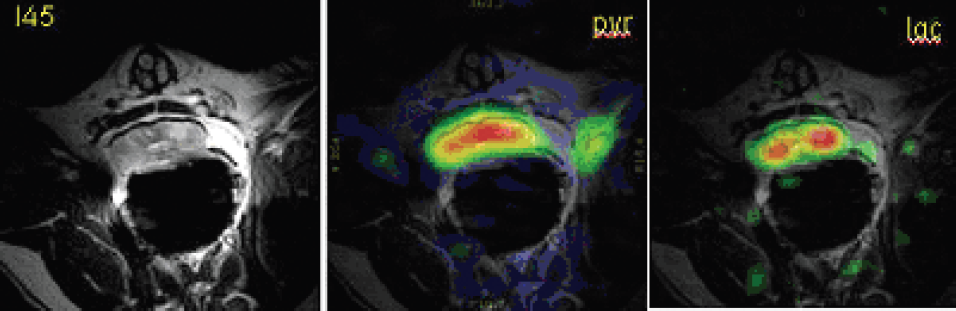
T1 high resolution sequences showed a highly significant plaque enhancement 2 hours after B22956 (Figure) versus Gd-DTPA in the atherosclerotic group (39.75% vs 9.5%, p < 0.0001). There was no difference between the 2 compounds in the control group (15% vs 15%, p = ns). With B-22956, CNR increased linearly up to 90 mn and then reached a plateau at 120 mn. The highest MR signal intensities after injection of B-22956 were found predominantly in the adventitia.

Axial T1-weighted MR images (TR: 800 ms; TE 5.6 ms; received bandwidth: 488 Hz per pixel; slice thickness: 3 mm; acquisition matrix: 256 × 256; field of view: 120 m:100 mm; number of signal averages: 4) of atherosclerotic rabbit abdominal aorta: Precontrast imaging (A) and 2 hours after B-22956 (B).
Abstract ID: 074
We have previously described transcriptionally targeted adenoviral (adv) therapies for Epstein-Barr Virus (EBV) positive cancers, and have utilized them in a locally administered context. These vectors feature an EBV-specific enhancer element attached to a minimal-CMV promoter, denoted as oriP. To explore the utilization of these therapies systemically, we have constructed three vectors to examine kinetics, specificity, and biodistribution: a) adv5.oriP.luc, expressing firefly luciferase under EBV-specific control; b) adv5.SV40.luc, expressing luciferase constitutively; and c) adv5.oriP.E1A.oriP.luc, a conditionally replicating adv, expressing both luciferase and the adv replication gene, E1A.
Longitudinal bioluminescence imaging (BLI) was conducted on tumour-bearing SCID mice (C666-1, EBV-positive human nasopharyngeal cancer), treated with 3times108 ifu of the above adv vectors, injected intravenously. At 72 hrs, adv5.oriP.luc demonstrated 8.4X higher tumour signal than adv5.SV40.luc, and adv5.oriP.E1A.oriP.luc was 26.7X higher; however, liver signal was also produced by leaky expression from the minimal-CMV promoter and natural adenovirus tropism to the liver. While the adv5.oriP.luc liver signal was 6.2X lower than adv5.SV40.luc, the adv5.oriP.E1A.oriP.luc signal was 195X greater. This intense liver signal was associated with histological evidence of viral replication, necessitating further manoeuvres to improve biodistribution.
Several compounds were examined, including norepinephrine, serotonin, liposomal clodronate, and STI-571, to determine whether any of these measures could improve adv biodistribution. Each of these interventions was assessed using BLI in mice intravenously injected with 3times108 ifu of adv5.oriP.luc. Amongst all these compounds, STI-571 achieved the highest increase in tumour-to-liver ratio (≈7 fold), by improving tumour uptake of adv5.oriP.luc. This increased tumour signal was associated with a 59% reduction in tumour interstitial fluid pressure. While STI-571 might not reduce hepatic uptake of adv sufficiently, this is the first report whereby this compound could improve tumour uptake of adv vectors, providing a greater window for improved therapeutic outcome for adv cancer gene therapy.
Abstract ID: 075
Leukotriene D4 (LTD4), a pro-inflammatory mediator, can induce cutaneous vascular permeability and vascular smooth muscle contraction in the lung. Montelukast is a well-known LTD4 antagonist. The increase in plasma extravasation stimulated by an LTD4 ID injection has historically been quantitated by administering Evans Blue IV and measuring the extent of the inflammatory flare. The visually blue skin surrounding the LTD4 injection site is harvested and Evans Blue is chemically extracted from the tissue and its concentration is calculated from the optical density measured spectrophotometrically at 620 nm. Utilizing a CRI Maestro multispectral imager and Evans Blue (Ex. 640 nm, Em. 730 nm), a rapid non-invasive optical method was developed for determining protein extravasation and pharmacological effects of Montelukast. Depillarized rats were randomized into four treatment groups: vehicle control (distilled water), and Montelukast, at 3, 10, and 30 mg/kg (n=6/group), and were dosed orally 4 hrs. prior to imaging. Ten minutes before imaging, each animal was administered Evans Blue IV, anesthetized using ketamine/xylazine IP, and then given 500 ng of LTD4 or saline ID in three parallel dorsal sites. Using a Maestro multispectral imager, images were acquired and quantitated for total signal intensity (SI) for each LTD4 and Sham injected site. Montelukast resulted in a dose dependent inhibition of plasma extravasation at 3 (1.6%), 10 (43%), and 30 (77%) mg/kg. Moreover, these data were highly correlated with the traditional tissue extraction methods with superior sensitivity. These data suggest this novel imaging approach could be used to non-invasively evaluate compounds for the LTD4 active site while providing repeated measures for kinetic analysis. This method would also increase the reliability and reduce sample sizes required to evaluate new compounds for therapeutic action.
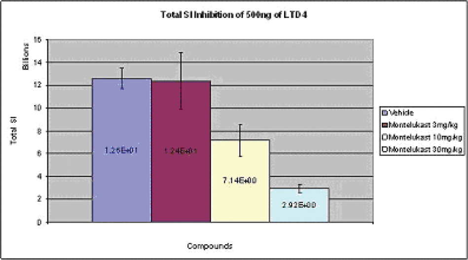
Abstract ID: 076
Concurrent 5: Session 14: Imaging in Cardiovascular Disease
Abstract ID: 077

Abstract ID: 078

Abstract ID: 079
Abstract ID: 080

Abstract ID: 081

Abstract ID: 082

Abstract ID: 083

Concurrent 5: Session 15: Advances in Optical Imaging
Abstract ID: 084
Fluorescence microendoscopy (FME) is emerging as a promising modality for cellular level imaging in live mammalian subjects. We have developed novel techniques for imaging the living nervous system using both one- and two-photon fluorescence excitation forms of FME. Our microendoscope probes are based on minimally invasive compound gradient refractive index (GRIN) lenses (350-1000 microns diameter) that provide micron-scale resolution. Such probes have allowed us to visualize cellular details in regions of the nervous system inaccessible to conventional microscopy. For example, we present the first views within the cochlea of live mammalian subjects of the auditory hair cells that transduce acoustic stimuli into electrical nerve impulses. However, the use of FME to date has been limited to acute studies of anesthetized subjects. We present two developments intended to extend FME usage to chronic imaging in anesthetized subjects over prolonged time periods, as well as to cellular level brain imaging in freely moving subjects. First, we present techniques for long-term FME studies that have enabled imaging of pyramidal neurons in live mice months after an initial surgery. Second, we present a compact, two-photon fluorescence microendoscope that is intended for brain imaging in freely behaving adult mice. This device is based on a flexible photonic bandgap fiber for near distortion-free delivery of ultrashort excitation pulses, a compound GRIN endoscope probe, and a DC micromotor for remote adjustment of the image plane. The imaging head has a mass of only 3.9 grams and provides micron-scale resolution. We have used such portable two-photon microendoscopy to visualize hippocampal blood vessels in the brains of live mice. We are now developing two-photon microendoscopy devices based on silicon micromachined scanning mirrors. Together, the development of long-term and portable FME imaging techniques should promote a broad range of biomedical applications, including clinical diagnostics and basic research in laboratory animals.
Abstract ID: 085
Human surgery is typically performed without the use of intraoperative image guidance because clinically viable technology would require high sensitivity, high specificity, and real-time operation. Invisible near-infrared (NIR) light in the 700–900 nm range has the potential to provide such guidance: tissue autofluorescence is relatively low, photon penetration is relatively high, real-time operation is possible, and the use of invisible light does not alter the appearance of the surgical field.
Three things are required for intraoperative NIR fluorescence imaging to become a reality: 1) imaging systems compatible with human surgery, 2) NIR fluorescent contrast agents specific for the task at hand, and 3) tools to train surgeons in NIR fluorescence image-guided surgery. This talk will describe recent advances in all three areas.
Under an NIH-funded Bioengineering Research Partnership (BRP), and in collaboration with GE Global Research, we have re-engineered our core intraoperative imaging system for multi-wavelength and ratiometric operation. We have also developed a unique high power, low-profile, multi-wavelength wide-field light source for this application.
We have developed robust chemical methods for the development of targeted and non-targeted NIR fluorescent contrast agents and will describe an HPLC/mass spectrometry platform for the rapid identification and purification of such molecules.
Tissue-like phantoms containing NIR fluorescent inclusions will be described, along with the concept of “ICG equivalence.” The former facilitates the training of surgeons in image-guided surgery, while the latter permits inter-laboratory comparison of imaging system performance.
Finally, using this technology we will present multiple surgical examples from large animal model systems approaching the size of humans.
Abstract ID: 086
Ovarian cancer is the fourth most lethal malignancy in women and is now considered a chronically relapsing cancer, repeatedly chemoresponsive, and with an exciting repertoire of targeted therapies under investigation. We have developed a molecular imaging platform with the goal of detecting and quantifying key molecular pathways in microscopic intraperitoneal tumor using laparoscopy. Preclinical experiments demonstrate the detection of as little as 0.3 picoM of the non-targeted NIRF agent indocyanine green (ICG) following excitation at 805 nm at a working distance of 9 cm using a hand held high quantum yield ICCD camera sensitive to ICG fluorescence emission at a wavelength of 835 nm in the near infrared with a resolution to 222 microns. This equipment is currently being tested in a clinical trial using ICG in women undergoing second look laparoscopy to examine their peritoneal cavity for small volume intraperitoneal tumor, and to date 7 of 20 patients have been studied. Technical challenges (safety issues, camera, laparoscopic platform, illumination, and image acquisition, resolution, and processing) have been overcome, and subsequently the imaging has been exciting. Following injection of up to three separate 8 mg boluses of ICG, intraperitoneal “hotspots” of near infrared fluorescence clearly identify visible tumor nodules. Furthermore, areas of peritoneum that appeared normal in white light and emitted IR light after ICG injections have been demonstrated to contain occult ovarian cancer in two biopsies, confirming proof of concept. Apparently false positive ICG signal has been seen in association with peritoneal trauma, and abnormal vessels. Image acquisition through a digital system will allow real time image integration. Second generation imaging equipment, laparoscopes, and imaging agents are being designed that will be used in clinical trials of enzyme activated near infrared fluorescent probes prototyped in the MGH Center for Molecular Imaging Research.
Abstract ID: 087
Abstract ID: 088
Fluorescence Molecular Tomography (FMT) is a new powerful near-infrared imaging modality that enables 3D quantitative determination of fluorochrome distribution in tissues of live small animals. Non-invasive, quantitative imaging plays a major role in developing tissue-engineering platforms for clinical applications. However, so far only 2D optical systems were available. We have previously shown that by implanting genetically engineered mesenchymal stem cells, conditionally expressing the osteogenic gene, BMP-2 (C9 cells) we could regenerate bone defects in various sites, in vivo. We hypothesized that engineered bone remodeling could be non-invasively and quantitatively monitored in 3D with FMT. C9 cells were implanted in the thigh muscle and radius non-union bone defect sites in C3H/HeN mice. Real time imaging of bone remodeling was performed after systemic administration of the fluorescent diphosphonate imaging agent, OsteoSense™ on Day 7, 14 and 21 post-implantation. In addition, we monitored and quantified bone formation in the implantation sites using micro-CT. Our results demonstrated an increase in OsteoSense signal on day 14 followed by a decrease on day 21-post implantation in the thigh, while the signal was still increasing in the radii on day 21. Micro-CT analysis revealed a large mass of matured bone formed in the thigh muscle and in the radius segmental defect on day 21. Correlation of micro-CT and FMT data revealed that the process of bone remodeling was almost completed during a 21-day period. This is the first report that demonstrates the feasibility of non-invasive, 3D, real-time, molecular imaging of the in vivo bone formation process. These findings demonstrate the effectiveness of FMT as a functional platform for molecular imaging in the field of bone regeneration and tissue engineering.

Abstract ID: 089
Sentinel Lymph Node (SLN) mapping and biopsy are important diagnostic tools to establish tumor staging and therapy for numerous cancer types. Key requirements of this procedure include the ability to reliably deliver a diagnostic agent to the lymphatic vasculature draining the tumor site, and to readily locate and visualize that agent after administration. This abstract reports a method combining a novel microneedle-based lymph delivery system and SLN optical imaging using indocyanine green (ICG) as a near-IR (NIR) contrast agent. ICG was delivered to the shallow swine dermis using a microneedle of 1000um length and visualized under fiber-optic tungsten lamp illumination with a 750 nm filter. Video images were captured using a CCD video camera with a 790 nm long pass filter and analyzed using frame by frame time analysis to quantify flow rate and time to target. This method resulted in rapid lymphatic trafficking and uptake in peripheral superficial nodes within seconds. Both the lymphatic drainage path and nodal locations, including inguinal, prefemoral, cervical, and popliteal chains, are readily visualized, and remain visible for several hours post-administration (Figure 1). Node-specific targeting and SLN accumulation was confirmed by post-administration biopsy. Microneedle based delivery resulted in SLN targeting 100% of the time and was 3X faster than traditional dermal injection techniques, which also exhibited a 25% failure rate. T-cell specific fluorescently labeled antibodies were similarly delivered in mice and exhibited specific binding to intranodal targets. SLN targeting was also demonstrated with other agents including blue tissue dyes, and ICG- labeled dendritic cells. This method potentially offers more rapid, reliable, and accurate SLN detection with reduced surgical morbidity after resection when compared to the traditional method of gamma counting injected radiocolloids.

Abstract ID: 090
Fluorescence molecular tomography is a novel imaging technology that resolves the bio-distribution of fluorescent reporters and can enable the visualization of molecular processes in small animals in vivo. Image resolution in FMT is limited by the high scattering of near-infrared light through biological tissue. We showed previously that imaging using high spatial-sampling of early-arriving photons can improve resolution of optical attenuation imaging through diffuse media since these photons have preferentially propagated along the least diffusive path. In this work, we apply this concept to FMT and demonstrate its performance in small animals in-vivo for the first time.
We utilized a pulsed, femtosecond laser and an ultra-fast time-gated camera to image early transmitted photons. Samples were suspended in a chamber filled with index-matching fluid and rotated through 360– during scanning. Imaging was first performed on complex fluorescent phantoms to verify correct operation of the system. In vivo experiments were then performed on nude mice orthotopically injected with Lewis Lung Carcinoma (LLC) cells one week prior to imaging with a Cathepsin-B activatable probe (Prosense 750, VisEn Corp.).
Tomographic reconstructions of early-photon data sets were performed on the above experimental models using a number of forward models and inversion algorithms. The reconstructed images exhibited higher spatial accuracy than corresponding frequency domain approaches and the results were confirmed using X-ray CT imaging and histology. Figure 1 shows an example reconstruction of an LLC tumor. This work represents the first in vivo implementation of FMT using early-photons.

X-ray CT image and FMT reconstruction of a distributed LLC tumor implanted in the right lobe of the lung of a nude mouse. Supported by the mouse imaging program at CMIR
Concurrent 6: Session 16: Imaging in Drug Discovery
Abstract ID: 091
The hollow fiber assay is developed for a preliminary screening of novel anti-cancer compounds prior to assessment in more complex tumor models. Bioluminescent imaging has been widely used to non-invasively evaluate drug efficacy of novel anti-cancer drugs and to image molecular pathways in tumor xenograft models. In order to enhance screening and evaluation of novel anti-cancer drugs, we have applied bioluminescent imaging to in vivo hollow fiber assay. Hollow fibers filled with MatBIII or MCF-7 tumor cells stably transfected with luciferase were subcutaneously implanted into nude mice. Proliferation of those cells in hollow fibers was determined with Xenogen IVIS®200 system. To evaluate the anti-tumor activities of known anti-cancer drugs, taxotere or camptosar was administered intraperitoneally in nude mice bearing hollow fibers. The growth of tumor cells, determined by bioluminescent imaging, was inhibited by both taxotere and camptosar. Furthermore, to investigate whether tumor cells inside hollow fibers communicate with the host mice, angiogenesis surrounding hollow fibers was determined by fluorescent imaging in vivo (AngioSense750, VisEn Medical) and detected by CD-31 immunostaining ex vivo. Fluorescent imaging showed that angiogenesis around hollow fibers developed 2 weeks after the implantation of hollow fibers. CD-31 immunostaining further confirmed the development of angiogenesis surrounding hollow fibers. These results demonstrate that tumor cells within hollow fibers are able to communicate with the host mice, and bioluminescent imaging can be used for rapid and accurate evaluation of anti-tumor activities in hollow fiber mouse models for preclinical drug screening.
Abstract ID: 092
University of Colorado Health Sciences, Denver, CO, USA. Contact e-mail:
Tyrosine kinase (TK) activity of epidermal growth factor receptor (EGFR) triggers oncogenic cell proliferation in a variety of tumors, including colorectal cancer. On the other hand, cancer related release of VEGF leads to an increase formation of abnormal blood capillaries (angiogenesis). A novel dual tyrosine kinase inhibitor (TKI) for EGF-R and VEGF-R, alone and in combination with a chemotherapeutic agent CPT11, was tested in a nude xenograft mouse model for human colon cancer HT29 cells. Proton density weighted (tumor dimension and necrosis) and gadolinium based dynamic contrast enhanced (DCE-MRI, tumor perfusion and permeability) scans were performed in the base-line, 10 days and 30 days after the treatment began. A compartmental pharmacokinetic model in which the tissue clearance of gadolinium was related to its transcapillary exchange in tumor and adjacent muscle tissue was constructed, based on T1-signal intensities. CPT11 partially prevented xenograft growth (tumor size at day 30: 0.15÷1.53 cm3 versus 0.56÷1.98 cm3 in non-treated group), but had no effect on gadolinium clearance, capillary surface factor or ktrans (Figure 1 represents T1-curves for baseline, 10 days (C1) and 30 days (C2) of the treatment). The novel TKI significantly decreased xenograft growth (0.16÷0.44 cm3), and also decreased ktrans values (from 0.45 to 0.32 min−1) as well as capillary surface factor (from 2877 to 1382). Gadolinium clearance was also partially improved. The combination group (CPT11+TKI) showed the best decrease rate in tumor proliferation and angiogenesis. In summary, the dual inhibition of both EGF-R and VEGF-R led to a significant decrease in colon cancer growth (EGF-R effect) as well as possessed significant anti-angiogenic properties (VEGF-R effect) in an experimental animal model.

Abstract ID: 093
Regulation of integrin functions plays critical roles in the adhesion and angiogenesis of endothelial cells. The human CD99 protein, which is a 32kD integral membrane protein expressed in most human cells, is supposed to be involved in controlling the activity of β1 integrin. In this study, the effects of CD99 signals on angiogenesis and tumor growth were investigated. We observed that crosslinking CD99 prevented human umbilical endothelial cells (HUVEC) from adhering to extracellular matrix proteins through β1 integrin-dependent mechanism. In addition, CD99 activation inhibited basic fibroblast growth factor (bFGF)-induced tubular morphogenesis of HUVECs. In parallel, it suppressed the vascular endothelial growth factor (VEGF)-induced angiogenesis in the chick embryo chorioallantoic membrane (CAM) assay. Through in vivo bioluminescence imaging, we showed that a 11-mer peptide from CD99 ligand suppressed the growth of mouse melanoma cells, B16F10, in the nude mouse xenograft model. These results suggest that a CD99 peptide has anti-tumoral effects in vivo through the inhibition of angiogenesis.
Abstract ID: 094

Abstract ID: 095
We have implemented a high frequency microultrasound imaging strategy to investigate microvascular perfusion and VEGFR2 (flk-1) expression in a mouse model of tumor growth. This technique enabled realtime visualization of actively perfused microvessels, and quantification of targeted contrast agents bound to VEGFR2 at spatial resolution superior to that of conventional ultrasound imaging. Five ×105 human melanoma (MeWo) cells were intradermally implanted in 6–8 week old athymic nude mice and grown for 3.5 weeks. TargestarB ultrasound contrast agents were coated with an anti-VEGFR2 monoclonal antibody or an isotype control antibody using biotin-streptavidin coupling chemistry and administered in a bolus (5times107 agents) via a jugular cannula. Contrast agents were detected using the Vevo 770™ micro-imaging system at a frequency of 40 MHz, which provided axial and lateral resolutions of 40 and 90 micrometers, respectively. A high acoustic pressure pulse sequence induced destruction of contrast agents within the beam elevation, and digital subtraction of the post- from pre-destruction sequences enabled derivation of the acoustic signal corresponding to retained contrast agents. The actively perfused microcirculation was visualized using an imaging technique known as maximum intensity persistence (MIP), which tracks the motion of intravascular contrast agents as they traverse the microcirculation. A bolus of TargestarP non-targeted ultrasound contrast agents was administered through the jugular cannula, and MIP images were formed in realtime using the Vevo 770™. We observed an approximately 4-fold greater retention of VEGFR2-targeted contrast agents relative to control agents within tumors (p< 0.01). MIP imaging revealed a relatively homogeneous perfusion pattern in the periphery of all tumors examined, while several tumors exhibited reduced perfusion within the central region. Lobular patterns of hyperperfused tumor tissue were also observed. Contrast-enhanced high frequency microultrasound imaging offers a powerful platform for assessing actively perfused regions in experimental tumors, as well as imaging expression of target molecules.
Abstract ID: 096


Abstract ID: 097
The endothelin (ET) axis, including the three ET peptides ET-1, ET-2 and ET-3 and their receptors ETA and ETB, is known to be a crucial factor in tumor angiogenesis and invasiveness. Recent clinical data suggest that specifically the expression of ETAR is linked to a poor clinical outcome in breast cancer patients.
Here we characterised a recently synthesised ETA receptor affine non peptidic near infrared fluorescent photoprobe (ETAR-Cy 5.5) in vitro and in vivo. While in vitro cell binding assays showed high amounts of cellular fluorescence in ETAR-positive MCF-7 cells, ETAR-negative MDA-MB 435 cells showed little to no celluar fluorescence confirming corresponding western blot analyses. Binding of ETAR-Cy 5.5 could be blocked by predosing with a corresponding anti-ETAR antibody or non-peptidic ETAR antagonists PD 156707. In vivo imaging of tumor xenografts by FRI and FMT (2 nmol ETAR-Cy 5.5 i.v.) showed high fluorescence signal yields for both MDA-MB 435 and MCF-7 xenografts. The in vivo binding specificity could be verified by predosing experiments with unmodified ETAR antagonist, which resulted in a significant decrease of tumor fluorescence. Protein expression analysis of whole tumor tissue revealed that in MDA-MB 435 cells mainly murine (e.g. derived from endothelial cells) ETAR is present while MCF-7 tumor xenografts express both the human and murine form of ETAR.
This leads to the conclusion that in vivo ETA receptor imaging is feasible using an ETA receptor affine non peptidic near infrared fluorescent photoprobe (ETAR-Cy 5.5). This imaging paradigm may be helpful for non-invasive chraracterisation of breast tumor tissues and may thus facilitate patient selection for novel ETAR antogonist therapies. In tumor xenograft models ETAR-Cy 5.5 can visualise both the ETAR expression of host (e.g. of endothelial cells) and of tumor tissue.
Concurrent 6: Session 17: Applications of Multimodality Imaging
Abstract ID: 098
Combined PET and MR imaging promises new possibilities for molecular imaging studies in humans and small animals. The first of a number of scanners being developed by both academic groups and scanner manufacturers are starting to appear and we can expect to see initial imaging results in the near future.
PET-MR follows rapidly on from the introduction of combined PET-CT systems that have proved highly succesful for clinical imaging. Many of the issues for PET-MR however are very different. Whilst PET and CT utilize similar technology, combined PET and MR requires significant modifications to the standard systems in order for the two scanners to work together without interference. Whilst combining PET and CT provides advantages in image registration and imaging time at minimal added cost, potential advantages of combined PET and MR, certainly for clinical imaging, are less clear. Accurate image registration with no additional radiation dose will be important where MR is the complementary anatomical imaging modality, however more exciting possiblilties exist for research applications. It will be possible to obtain temporally correlated functional data, for example fMRI and neuroreceptor ligand uptake, in response to interventions such as drug administration in single subjects.
Whilst sequential acquisition of PET and MR images is technically more straightforward, most systems under development aim to provide truly simultaneous PET and MR imaging. Diverse solutions to achieving this are being tried ranging from the development of MR-compatible PET detectors to purpose designing a PET-compatible MR magnet that can accommodate an only slightly modified PET design. The approach we have adopted has been to use optical fibres to build a small MR-compatible PET scanner capable of operating within a range of experimental MR systems with fields up to 7T.
This system and other systems currently under construction and evaluation will be described.
Abstract ID: 099


Representative CT/FDG-PET and multicontrast/dynamic MRI images of the carotid arteries of a patient with known carotid artery disease. The red arrows on the CT/FDG-PET indicate the MR slice used for cluster analysis and DCE-MRI parameters evaluation.
Abstract ID: 100
The multifunctional growth factor scatter factor/hepatocyte growth factor (HGF) and its tyrosine kinase receptor, c-MET, is implicated in the genesis, and malignant progression of a number of human malignancies, including glioblastomas. In this study we used an MRI molecular targeting agent to specifically tag the extracellular cell surface receptor, c-Met, with an anti-c-Met antibody linked to a Gd-DTPA-albumin contrast agent in an intracerebral implantation rat C6 glioma model. Gadolinium (Gd)-DTPA (diethylenetriaminepentaacetic acid)-albumin anti-c-MET was intravenously administered, and found to detect over-expression of c-MET in the C6 glioma in rat model, as determined by an increase in MRI signal intensity in T1w images and a corresponding decrease in regional T1 values. Specificity for the binding of the molecular targeted anti-c-MET contrast agent was determined using rat glioma cell cultures, and immunofluorescence confocal microscopic imaging of the targeting agents within neoplastic brain tissue. Gd-DTPA and Gd-DTPA-albumin administered controls indicated no non-specific binding of the targeting agent. Use of the molecular targeting agent in conjunction with MRI, as presented in this study, can be used to visualize in vivo over-expression of c-MET during glioma formation.

(Right-top) Rat glioma pre-contrast MRI, (right-bottom) post-contrast with Gd-DTPA-albumin-anti-c-MET, and (left) immunofluorescence image of targeting agent in glioma tissue following staining with a streptavidin-fluorophore.
Abstract ID: 101
The effect of hypoxia on the integrity of the extracellular matrix (ECM) of tumors is relatively unexplored. Here for the first time using novel MRI analyses in conjunction with fluorescence microscopy, we have related transport of macromolecules through the ECM to tumor hypoxia, since macromolecular movement through the ECM provides an index of ECM integrity. We conducted MRI on two anesthetized MDA-MB-231 tumor-bearing mice pre-selected for different tumor sizes. GFP expression of MDA-MB-231 cells was under the control of a hypoxia response element (HRE) promoter to visualize hypoxia within imaged slices. Albumin-GdDTPA (60mg/ml) was administered i.v. and MR concentration-time data analyzed using a multiple regression approach [1] to determine draining and pooling rates within tumor voxels (Fig 1). Additionally, the vector gradient of the contrast agent concentration within each voxel (Fig 2) was computed. The larger of the two tumors exhibited hypoxic regions (evident from the GFP image). Consistent with the possibility of ECM remodeling in response to hypoxia-induced upregulation of cytokines such as MMPs, contrast agent drainage rates were much higher in the larger tumor compared to the smaller tumor. Additionally, gradient maps enabled visualization of the magnitude and phase of these transport events within hypoxic and non-hypoxic regions of the ECM (Fig 2). Fluorescence microscopy was used to characterize CD34 as well as collagen IV staining in hypoxic and non-hypoxic regions (Fig. 3). Such integrated imaging techniques enable better understanding of the role of the tumor microenvironment in remodeling the ECM, and facilitate the design and evaluation of therapies that alter the ECM for enhanced drug delivery.
Acknowledgements: Supported by NIH P50 CA103175.

Abstract ID: 102
Human breast tumor cells (106 stably luciferase transfected MDA-MB-231-luc) were injected IV in a nude mouse. The animal was imaged repeatedly by LET and MRI over several weeks as the lung-colonizing metastases appeared and progressed in size and number. Following D-luciferin injection (450 mg/kg, SQ) in the anesthetized mouse, a set of 20 images, 18– apart, was obtained using 1 min exposure starting 5 min post-injection. Data reconstruction provided a 3D model of the lung tumors. The algorithm was also applied to images obtained using light reflected from the skin under external illumination. The imaging time varied from 300 s/angular position for the first imaging sessions that were characterized by low bioluminescent signal, to 30 s/angular position for the later sessions when the bioluminescent signal was large. MRI scans covering the chest of the mouse were acquired on a 4.7 T Varian scanner using a respiratory gating unit. We obtained contiguous proton density weighted spin-echo MR coronal slices with the following parameters: TE = 12 ms, FOV= 3.2 cm × 6.4 cm, slice thickness = 1 mm, matrix= 64 × 128, 4 averages. The LET first detected a bioluminescent signal 22 days after cell implantation, and revealed the growth and spread of the lung metastases at weekly intervals. The tumors were first detected by MRI 46 days after inoculation. The smaller tumors from the left lung shown on the LET images were still not observed by MRI. The position and relative sizes of the tumors are consistent between the two imaging modalities. Nevertheless, LET was able to detect the tumors 17 days earlier later than MRI, when the tumors were much smaller.

Abstract ID: 103
Concurrent 6: Session 18: Imaging of Cell Trafficking
Abstract ID: 104

Abstract ID: 105
Animal studies suggest that stem cells may be able to home to sites of myocardial infarction (MI) and assist in tissue regeneration. Recently, iron-oxide labeled stem cells have been used for in-vivo tracking of stem cells using MRI. However, due to negative contrast generated by iron oxides, differentiation between signal losses caused by iron from native low signal in tissue or signal loss caused by other artifacts is problematic.
It may be preferable to have a technique that produces positive contrast in the presence of iron. The aim of the current study was to test a positive contrast MR technique using reduced z-gradient rephasing (GRASP) for dynamically tracking stem cells in an in-vivo model of mouse myocardial infraction.
Ferumoxides and protamine sulfate were complexed and used to magnetically label embryonic stem (ES) cells. Experiments to test feasibility of MR tracking of these labeled ES cells within the mouse heart were performed on mice with induced MI using a direct coronary ligation model and controls. After infarction, 500,000 ES cells were injected in the border zone of the infarct. MR imaging was performed on a 9.4T scanner using conventional T2* GRE sequences (negative contrast) and positive contrast GRASP technique before MI and two weeks after MI. Following imaging, mice were sacrificed, and histology performed. Perl's staining was used to confirm iron within the myocardium.

In vivo images of a SCID mouse with myocardial infarction induced by coronary ligation and then transplanted with stem cells labeled with ferumoxides. (A) Prestem cell transplantation image (conventional T2*GRE image). (B) Poststem cell injection (conventional T2*GRE image). Dark spot on image indicates location of stem cells (arrow). (C) Prestem cell injection GRASP image of mouse heart. (D) Poststem cell injection GRASP image. Bright spot indicates locaton of stem cells (arrow). (E) Color-coded GRASP image superimposed on conventional T2*GRE image. (F) Histology of imaging slice (H&E staining). (G) Blue spots seen on Perls staining indicating presence of iron at the location (zoom ed view of box in F). Good correlation between MRI and histology is seen.
Figure shows sample images obtained. Excellent correlation was observed between signal loss seen on conventional T2* images and bright areas on GRASP and with the presence of iron on histology.
This demonstrated the feasibility of in-vivo stem cell imaging with positive contrast MRI.
Abstract ID: 106
Abstract ID: 107
Abstract ID: 108
The aim of this study was to track iron-oxide labeled adult mesenchymal stem cells using MR imaging at 9.4T and evaluate their potential benefit using PET in a rat model of myocardial infarction.
Iron-oxide labeled rat bone marrow derived stromal cells (IO-rBMSC; 4-5times105) were implanted into the infarcted area, immediately following myocardial infarction in male Sprague Dawley rats (n=4). In 4 animals, as control, the same procedure was performed using labelled dead cells in infarcted hearts (n=2), and labelled cells in non-infarcted hearts (n=2). Rats were imaged on day 2–3 and up to 6 weeks post-cell implantation using MRI and PET. A gradient echo cine-MRI was performed at 9.4T (Varian, USA). PET scans were carried out on a dedicated small animal PET scanner after an intravenous bolus injection of [18F]FDG (260-300 μCi). Scans were acquired over 65 minutes.
Signal voids caused by the IO-rBMSC were detected by MRI in the left ventricular wall of all hearts 2 days post-cell implantation (Figure 1a). PET showed an increase in [18F]FDG time versus radioactivity curves at 6.2±1.7 days post-cell implantation (Figure 1b) in infarcted regions implanted with rBMSC compared with infarcted regions implanted with dead rBMSC. However, by day 31±4.9, there was not only loss of MRI signal but also loss of [18F]FDG uptake in the infarcted area in rats implanted with IO-rBMSC.
In conclusion, the combination of MR and PET imaging can be used to track stem cells and to assess their therapeutic effects. This study shows that there are short term therapeutic benefits of rBMSC implantation for cardiac repair.

a) Short-axis MR image 2 days post-cell implantation; b) time versus mean normalized uptake value (NUV) for [18F]FDG.
Abstract ID: 109
The purpose of the study was to determine the factors responsible for the migration and homing of magnetically labeled endothelial progenitor cells (EPCs) to the sites of active angiogenesis. Two models were used: 1) magnetically labeled EPCs were administered in nude mice bearing 0.2 cm rat glioma in right flank; 2) magnetically labeled EPCs were mixed with tumor cells and implanted in flank of nude mice. EPCs were labeled with ferumoxides-protamine sulfate complexes. At specific time points, animals were euthanized, perfused and the tumors (1-1.5 cm) with surrounding tissues were collected. Ex vivo MRIs were obtained using a 7 Tesla MRI system. Serial sections of the tumor were made and consecutive sections were stained for DAB enhanced Prussian blue (PB), platelet derived growth factor (PDGF), hypoxia inducible factor 1α (HIF-1α), stromal derived factor 1 (SDF-1), matrix metalloproteinase 2 (MMP-2), VEGF and endothelial markers CD31 and vWF. The MRI demonstrated hypointense regions at the periphery of the tumors where the PB+-EPCs were positive for endothelial cell markers. At sites of PB+-EPCs, both HIF-1α and SDF-1 were strongly positive and PDGF and MMP-2 showed generalized expression in the tumor and surrounding tissues. Tumors cells expressed VEGF however no significant expression of VEGF associated with PB+-EPCs. Overall our studies show that labeled EPCs migration seemed to be related to HIF-1α and SDF-1 expression. We demonstrate the utility of SPIO labeled cells, which can be used as probes for MRI as well as marker for histological identification of administered cells similar to fluorescent dyes or reporter genes.

Expression of different angiogenic and chemoattractant factors at the sites of migrated labeled EPCs in tumors (consecutive sections).
Abstract ID: 110
Long-term cellular tracking will be important to non-invasively determine cell survival and function following transplantation in cell-based therapies. Tracking agents applied in vitro suffer from dilution effects; however, cells that sequester labeled substrates in vivo by stable gene over-expression will maintain the ability to sequester the substrate. This work evaluated the stability of 131I FIAU in canine bone marrow stromal cells (BMSC) over-expressing herpes simplex virus thymidine kinase (HSV-tk) and autologously transplanted into normal canine myocardium.
Dual radioisotope SPECT was conducted on a female dog for 46 hrs following injection of 5.4times106 tk+ BMSCs into the myocardium. Four weeks prior to transplantation, bone marrow was harvested, transfected with tk-gfp cDNA, and culture expanded. On transplantation day, cells were labeled with 111In tropolone and 131I FIAU in vitro to compare contrast agent stabilities within myocardially transplanted BMSCs. Region of interest analysis was conducted on corrected SPECT images to generate time activity curves (TAC).
BMSC viability was 98% prior to injection and expressed GFP. Fig 1. shows registered SPECT/CT images of BMSCs labeled with 131I FIAU. Biological half-lives were 19 hrs and 39 hrs for 131I and 111In, respectively.
SPECT provides the ability to image thymidine kinase over-expression and uniquely allows simultaneous acquisition of multiple cellular characteristics. 131I FIAU demonstrated faster washout kinetics compared to 111In suggesting that 131I FIAU is less stable than 111In, therefore 131I efflux is greater than that attributed to cell death alone.

SPECT/CT (A) transaxial and (B) coronal slices of BMSCs labeled with 131I FIAU and transplanted into normal canine myocardium. (FD=fiducial marker; TC=transplanted cells).
Plenary III: Session 20: The Future of Imaging Using Nanotechnology
Abstract ID: 111
Recent advances in nanoscience and nanotechnology have opened exciting new opportunities for disease imaging and targeting at the molecular level. The basic rationale is that nanometer-sized particles such as semiconductor QDs have novel optical, electronic, and structural properties that are not available from either individual molecules or bulk solids. When linked with tumor targeting ligands such as monoclonal antibodies, peptides, or small molecules, these nanoparticles can be used to target tumor antigens (biomarkers) as well as tumor vasculatures with high affinity and specificity. In the “mesoscopic” size range of 10–100 nm (diameter), quantum dots and polymeric nanoparticles also have more surface areas and functional groups that can be linked to multiple diagnostic (e.g., optical, radioisotopic, or magnetic) and therapeutic (e.g., anticancer) agents. Here we report a new class of multifunctional nanoparticles by combining diagnostic and therapeutic agents for cancer targeting and imaging. We also report self-assembled nanoparticles that are biodegradable and nontoxic and could complement the properties of QD imaging probes. Focusing on primary and metastatic breast cancer cells, we have prepared and validated peptides (protein fragments) for selective targeting and imaging of over-expressed cancer biomarkers. In particular, we have used the amino terminal fragment (ATF) of plasminogen to target the urokinase plasminogen activator receptor (uPAR), and a single-chain antibody fragment (ScFv) to target the epidermal growth factor receptor (EGFR), both of which are signatures of human breast cancer and several other cancer types.
Abstract ID: 112
Magnetic and magnetofluorescent nanoparticles have become important materials for biological applications such as sensing, separation, and imaging. These nanomaterials are often covalently modified with binding proteins such as antibodies or proteins to achieve target specificity but can face regulatory hurdles when clinical translation is contemplated. We hypothesized that the multivalent attachment of small molecules to fluorescent magnetic nanoparticles could lead to amplified binding and new, unknown biological properties of such nanomaterials. Therefore, we have created nanoparticle libraries that achieve specificity through multivalent modification with small molecules. We explored different synthetic routes to attach small molecules with anhydride, amine, hydroxyl, carboxyl, thiol, epoxy, azide and alkyne handles. By taking advantage of the fluorescent moiety on the nanoparticles, we were able to rapidly screen the library against a variety of cell lines. To date we have discovered a series of novel nanoparticles with specificity for either endothelial cells, activated human macrophages or pancreatic cancer cells. The method and described materials could have important applications for the development of functional nanomaterials for biology, functional differentiation of cell lines, and targeting.
Abstract ID: 113
The heat and vibration produced by ultrasonic waves enhance the delivery of drugs by altering the biodistribution and physical characteristics of delivery vehicles. With optimized parameters, ultrasound can deflect a vehicle to a vessel wall, fragment the vehicle, produce a phase transition in a lipid membrane, or increase cellular uptake of delivery vehicles. In addition, vascular permeability can be increased by ultrasound through multiple mechanisms. We are developing new approaches to localize a drug by combining these effects with molecular targeting and optimized vehicle properties. Vehicles under evaluation include perfluorcarbon nanoparticles, liposomal nanoparticles, micron-diameter gas bubbles, and hybrid vehicles combining the properties of gas bubbles with nanoparticles. Local delivery of cargos including DNA plasmids, siRNA, and chemotherapeutics has been shown to be feasible. Ultrasound instrumentation must also be advanced to accomplish these studies and we describe the development of very wideband transducers and new pulse sequencing methods. Given the complex set of mechanisms combined in such studies, correlative imaging techniques to assess the biodistribution of the vehicle and drug and therapeutic efficacy are important if these techniques are to be optimized efficiently. Here, we combine positron emission tomography and optical probes to evaluate the resulting biodistribution and therapeutic efficacy of the resulting strategies and survey the in vitro and in vivo performance of delivery techniques.
Acknowledgements: NIH CA 103828