
Abstract

Abstract ID: 464 Poster board space: 23
We have reported that genetically engineered bioluminescent E. coli specifically target cancers. In this study, our group exploited that genetically engineered bioluminescent E. coli and Salmonella typhimurium selectively target and proliferate in the breast cancer in vivo by employing optical bioluminescence imaging technique. pUC19 plasmid encloning Lux/GFP, was transformed into E. coli strains (DH5α) generating bioluminescent E. coli. To generate bioluminescent S. typhimurium, lux transposon cassette was integrated onto its chromosome. Breast cancer models were generated by direct inoculation of 4T1 (highly metastatic cells) and 4T7 (negative control) murine breast cancer cell lines into the abdominal mammary fat pad of BALB/c mice. Bacteria were intravenously injected into the tumor bearing mice. We found the minimal number of the subcutaneously injected bacteria required for imaging by cooled CCD camera (IVIS100, Xenogen) was 1times106 for DH5α and 1times103 for S. typhimurium, and the intensity directly correlated with the number of bacteria up to 109 colony forming unit, CFU. The lowest dose of intravenously injected DH5α and S. typhimurium to visualize cancer was 1times107 and 1times106 CFU, respectively. The imaging signal from DH5α was detected initially in the liver but diminished from the next day. The bioluminescence of DH5α and S. typhimurium was observed in the breast cancer from 2nd day after injection. The bacterial bioluminescence was observed in the liver, spleen, and bones of established metastatic models with 4T1 cells. All the metastatic lesions were confirmed histologically. E. coli and S. typhimurium strongly targeted primary breast cancer as well as metastases. Chromosome-based lux expression strain (S. typhimurium) revealed stronger bioluminescence than plasmid-based lux expressing strain (DH5α). Live attenuated strains are applied for developing cancer targeting delivery vehicle.

Abstract ID: 465 Poster board space: 24
Levels and location of protease activity are altered in response to biological insults conferred by infection, malignant growth and autoimmune responses. Monitoring these changes, in vitro and in vivo, is useful for early detection, assessing the extent of disease progression and measuring therapeutic outcome. In vitro assays for certain proteases have been developed with different reporting techniques: proteolytic release of p-nitro aniline for UV detection, proteolytic release of fluorescent dyes for fluorescence detection and mass spectrometry detection of proteolytic cleavage products. There are, however, various problems associated with the current detection techniques: low sensitivity of UV absorption, autofluorescence of cells or fluorescent interference from chemical and natural products, and the vacuum and other expensive and cumbersome parts required for mass detection.
The use of bioluminescence imaging offers the advantage of greater sensitivity both in vitro and in vivo. At the heart of this modality there are luciferase enzymes that generate visible light (i.e. ≈560 nm) through the oxidation of a specific substrate in the presence of oxygen and usually a source of energy (such as Mg2+ ATP). This is an ideal read out for the assessment of biological processes in living tissues and since there is no need for external excitation light, the signal-to-noise ratios are extraordinarily high.
We have used a modified luciferin substrate, aminoluciferin. The -NH2 group of aminoluciferin can be conjugated to specific peptide sequences that can be recognized and cleaved by target proteases. We have refined the synthesis of the aminoluciferin precursor to enable rapid production of bioluminescent protease probes, which will lead to the possibility of very sensitive multiplexed assays. Accelerated protease assays that are robust and sensitive will greatly enhance our ability to use protease activity as a measure of disease states both in vitro and in vivo.
Abstract ID: 466 Poster board space: 25
Pancreatic ductal adenocarcinoma (PDAC) is a highly aggressive cancer with 5 year survival rates of less than 5%. The development of novel molecular markers and imaging probes for incipient PDAC and pancreatic intraepithelial neoplasia (PanIN) would enable earlier detection and facilitate rational treatment. Furthermore, the identification of biomarkers for specific disease stages or particular genetic lesions would likely have significant utility in stratification of patients, in tailoring treatment protocols and in evaluating the efficacy and specificity of targeted therapies. Small peptides have considerable potential as imaging agents based on their lack of immunogenicity, short plasma half-life and high affinity. Therefore, we decided to use phage display selection procedures and primary cell cultures derived from mice that produce genetically accurate pancreas-specific lesions to aid in the identification of a human disease relevant diagnostic agent. We identified two motifs that target mouse PDAC cells over normal pancreatic duct cells 13.5 fold in vitro. In addition, the identified sequences distinguished human PDAC cells over normal pancreatic duct cells. These peptides were also able to specifically detect human PDAC in ex vivo biopsy specimens. Moreover, we used these peptides as imaging agents, in conjunction with intravital confocal microscopy, to detect emerging tumors and PanINs in an engineered mouse model of PDAC (Kras activation and Ink4a deletion). These specific and sensitive probes may promote earlier diagnosis and more astute management of this disease in humans.
Abstract ID: 467 Poster board space: 26
Thomas Steinberg, Julie Nyhus,
Functional labeling is the ability to label a probe to maximally retain its native targeting properties while minimizing the pharmacokinetic perturbations due to the labeling agent. Reactive fluorescent dyes are commonly used to label biopolymers and small molecules to produce optical probes. Many labeling protocols and commercially available labeling kits are developed with the intent of producing maximal fluorescent signal by conjugating multiple fluorophores (>3) to each probe; this increases the likelihood of the labeling agents to affect the pharmacokinetics of the optical probe thus influencing the probe's intended performance. We have developed a simple, convenient and rapid method of labeling probes with amine reactive fluorescent dyes so as to limit the degree of labeling (DOL) to an average of two fluorophores (or less) per probe. Data will be presented to demonstrate the capability of this labeling system to label a variety of proteins of various molecular weights and amounts at an average DOL of 2 (or lower) with no optimization of the labeling protocol. We will also demonstrate that probes labeled at an average DOL of 2 (or lower) are sufficiently labeled to function as in vivo imaging optical probes.
Abstract ID: 468 Poster board space: 27
Optical-based in vivo imaging of vasculature is an emerging modality for studying vascular structure, function and angiogenesis. Vascular imaging provides information about the number and spacing of vessels, permeability of the vasculature and functional abnormalities of the vessels. It is of particular importance in tumor biology where imaging can be used to assess the efficacy of anti-angiogenesis drugs. Imaging agents that fluoresce in the near IR are particularly useful as vascular contrast agents as they avoid the signal interference due to hemoglobin absorbance. Vascular contrast agents are transient imaging reagents with imaging time windows ranging from a few minutes to several hours, depending on the properties of the probe and the target. The choice of a contrast agent is dictated by the experimental model and the imaging protocol. We will present data demonstrating the performance of a variety of vascular agents: fluorescently labeled proteins, fluorescent Microspheres and Qdot® nanocrystals.
Abstract ID: 469 Poster board space: 28
We describe two novel cancer targeting fusion proteins consisting of an engineered antibody fragment fused to Renilla luciferase or Gaussia luciferase for in vivo optical imaging of carcinoembryonic antigen (CEA). The engineered anti-CEA T84.66 diabody fragment (Db) has previously exhibited high level tumor targeting in biodistribution and microPET imaging studies using a CEA-positive tumor model. Fusion proteins were generated using RLuc8, which was optimized for in vivo use by the incorporation of 8 amino acid substitutions, and GLΔ15, a 15 amino acid N-terminal truncation of GLuc. A bioluminescence ELISA demonstrated that purified Db-RLuc8 and Db-GLΔ15 could simultaneously bind to antigen with high affinity and emit light. Additionally, the fusion proteins exhibit in vitro enzymatic stability suitable for in vivo use (t? >180 h). In vivo optical imaging of tumor bearing mice demonstrated specific targeting of both Db-RLuc8 and Db-GLΔ15 to CEA-positive xenografts with Db-RLuc8 displaying a tumor to background ratio of 6.0 ± 0.8 in CEA-positive tumors at 6 h after intravenous injection, compared to CEA-negative tumors at 1.0 ± 0.1 (p = 0.001, n=7) and Db-GLΔ15 displaying ratios of 3.8 ± 0.4 and 1.3 ± 0.1, respectively, at 4 h (p = 0.001, n = 8). MicroPET imaging using 124I-diabody-luciferase fusion proteins confirmed the optical signal was due to antibody-mediated localization of luciferase and dynamic microPET scanning showed the clearance of each. Ultimately, a comparison of the two fusion proteins shows that although the Db-GLΔ15 is brighter and more stable in vitro, the Db-RLuc8 demonstrated better tumor targeting in vivo with a brighter signal. These two luciferases, fused to biospecific sequences such as engineered antibodies, can be administered systemically to provide a novel, sensitive method for optical imaging based on expression of cell surface targets in living organisms.
Abstract ID: 470 Poster board space: 29

Abstract ID: 471 Poster board space: 30
Cong Li, Tiffany Greenwood, Zaver Bhujwalla,
Imaging lysosomes in breast tumors in vivo has great potential for assessing the metastatic and invasive potential of breast cancers since lysosomes play a major role in cancer invasion and metastasis by mediating protease routing, regulation, and secretion. We have synthesized two novel near-infrared (NIR) optical imaging probes for noninvasive imaging of lysosomes. These two probes, whose chemical structures are shown in Figure 1, contain an NIR emitting indocyanine moiety covalently bound to glucosamine through two different extended carbon chain linkers. Compounds

Abstract ID: 472 Poster board space: 31
The use of fluorescent dyes and fusion proteins, such as GFP, is widespread for the intracellular labeling of specific biomolecules for live cell imaging. These organic and biochemical fluorophores are, however, susceptible to photobleaching, making them inadequate markers for extended time-lapse fluorescence microscopy. We seek to overcome this limitation by targeting semiconductor quantum dots (QDs) to specific intracellular proteins. The QDs photobleach minimally, and multi-colored QDs can be simultaneously imaged, making them suitable for tagging and tracking multiple proteins or pulse-chase experiments. To covalently link QDs to proteins of interest, we constructed fusion proteins containing a DsRed-actin sequence fused with the commercially available SNAP tag. The SNAP tag is a 22 kDa protein that is an engineered form of the human O6-alkyl-guanine-DNA-alkyltransferase (AGT). Endogenous AGT repairs alkylated DNA by transferring the alkyl residue from the O6 position of guanine to the reactive cysteine at its active center. The SNAP protein is also able to transfer residues from para-substituted benzylguanines, resulting in the covalent attachment of the substituted group (such as a QD) to the SNAP tag. SNAP-DsRed-Monomer-Actin fusion proteins were expressed in E. coli and purified in order to perform in vitro polymerization experiments to prove the functionality of the fusion proteins, which was assessed both by polymerizing actin and then adding QDs for tagging of the filaments and by attempting to polymerize the actin already labeled with the QDs. The use of the DsRed-Actin fusion allowed for continuous monitoring of both the actin and the QDs without the need for fixing and staining. In addition, the pore-forming bactotoxin streptolysin-O (SLO) was used to deliver ben-zylguanine-conjugated QDs into 3T3 cells transfected with pSNAP-DsRed-Monomer-Actin. The feasibility of using this covalent method to label intracellular proteins in live mammalian cells was assessed.
Abstract ID: 473 Poster board space: 32
Abstract ID: 474 Poster board space: 33
Optical fluorescence imaging has great advantages for biomolecular imaging, not only because its sensitivity is quite high, but because the signal of the imaging probe can be greatly amplified only under certain conditions. However, only a very limited range of biomolecules can currently be visualized because of the lack of flexible design strategies for fluorescence probes. At present, design is largely empirical. Recently, we could establish the first and totally rational design strategies of novel fluorescence probes by using the concept of photoinduced electron transfer (PeT), and succeeded to develop various functional fluorescence probes. One of the successful examples was our beta-galactosidase probe (TG-betaGal) which was reported in the previous SMI meeting.
Reactive Oxygen Species (ROS) have recently attracted considerable attention as mediators which cause many diseases or intermediate signal transductions. In order to evaluate the biological functions of ROS, we thought it essential to develop fluorescence probes which can detect one type of ROS selectively. We designed type-specific fluorescence probes for each ROS by using our design strategies with PeT, and developed some probes such as DAMBO, HPF and DPAX for nitric oxide, highly reactive oxygen species and singlet oxygen, respectively. Further, very recently, we developed a novel fluorescence probe, MMTR, quite specific for hypochlorite. MMTR itself has a tetramethylrhodamine platform, but little absorbance in visible region and little fluorescence. When hypochlorite was added to a colorless solution of MMTR, absorbance in visible region and fluorescence were enhanced. This is because spirodihydrothiophen in MMTR was oxidized by hypochlorite and the MMTR lost its closed-form favorability. With MMTR, we could observe marked and selective fluorescence enhancement both in myeloperoxidase system and living neutrophils. Our new probes should play important roles in ROS research.
Abstract ID: 475 Poster board space: 34
Developments of multimodality imaging technology that combines functional information and anatomical information are significant for early detection and screening of cancers. This molecular multimodality imaging strategy relies on the successful synthesis of fusion probes. Bombesin (BBN) is a 14-amino acid peptide that shows high affinity for the BB2 receptor subtype of the bombesin receptor family. BBN analogs have been used as radionuclide-tracers for in vivo PET and SPECT imaging and treatment of tumor models in animals. Based on this work, we are adapting these peptides for molecular optical imaging modality applications. In this study, we report developments of a series of new Alexa Fluor 680-BBN and Alexa Fluor 750-BBN conjugates. Fluorescence spectra show the efficient excitation wavelength band and emission band of the conjugates. In order to assess the binding affinity (IC50) of the Alexa Fluor-BBN conjugates for the BB2 receptor, in vitro competitive cell-binding assays were performed using PC-3 prostate and T-47D breast cancer cells. These new peptide conjugates demonstrated high specificity and affinity for the GRP receptor. In vitro cell-binding studies were performed and the degree of cell-associated activity was determined by confocal fluorescent microscopy to assess the degree of uptake, internalization, and blocking of the conjugates in PC-3 and T-47D cells. Blocking studies, in which high levels of cold BBN were administered to the cells prior to the Alexa Fluor-ligands, reduced the uptake/retention in normal BB2 receptor-expressing PC-3 and T-47D cells. In order to assess the in vivo uptake of the Alexa Fluor conjugates, we evaluated AF 680-β-Ala-BBN[7–14]NH2 in human breast T-47D cancer cell xenografted SCID mice. The results clearly demonstrate the effectiveness of these new conjugates for targeting the BB2r with very high selectivity and affinity.
Abstract ID: 476 Poster board space: 35
Positron emission tomography (PET) is widely used for oncologic diagnostics, has inherently high sensitivity and is often practiced with the radiotracer molecular imaging agent 2-[18F]fluoro-2-deoxy-
In vivo imaging studies were performed in two xenograft mouse models bearing well-established tumors (SW480 human colon cancer and HT1080 human fibrosarcoma). Biodistribution of the potential metaboli-cally-targeted probe was monitored in real time for up to seven days postinjection of 20nmoles of NIR-glucosamine using the Xenogen IVIS 200; harvested tissues were also imaged. Time activity curves generated for tumor and normal regions demonstrate the clearance profiles and accumulation of NIR-glucosamine. Observable accumulation in the tumor that persists for at least seven days suggests that the NIR-glucosamine may be taken up and trapped in the tumor cells, similar to the fate of 18FDG. In vitro assays and confocal fluorescence microscopy provide further evidence of cellular uptake characteristics, internalization, and insights regarding the transport mechanism. Results from ongoing comparison studies on the uptake kinetics of NIR-glucosamine with that of 18FDG will be presented. Ultimately, we demonstrate that NIR-glucosamine can serve as an optical imaging agent of metabolism, devoid of ionizing radiation with a significantly longer length of detection that is compatible with high-throughput screening and small animal in vivo imaging.
Abstract ID: 477 Poster board space: 36
Olga Pena, Goar Smbatyan, Jeffrey Peterson, Rex Moats, Yves DeClerck,
Neuroblastoma (NB) is the second most common solid tumor in children and a cancer that frequently metastasizes to the bone. Although when localized, NB can be cured in 90% of cases, when locally invasive or metastatic the overall prognosis is much poorer. Methods of early detection of bone metastases might have significant impact on morbidity and mortality by enabling earlier treatment initiation. The understanding of the role of osteoclasts in the formation of osteolytic lesions in cancer metastasis has led to the use of bisphosphonates as therapeutics in patients with cancer bone metastases. We have developed a NB bone invasion model using human NB cells injected directly into the femurs of nude mice and have used multi-modality imaging in an attempt to identify these lesions at early time points. The near infrared (NIR) fluorescent bisphosphonate derivative OsteoSense™ (VisEn Medical, Boston, MA) has been shown to detect areas of increased bone growth and resorption in vivo. In a preliminary experiment to test the use of this probe in our NB bone invasion model, 6-and 16-week old female mice were injected with firefly luciferase expressing human neuroblastoma CHLA-255 cells or PBS (control) in the right femur and imaged 3, 4, and 5 weeks after injection. Three techniques were used to image injected animals: high resolution plain-film X-ray, D-luciferin-induced bioluminescence, and OsteoSense-mediated fluorescence. Osteolytic lesions were radiographically detectable in mice injected with tumor cells by the 3rd week along with a clear D-luciferin-induced signal. OsteoSense-generated fluorescence was also detected in the right femur as well as in normal bone (spine, skull, and extremities where growth occurs). These latter signals were stronger in younger mice when compared to the older mice. These results suggest that the NIR-probe OsteoSense may be useful for noninvasively detecting areas of bone changes in NB bone lesion models.
Abstract ID: 478 Poster board space: 37
Early detection of metastases would significantly impact the individualized treatment plan for cancer patients with reductions in both morbidity and mortality. Despite numerous advancements, current methods of early screening for metastases, especially those with a predilection for bone, do not provide adequate sensitivity. In our research, we aim to develop a target-specific, optical contrast agent for use in the detection of bone-seeking breast cancers.
We have synthesized a peptide analog of interleukin-11 (CGRRAGGSC), labeled with an optical agent, identified through phage display to target the interleukin-11 receptor alpha (IL-11Rα). IL-11Rα is expressed on both osteoclastic progenitors as well as on certain metastatic cancer cell lines. Specifically, we have conjugated the peptide with a near-infrared dye generated in-house to allow for fluorescence-based optical imaging. Breast cancer cell lines, MDA-MB-231 and subline 1833, which both express IL-11Rα, were utilized for the in vitro and in vivo studies. These cell lines were cultured in DMEM/F12 with 10% FBS before inoculation into the animal and allowed 3–4 weeks to grow subcutaneously. The receptor-positive tumor nodule was imaged immediately after tail vein, i.v. injection of CGRRAGGSC-NIR dye for 40 minutes and again at 24 hours. Fluorescence was accomplished by illumination with a 780 nm laser diode and the resulting light emission at 830 nm was captured by an image intensifier lens coupled to a charge-coupled device camera outfitted with band-pass and holographic filters.
In vitro studies demonstrated that the imaging agent binds to the IL-11Rα-positive metastatic breast cancer cell lines. Furthermore, the imaging compound accumulated within the tumor of the animal models. Our results demonstrate the feasibility for employing the synthesized conjugate targeting IL-11Rα and thus provides the basis for its deployment for assessing metastatic lesions in preclinical models.
Poster Session II: P08: Nanotechnology
Abstract ID: 480 Poster board space: 40
The byproducts of atherosclerotic vascular disease are the leading cause of mortality worldwide. It is therefore essential to be able to diagnose and treat atherosclerosis prior to the onset of clinical symptoms. The macrophage has emerged as a key biological, imaging, and therapeutic target for inflamed plaques. Thus, we have developed a magnetofluorescent nanoparticle (MFNP) functionalized with a potent photodynamic therapy agent, 5-(4-carboxyphenyl)-10,15,20-triphenyl-2,3-dihydroxychlorin (TPC), to target and locally eliminate tissue macrophages in lesions, such as those which occur in stent re-occlusion.
Alexa Fluor 750 was initially conjugated to the dextran-coated particle in a ratio of 3 fluorophores per particle. In order to impart upon the nanoparticle the ability to generate singlet oxygen, TPC was covalently conjugated to the primary amines of the MFNP, resulting in a concomitantly therapeutic nanoparticle (TNP). TNP were stable for months without precipitation, and were detectable by MRI and fluorescence imaging. The UV-vis absorption spectrum of the TNP shows characteristic features of all three components, and was used to estimate the number of TPC per particle (n = 30). It is notable that the roughly 100 nm difference between the longest wavelength absorption for TPC and that of AF750 minimizes energy transfer when the particles are excited at 650 nm (the therapeutic wavelength). This feature should avoid cell killing while imaging the spatial distribution of the agent within cells (750 nm excitation). Preliminary in vitro experiments have demonstrated multimodal detection and exquisite phototoxicity to macrophages when illuminated by appropriate light. Since these TNP are expected to preferentially localize within macrophages at sites of inflammation, they may prove further useful in the treatment of other inflammatory conditions, as outlined below.

Abstract ID: 481 Poster board space: 41
Yuetang Wang1, Xiaoxia Wen1, Jeffrey Leon2, William Harrison2, Joe Bringley2, Tiecheng Qiao2, Juri Gelovani1,
Recent advances in nanotechnology are likely to accelerate the discovery of new near-infrared fluorescence (NIRF) imaging agents for molecular imaging applications. In this study, we used both optical and nuclear imaging to evaluate the imaging property and biodistribution of 3 novel NIRF nanoparticle preparations: PEGylated polymer-shelled silica nanoparticles, PEGylated nanolatex particles, and cross-linked PEG nanogel. All nanoparticles were encapsulated with a NIRF dye (ex/em: 740-760/766-785 nm) and conjugated with aminobenzyl DTPA for In-111 chelation. The media diameters of silica, nanolatex, and nanogel were 100 nm, 21 nm, and 21 nm, respectively. Gamma scintigraphy and NIRF optical imaging were performed at various times after intravenous injection of nanoparticles at doses ranging from 2- to 4times1013 particles/g b.w. in nude mice bearing subcutaneously inoculated human MDA-MB-468 tumors. All particle preparations could be readily imaged with NIRF at the injected dose. Both γ-scintigraphy and optical imaging revealed that most silica nanoparticles were rapidly distributed to the liver and the spleen. In contrast, nanolatex and nanogel particles showed sustained blood pool activity. The blood activities at 48 hr postinjection, expressed as percent of injected dose per gram of tissue, were 0.03±0.006%, 1.39±0.08%, and 4.33±0.23% for silica, nanolatex, and nanogel, respectively. The uptakes in the liver and spleen were reduced from 47.6±5.0% and 120.3±31.9% for silica nanoparticles to 22.3±3.5% and 10.0±1.1% for nanolatex, and 25.5±2.3% and 5.0±0.8% for nanogel, respectively. Prolonged blood pool activity resulted in significantly increased tumor uptake for nanolatex and nanogel owing to enhanced permeability and retention effect. The tumor uptakes of silica, nanolatex, and nanogel were 0.33±0.06%, 2.02±0.09%, and 2.79±0.26, respectively. Our data suggest that particle size and deformality are important factors governing the biodistribution pattern of nanoparticles, and that both nanolatex and nanogel particles may be suitable carriers for targeted optical imaging (supported by NIH grants R01 CA119387).
Abstract ID: 482 Poster board space: 42
In view of the need for biocompatible and non-immunogenic nanosystems, we recently developed a new nanoplatform that allows lipoproteins to be redirected from their normal destination and co-opted to carry molecular imaging probes or toxic payloads to a variety of cancer cell types through targeting of their specific, unique surface tumor markers. For proof-of-concept, folic acid (FA) was used as a tumor marker to redirect low-density lipoprotein (LDL) from LDL receptors (LDLR) to folate receptors (FR). Specifically, FA was conjugated to side-chain Lys residues of LDL apoB-100 protein. Near infrared fluorophore (DiR, ex: 748nm, em: 782nm) was intercalated to LDL phospholipids monolayer to visualize the uptake of this nanoparticle (DiR-LDL-FA) by tumor cells in vitro and in vivo. Using confocal microscopy in KB (FR+), HT 1080 (FR−) and HepG2 (LDLR+) cells, we demonstrated that DiR-LDL-FA no longer binds to LDL receptor (no uptake in HepG2 cells), instead, strong fluorescence was found in KB (FR+) cells but not in HT 1080 (FR−) cells. To confirm the LDL redirecting in vivo, DiR-LDL-FA was intravenously injected into mice with size matched KB and HT1080 tumors on the opposite flanks. Significant fluorescence enhancement in KB tumor over HT 1080 tumor was observed using the Xenogen imager. This result was consistent with ex vivo biodistribution studies by measuring the fluorescence of the harvested tumors and other organs. To further validate the FR-mediated nanoparticle uptake, 30 folds excess of free FA was pre-injected into mice 5 min before DiR-LDL-FA, resulting diminished fluorescence signal in KB tumor. In addition, no difference in fluorescence intensity was observed between two tumors. This indicates that the preferential uptake of DiR-LDL-FA in KB tumor by FR was blocked. In conclusion, conjugating LDL with FA successfully redirected LDL from LDL receptor to FR, as we demonstrated both in vitro and in vivo.
Abstract ID: 483 Poster board space: 43
Xiang Hong Peng1, Andrew Yang2, XiaoXia Wang1, Zehong Cao1, Chunchun Ni1, William Wood1, Shuming Nie1, Hui Mao1,
Urokinase plasminogen activator (uPA) is a protease regulating matrix degradation, cell motility, metastasis and angiogenesis. Cellular receptor of uPA (uPAR) is highly expressed in most cancer cells, intra-tumoral fibroblasts and endothelial cells. We developed amphiphilic polymer-coated SPIO nanoparticles that conjugate with the amino-terminal fragments of uPA (ATF, 135 amino acids) via carboxyl groups. This ATF-SPIO nanoparticle contains 8 to 10 ATF fragments (Fig. 1A) with strong T2 effect on MRI. We demonstrated that the targeted SPIO nanoparticles specifically bound to and were internalized by mouse mammary carcinoma cells, resulting in significant shortened T2 measured by MRI. Presence of ATF-IO was confirmed with positive Prussian blue staining in the tumor cells. We found that interaction of ATF-SPIO with uPAR could decrease proliferation and invasiveness of the tumor cells. Furthermore, delivery of ATF-SPIO nanoparticles through the tail vein led to selective accumulation of the SPIO particles in subcutaneous and peritoneal tumor lesions in a mouse mammary tumor model as demonstrated by Prussian blue staining of frozen tissue sections obtained from tumor and normal tissues. In vivo MRI revealed significant decreases of T2 signal in tumor areas around 24 hrs after systemic delivery of the targeted SPIO nanoparticles (Fig. 1B). Liver and spleen uptake of ATF-SPIO is reduced compared to SPIO without conjugated to ATF, evidenced by observing less MRI signal decreases and T2 reduction in those organs. Current effort focuses on the development of dual functional SPIO nanoparticles with tumor targeting ligands and chemotherapy drugs for the detection and treatment of breast cancer.

Targeted-SPIO nanoparticles MR imaging of breast cancer. A. ATF peptide conjugated SPIO nanoparticle. B. In vivo MR imaging of a mouse mammary carcinoma (4T1) growing subcutancously after systemic delivery of ATF-SPIO nanoparticles.
Abstract ID: 484 Poster board space: 44
We report the fabrication of multifunctional silica porous nanocapsules that have magnetofluorescent properties as well as delivery capacity. The silica nanocapsules containing quantum dots (QDs) and magnetic nanoparticles can be successfully synthesized by a single system emulsion-mediated process. During the emulsion mediated process, hollow type silica nanoparticles having holes on their surfaces can be generated. The visible or near-infrared (NIR) emitting QDs embedded in silica shell renders the silica nanocapsules to be monitored by in vitro or in vivo fluorescence imaging technique, and the magnetic nanoparticles allows for magnetically active property. We also show that the silica nanocapsules can be loaded with small or large molecules, and finally the holes on their surfaces can be encapsulated by polyelectrolytes. This demonstrates that the silica nanocapsules can be used as multifunctional platform for guided delivery of small or large molecules with optical imaging tracking.
Abstract ID: 485 Poster board space: 45
The lymphatic system is difficult to study due to its difficult accessibility. An in vivo non-invasive 2-color optical lymphangiographic technique to separately visualize dual lymphatic flows to the axillary lymphatics from the breast and from the ipsilateral extremity may help to better understand the mixing of these two lymphatic basins. Ten athymic mice received injections of quantum dot (Qdot) optical agents into the middle phalange of the left upper extremity (Qdot 800) and into the left breast (Qdot 705). Immediately after injection, wavelength-resolved spectral fluorescence imaging was carried out. The different colors of the Qdots allowed real time imaging of the lymphatic flow. The lymphatic drainage territory of each contrast agent was assessed before and after removal of the skin. After in vivo lymphangiography, the axillary, lateral thoracic and cervical lymph nodes (LNs) were extracted for ex vivo fluorescence imaging and fluorescence microscopy to validate the in vivo imaging. Another 5 mice were injected agents at opposite sites. Two-color lymphangiography successfully visualized lymphatic vessels as well as the lymphatic drainage territory in all 10 mice. Eight of 10 axillary LNs received two different lymphatic flows simultaneously from the breast and from the upper extremity (Fig.). Ex vivo fluorescence imaging and fluorescence microscopy of resected LNs validated the lymphatic drainage found in the in vivo lymphangiography in all mice. The opposite injection did not show the lymphatic drainage from the mammary gland. Two-color imaging of the lymphatics using appropriate sizes of Qdots demonstrates a highly variable pattern of drainage from the breast and upper extremities into the axillary LNs. Qdot imaging provides an excellent means of studying lymphatic flow in vivo.

Abstract ID: 486 Poster board space: 46
Quantum dot nanocrystals offer desirable optical properties which make them suitable bioimaging probes, including size-tunable emission profiles, high emission intensity, and increased photostability relative to organic dyes. In order to enhance the scope of their application, strategies to rapidly deliver quantum dot payloads into cells are needed, for example, to image cells with optimal signal to noise ratios in tissue or to target and image intracellular structures. Here, we develop and test several quantum dot constructs with various natural and synthetic coatings hypothesized to permeate cell membranes and compare them to a commercially-available reagent for quantum dot loading in endosomes. A variety of cell lines and immune cells were incubated with surface-engineered quantum dot constructs, in the presence and absence of multiple inhibitors and co-localization markers of endocytic pathways. Temperature-dependency of internalization was also assessed. Fluorescence spectroscopy, fluorescence microscopy, cell viability assays, and flow cytometry were utilized to characterize quantum dot internalization. Our preliminary tests indicated that several quantum dot assemblies rapidly enter the cytosol without significant cytotoxicity. Labeled immune cells were readily visible in vivo using a rat retinal fluorescence imaging technique, and the high intracellular loading capacity of our nanocrystals enabled bioimaging with high signal to background ratios. We discuss the implications of cytoplasmic delivery of high molecular weight cargoes for molecular therapy as well as bioimaging applications.
Abstract ID: 487 Poster board space: 47
Different strategies have been pursued to eliminate cancer cells while avoiding the potential collateral damage to the healthy normal tissues. Targeting tumor cells or tumor vasculature through tumor specific recognition small peptide could be a promising pathway for the purpose. Previous reports have shown that RGD-4C peptide could bind specifically to αvβ3 and αvβ5 integrins, which were known to highly express in angiogenic neovasculature. In this study, we have fabricated aqueous nickel-Nitrilotriacetate modified Fe3O4 nanoparticles of 9 nm in diameter. The nanoparticles were able to perform self-assembly to the hexahistidine tagged RGD-4C peptides with controlled orientation through specific affinity between nickel ion and the 6-His tag. To demonstrate the specific targeting of the nanoparticles to oral cancer cells in vivo, we have performed iron staining in the fixed cultured cells and demonstrated the enhanced efficiency of the targeting compared to traditional random chemical crosslinking by atomic absorption spectroscopic quantification. The nanoparticles were then evaluated for their in vivo targeting and imaging in MRI. The atomic absorption analysis and histochemistry were also performed after the in vivo targeting. The results indicate that RGD-4C peptides modified iron oxide nanoparticles could bind specifically to the oral cancer cells induced in hamster orthotopically and showing tumor specific labeling in MRI in T2* weighed imaging sequence after intravenous injection of 5mg/Kg of the nanoparticles. The results from iron stain and atomic absorption spectroscopic quantification were consistent with the MRI image data.
In conclusion, here we have demonstrated the fabrication of a nanoparticle with modulated designed surface chemistry to bind 6-His tagged peptides and demonstrated their application as tumor specific MRI imaging contrast agent.
Abstract ID: 488 Poster board space: 48
Diverse bionanotechnology applications for viral particles are currently under intense investigation. In this study, we demonstrate that viral capsids can be utilized as nanoparticulate substrates for extensive chemical modification, enabling them to be tailored for diverse imaging applications. A nanoparticle magnetic resonance imaging (MRI) contrast agent was developed by conjugation of more than 500 gadolinium chelate groups onto a viral capsid. The high density of paramagnetic centers and slow tumbling rate of modified MS2 capsids provided enhanced T1 relaxivities up to 7200 mM−1s−1 per particle. A bimodal imaging agent was generated by sequential conjugation of fluorescein and Gd3+chelate. Viral nanoparticles can be prepared readily via biosynthesis through viral propagation or recombinant protein expression. In many cases, the viral capsids form through self-assembly of the coat protein. Spherical viruses have a genetically determined size, yielding a uniquely monodisperse platform for subsequent modification. Many are of an appropriate size (20-100 nm) to facilitate intracellular uptake. The exterior and interior surfaces of the capsid present a precise display of chemically addressable groups. Genetic engineering of viral coat proteins can be used to alter the pattern of reactive groups or to introduce peptide sequences suitable for binding targets of biomedical relevance. Furthermore, the proteinaceous nature of the viral coat provides advantages with regard to biocompatibility.

Abstract ID: 489 Poster board space: 49
Cell labeling with SPIO is becoming routine procedure in cellular MRI. Measurement of intracellular iron is a main element to determine the number of accumulated cells by MRI in quantitative studies. The purpose of this study was to investigate the most sensitive and reproducible method for the determination of iron concentration in magnetically labeled cells. We compared five different methods using UV-spectrometer and MRI. To obtain background spectrometric profile of different solutions, two different concentrations of hydrochloric acids (5 and 10 Mol/L), mixture of 100 mMol/L citric acid plus ascorbic acid and BPS, mixture of 5 Mol/L hydrochloric acid plus 5% ferrocyanide were prepared and spectrometric profiles were created by an UV-spectrometer to determine the background and peak absorption wave-length of the solutions. Spectrometric profiles of all the solutions containing 5 μg/ml iron oxides (ferumoxides) were also created to determine the peak absorption wavelengths using the same spectrometer that would indicate the absorption for soluble iron. The measured peak absorption wavelengths for hydrochloric acid, mixture of citric acid solution and hydrochloric acid plus ferrocyanide are 351, 535 and 700 nm, respectively. After determination of the peak, different known iron concentrations were measured and standard curves for each method were prepared. We also used R2 maps generated from T2-weighted images for iron measurement. To this end, NMR tubes containing different concentrations of iron oxides dissolved in hydrochloric acids (10 Mol/L) were prepared. MRI was performed using a 3 T clinical system to get T2-weighted images and R2 maps. Our data for spectrometric methods show that a mixture of 5 Mol/L hydrochloric acid plus 5% ferrocyanide and only 5 Mol/L hydrochloric acid have the highest sensitivity and accuracy even for low iron concentrations (R2=99.9%). The result also shows that MRI is applicable to accurately measure iron concentrations above 2.5 μg/ml (R2=91%).
Abstract ID: 490 Poster board space: 50
Semiconductor luminescent nanoparticles, also named quantum dots (QDs), as fluorescent imaging contrast agents enable visualization of phenotypic expression of key targets in gene therapy. We have designed and synthesized CdTe QDs, and evaluated the bioactivity of the QDs potential for gene therapy of prostate cancer. The QD sizes were controlled by using thiol compounds. The green, orange and red emitting CdTe QDs stabilized by thioglycolic acid (TGA) were around 4, 6 and 8 nm and prepared by the reaction of precursors containing cadmium perchlorate hydrate [Cd(ClO4)2·H2O] and hydrogen telluride (H2Te) via a solution route (Fig. 1a). The QDs show very bright colors dependent on the sizes as the fluorescence images recorded with fluorescence imaging system are shown in Fig. 1b (10μg). The fluorescence spectra (Fig. 1c) indicate that the QDs have high quantum efficiency, size dependent fluorescence and relatively broad excitation wavelength and narrow emission wavelength. The good stability and water solubility of the QDs make them ideal for biological application such as fluorescence labeling and imaging. We designed and developed surface chemistry of QDs allowing the easy linkage of QDs with tumor targeting moieties to target cancer-specific receptors, tumor antigens and tumor vasculatures with high affinity and precision. These serial CdTe QDs can be used as sensitive probes for classifying tissue biopsies, and as high resolution contrast agents for molecular imaging. They hold massive multiplexing capabilities for the detection of many cancer markers simultaneously and will promise to provide researchers with unprecedented power to visualize biological processes by targeting specific receptors or antibody on the cell surface, whereas internal labeling revealed gene expression.

Fluorescence colors of CdTe QDs with different sizes.

Fluorescence imaging of green, orange and red emitting CdTe QDs.

Abstract ID: 491 Poster board space: 51
Abstract ID: 492 Poster board space: 52
Multi-spectral reflectance imaging has demonstrated the ability to improve diagnostic imaging in several clinical settings. Here, we describe the development of novel optical contrast agents to further extend this imaging modality to provide molecular specific reflectance imaging in vivo. Our optical contrast agent is based on the development of metal nanoparticles of gold and silver for spectrally tuned reflectance contrast.
In this research, spectral properties of metal nanoparticles of spherical and anisotropic geometry are evaluated in diffuse and confocal reflectance modes using model systems of increasing complexity. To evaluate potential image contrast which can be achieved with these contrast agents, we measured diffuse reflectance spectra of nanoparticles in water and gelatin, and measured diffuse reflectance and confocal reflectance images of multilayer SiHa cell phantoms. Results of normalized scattering from both spherical and nanorod shaped gold and silver contrast agents targeted to EGFR expression in SiHa cells are shown in Figure 1. The data clearly highlight the unique spectral features of these contrast agents and illustrate highly scattering properties of spherical silver nanoparticles and nanorods as compared to gold-based nanoparticles. Results also indicate that subnanomolar concentration of these nanoparticles in optically thick tissue phantoms is sufficient for detection of spectral signatures of these particles in diffuse reflectance. Analysis of confocal imaging data shows significant signal to noise ratio in molecular targeted contrast as compared with nonspecific IgG targeted control images. In summary, the tandem development and characterization of nanomaterials with unique optical properties and novel approaches to image these agents has the potential to lead to inexpensive approaches to molecular imaging of tumor biomarkers and to significantly improve the diagnosis and detection of early stage neoplasia.

Abstract ID: 493 Poster board space: 53
To develop optical probes capable of in vivo imaging of deeply seated tumors, we are interested in naphthalocyanine-based near-infrared (NIR) dye, which has extremely high optical absorbance (ε = 3.7times105 M−1cm−1) at 770–790 nm where tissue has greatest transmittance. Treatment of certain cancers that over-express the low-density lipoprotein (LDL) receptors (LDLR) has been possible using naturally occurring LDL: lipid-based nanoparticles (≈22nm) that ferry cholesterol through the bloodstream and which, importantly, are nonimmunogenic. To facilitate the LDLR targeting, a tetra-t-butyl silicon naphthalocyanine bisoleate (SiNcBOA) conjugate was synthesized and successfully reconstituted into the LDL lipid core (r-SiNcBOA-LDL) with high payload (100:1). As determined by electron microscopy, reconstitution of SiNcBOA into LDL doesn't change the size of the particle (21.1±3.4nm vs. 20±2.7nm). LDLR-mediated uptake of r-SiNcBOA-LDL by tumor cells was demonstrated by confocal microscopy. Intensive fluorescence was observed in LDLR over-expressing HepG2 cells but not in LDLR less-expressing ldlA7-SRBI cells. Inhibition of this fluorescence by over excess of native LDL verified crucial involvement of LDLR. Preferential uptake of r-SiNcBOA-LDL by tumor vs normal tissue was also validated by in vivo optical imaging technique. After r-SiNcBOA-LDL intravenous injection into the HepG2 tumor bearing mouse, significant absorption enhancement was observed in tumor compared to the surrounding muscle tissue with a tumor to muscle ratio reaching 8:1 at 2 hr post-injection. On the contrary, no absorption difference can be detected between tumor and muscle when using r-SiNcBOA-AcLDL as a control, since acetylated LDL is known to lack LDLR binding specificity. In summary, we have designed and synthesized a novel NIR optical probe, naphthalocyanine-reconstituted LDL nanoparticle. LDLR targeting specificity of r-SiNcBOA-LDL was validated both in vitro and in vivo, demonstrating the feasibility of using this NIR probe for noninvasive cancer imaging. The application of this agent for photodynamic therapy is currently under the investigation.
Abstract ID: 494 Poster board space: 54

Fluorescent and corresponding bright field confocal images of KB (FR positive) and HT1080 (FR negative) cells intubated with FA-(DiR-BOA)rHDL (7.6 μM) for 3 hrs. The inhibition study was performed with 130 fold excess of Folic acid.
Abstract ID: 495 Poster board space: 55
Optically coded nanoparticles such as Quantum Dots and FRET nanobeads have recently become popular as biological probes due to their small (5-40 nm) size, superior brightness and photostability, and suitability for bioconjugation. Using nanoparticles as probes, significant advances in fields ranging from single molecule biophysics to in vivo imaging have been reported. These nanoparticles hold exceptional promise in the detection of extremely-low levels of small DNA fragments for early disease diagnosis.
Here, we report application of dual-color nanoparticle probes for ultrasensitive detection and counting of unlabeled DNA molecules in a fast, scalable, imaging-based format. In our scheme, complementary DNA probes conjugated with red or green color nanoparticles are allowed to hybridize with unlabeled target before imaging. When a specific target is present, a DNA sandwich is formed, confining the two nanoparticles in a diffraction-limited spot, resulting in a color-change. Next, a fast image processing algorithm is used to locate nanoparticles with very high accuracy. Since the two nanoparticles have fluorescent emission at different wavelengths and have high photon flux, we can separate them based on color and localize the center of the nanoparticles with an accuracy of less than 1 nm. By calculating the distance between the nanoparticles of different colors, we can detect if short-distance nanoparticle pairs have been formed, indicating presence of the target.
Using this assay, we detect and count unlabeled DNA molecules, and measure the length of small DNA fragments in dilute samples. The approach presented here can easily be automated and extended to proteins and viruses. Novel nanoparticle probes combined with fast imaging and data processing should enable single biomolecule detection even in complex, heterogeneous samples with low target concentration, enabling early disease diagnosis and screening.
Abstract ID: 496 Poster board space: 56
Fluorine based nanoparticles are a new MRI contrast agent used for molecular and cellular imaging at MRI field strengths from 1.5 to 11.7T. They are typically perfluorocarbon emulsions with a diameter of 100 to 250 nm and 19F concentration of 12.14M. The size of these particles restricts their use to targets within the vasculature or cell labeling. They do not clear renally. We have prepared dendrimer-based nanoparticles with an extremely high number of NMR identical F-19 (Figure 1) atoms, and a surface which can be functionalized. These particles mimic covalent micelles but with diameters less than 6nm. This platform is prepared in a convergent manner by making the appropriate dendrons, followed by coupling with the central core. We have prepared generation 0, 1 and 2 dendrimers with 9, 27 and 81 (or 18, 54, 162 if 3,5-Bis(trifluoromethyl)-substituted dendron is used) fluorine atoms (Figure 2). T1 values of the 19F signal depends on the generation where G0, G1 and G2 have the values of 1.05s, 0.87s and 0.71s at 11.7T, respectively. The G1 hydrodynamic radius is 1.4nm. The 19F concentration within the volume of the dendrimer is 3.9 and 7.8M for the p-CF3 and bis-m-CF3 respectively. In vitro and in vivo F-19 images have been obtained at 7T using a H-1/F-19 dual channel rf coil. As higher field strengths (3, 7, 9.4, and even 11.7T) for human MRI become common, contrast agents based on mechanisms other than dipole-dipole interactions may become useful. This is especially true for molecular and cellular imaging, as the efficiency of macromolecular paramagnetic contrast agents falls off with field strengths above 1.5T.

Representative 19F NMR spectrum taken at 11.7T and 37 °C in PB Buffer (Trifluoroacetic add at −76.55ppm was used as a calibrated reference).

Generation 1 Dendrimer (G1)
Supported by NIH R01 CA098717, P41 EB001977, and 5P30CA047904-18.
Abstract ID: 497 Poster board space: 57
This work is motivated by the desire to have a quick and simple method to estimate the amount of image contrast expected from varying concentrations of metallic nanoparticles in computed tomography applications. These nanoparticles may be comprised of a variety of elements. Some elements recently discussed in the literature include gold, silver, gadolinium and bismuth.
The simulation involves the use of commercially available scientific software. A simulation of the CT scanner has been programmed to properly represent the spectral content of the x-ray beam passing through the object being imaged. This includes the ability to vary the kVp and exposure (mAs) in the experiments as well as modification of the slice thickness. The simulation includes quantum noise as a means of determining detection limits for concentrations of metallic nanoparticles.
The validity of the simulation has been verified using a series of phantom experiments. The simulation has been extended to allow the importation of DICOM based CT images and thus to add contrast into regions of the image. In this manner, a priori predictions of the required concentrations may be obtained before animal or human studies. Another advantage of this approach is to be able to simulate varying kV/filter combinations to achieve optimal beam characteristics for dual-energy applications.
Figure 1 is an example of a simple water phantom with gold nanoparticles.
Figure 2 is an example of DICOM image data used in the simulation with simulated gold nanoparticle concentrations in the prostate.

Abstract ID: 498 Poster board space: 58
In-situ high-resolution 3D imaging of cell or tissue samples offers an important capability to biomedical researchers, particularly when the observation can be made at a resolution approaching molecular level and with samples that have undergone little morphological and functional changed from its natural living state, and in combination with the ability to image function specific feature with established labeling techniques. A tabletop x-ray microscope is being developed for in-situ 3D imaging of frozen-hydrated single cells or tissues using the natural phase contrast between water and organic structures containing protein, DNA or RNA, with no additional modification. The 3D imaging is performed with “virtual sectioning” where a number of pre-selected sub-regions within a cell or tissue culture with up to 100 microns thickness, or a cylinder-shaped specimen (e.g. a needle biopsy sample) with up to 100 microns in diameter can be studied at 30-nm resolution in 3D. Each region can be 10 um × 10 um × 10 um volume in size. This provides a unique window on the inter-cellular organization and architecture of complex biological structures. The microscope will also be able to function-specific features of a cell with labeling techniques commonly practiced with light or electron microscopy.
Abstract ID: 499 Poster board space: 59
There is significant clinical demand for new and novel methods for highly sensitive, accurate and economically viable detection and treatment of cancer. “Molecular Imaging” is one possible way to drastically change the way diseases are diagnosed and treated. Because nanoparticles bear size similarities to those of living cells, development and applications of biocompatible nanoparticles will bring a paradigm shift in the way diseases are diagnosed and treated. Despite the huge potential for the application of functionalized gold nanoparticles, non toxic gold nanoparticulate constructs and formulations that can be readily administered through intravenous mode site specifically for diagnostic molecular imaging through CT or for therapy via X rays or through the corresponding beta emitting isotopes of Au-198/199 are still rare. In this context, we have initiated studies on using gum arabic (or acasia gum) as a plant derived construct for stabilizing gold nanoparticles. Gum arabic is a widely accepted ingredient within the food and pharmaceutical industry. In addition to its non toxic properties, gum arabic has unique structural features that attracted our attention. Our results to date have demonstrated that the complex polysaccharides and protein structures within gum arabic can effectively lock gold nanoparticles to produce in vivo stable non toxic nanoparticulate constructs for potential applications in molecular imaging. This presentation will outline results of our studies encompassing (i) synthesis and stabilization of gold nanoparticles within the gum arabic matrix (GA-AuNP); and (ii) detailed in vitro analysis and in vivo pharmacokinetics studies of GA-AuNPs in pigs to gain insight on organ specific localizations of this new generation of gold nanoparticulate vectors and (iii) in vivo molecular imaging results in swine models via X ray contrast imaging.
This work has been supported by funds from the National Cancer Institute under the Cancer Nanotechnology Platform program (grant number: 1R01CA119412-01 PI Katti).
Abstract ID: 500 Poster board space: 60
Carbohydrate stabilized gold nanoparticles are of current interest in nanomedicine, because of their potential applications in molecular imaging. Monitoring the upregulation of carbohydrates near tumor sites serves as an important marker for molecular imaging modalities. Nanoparticulate probes have the potential for increasing the sensitivity of signals in molecular imaging techniques, thus enabling observations of the slightest variations in carbohydrate concentrations around tumor sites. Design and development of suitable nanoprobes to monitoring the carbohydrate levels which in turn can help monitor tumor growth by molecular imaging techniques is of vital significance. Biochemical events that ensue at the tumor sites will result in the consumption of sugar coating of the nanoparticles with consequential aggregation leading to better contrast in CT imaging modality. In sharp contrast, minimal/no aggregation will be expected at the healthy sites, thus causing no significant changes in contrast images. Toward the overall goal of developing carbohydrate stabilized gold nanoparticles probes for molecular imaging applications, we have developed novel pathways to generate and embed nanoparticles using a library of simple and complex sugar moieties. This presentation will discuss results of our in vitro and in vivo imaging studies utilizing sugar functionalized biocompatible gold nanoparticles.

Cartoon explaining the localization of gold nanoparticles in tumor site.
Abstract ID: 501 Poster board space: 61
Non-invasive in-vivo imaging of preclinical animal model is a rapidly growing field with new technologies and techniques being constantly developed. Quantum dots (QDs) labels with longer emission wavelengths in the NIR are more amenable to deep-tissue imaging because both scattering and autofluorescence are reduced as wavelengths are increased. We designed and synthesized two longer wavelength QDs: CdTe (≈8 nm, red emission) and CdHgTe (≈8 nm, near infrared emission) for the fluorescent imaging evaluation.
The CdHgTe QDs with near infrared emission were achieved by adding certain amount of mercury perchlorate solution to newly prepared CdTe QDs solution to gradually form CdHgTe QDs. We tested the toxicity of these QDs and found that after purification by getting rid of the free Cd2+ and Hg2+ ions in QDs solution via centrifuge, the QDs show less toxicity in culture cells. For in vivo study, 10μl (2.8mg/ml) CdTe and CdHgTe QDs were injected into Nu/Nu mice subcutaneously and fluorescence imaging was performed using a multi spectral CRI Maestro fluorescent imaging system with excitation filter range 575–605 nm, and emission filter 645 nm.
Based on wavelength selective imaging each could be detected separately when injected together in a mouse. Fig. 1a shows CdTe QDs and Fig. 1b shows CdHgTe QDs. Combined images and spectra are shown in Fig. 1c and 1d. The photophysical properties make these excellent probes for in vivo imaging and we are investigating their use as antibody conjugates for targeting and receptor expression.

Abstract ID: 502 Poster board space: 62
Abstract ID: 503 Poster board space: 63
Photoacoustic tomography (PAT) is a multi-modality imaging technique that may play a significant role in early detection and monitoring of breast cancer. There have been few published reports on the development of contrast agents optimized for photoacoustic imaging. We hypothesized that absorbing dye-labeled protein nanospheres would respond to laser stimulation by thermoelastically generated sound production. And furthermore that manipulation of laser pulsing and nanosphere physico-chemical composition could lead to control over nanosphere oscillation and frequency of detection. The primary goals of this study are to demonstrate the utility of labeled protein nanospheres for improved photoacoustic signal generation. Towards this aim we will explore the enhancement in signal amplitude gained by laser-driven protein nanosphere oscillation vs. that of the precursor material for nanosphere synthesis, i.e., photoacoustically active “monomers,” using a phantom vessel and either ultrasound or laser stimulation (Nd:YAG @ 532nm, 10Hz rep rate, 20 mJ/cm2). The photoacoustic contrast agent (PACA) used in the present studies, fitc elastin nanospheres suspended in aqueous solution, were synthesized using sonochemical methods. We report the development of stable, unimodal distribution of PACA 650 nanometers in diameter capable of emitting sizable echoes in response to ultrasound stimulation and an even greater increase in signal-to-background in response to laser stimulation. The increase in PA signal amplitude is in part due to the low density and great compressibility of the protein nanospheres subsequent to gas and/or low density liquid incorporation. In addition, the great elasticity and strong restoring forces of the elastin shell likely results in a sizable relative expansion ratio and therefore a large amplitude of response to increasing driving pressure. The size range at which we are able to achieve Monodisperse distribution also makes extravasation of nanospheres for imaging of tumor vasculature more likely and improves the chances of receptor targeted imaging.
Abstract ID: 504 Poster board space: 64
The dynamic contrast enhanced-MRI (DCE-MRI) generalized kinetic model allows for real-time measurement of tumor vascular permeability parameters of systemically administered contrast agents. Gadolinium-chelated polyamidoamine (Gd-PAMAM) dendrimers are macromolecular contrast agents of highly defined sizes. Larger and higher generation dendrimers have ample additional reactive surface functional groups that can be labeled with glioma targeting and anti-neoplastic agents for potentially effective systemic glioma therapy. Therefore we sought to determine the baseline glioma blood-tumor-barrier pore exclusion criteria to Gd-PAMAM dendrimers ranging from 3 nm (Generation 2, G2) to 13 nm (G8) in diameter using DCE-MRI.
Adult male Fischer rats with 10 to 13 day old implanted RG-2 intracerebral gliomas were imaged for 45–85 minutes, in a 3.0T MR scanner using a 7 cm solinoid rf-coil, following 0.03 mmol/kg intravenous bolus administration of G2, G4, G6, or G8 Gd-PAMAM dendrimer. 6 tumors in implanted rats were evaluated for each dendrimer generation. Permeability/transfer constant (Ktrans), extravascular volume (Ve) and fractional plasma volume (Fpv) were calculated by comparing tissue to plasma concentrations and curve-fitting them to a 3-parameter GKM solution. Our results demonstrate that intravenously administered G2 and G4 Gd-PAMAM dendrimers are permeable to the blood-tumor-barrier of RG-2 rat gliomas. Future work will focus on developing a multi-functional dendrimer that can be imaged with MRI and deliver glioma tumor specific therapeutics.
Abstract ID: 505 Poster board space: 65
Development of combined targeted imaging and drug delivery systems is an area of active research. The ability to directly target a therapeutic agent to a tumor site would minimize systemic drug exposure thus providing potential for increasing the therapeutic index of agents and improving treatment outcome. Photodynamic therapy (PDT) involves the uptake of a sensitizer by the cancer cells followed by photo-irradiation to activate the retained sensitizer. PDT using Photofrin® has several disadvantages including the time to irradiation (24 h) and prolonged cutaneous photosensitization. Delivery of a nanoparticle encapsulated photodynamic agent specifically to a tumor site could potentially improve the therapeutic benefit of this approach. In this study, we have developed a biodegradable polymeric nanoparticle with a surface localized 31-amino acid fragment peptide (F3) which targets angiogenic blood vessels through the nucleolin receptor. Multifunctional F3-targeted nanoparticles were synthesized with encapsulated PDT agent (Photofrin®) as well as either a fluorescent molecule or an MRI-detectable contrast agent (iron oxide) for in vitro and in vivo studies, respectively. In vitro studies of targeted nanoparticles revealed their ability to specifically bind and internalize to cell nuclei conferring photosensitivity. Significant MRI contrast enhancement was achieved in intracerebral rat 9L gliomas following intravenous nanoparticle administration allowing for determination of pharmacokinetics and distribution within the tumor. PDT of brain tumor rats receiving targeted nanoparticles were found to have a significant survival rate improvement compared to animals treated with non-targeted nanoparticles or Photofrin. This study reveals the versatility and efficacy of this multifunctional nanoparticle for the targeted detection and treatment of cancer.
Abstract ID: 506 Poster board space: 66
Abstract ID: 507 Poster board space: 67
Cell membrane can be a formidable obstacle for molecular imaging probes of all sizes. Here, we report the development of a single-walled carbon nanotube (SWNT) transporter that is versatile and highly efficient in intracellular delivery of reporter molecules and/or nanoparticles.
Various studies have discovered the ability of SWNTs to penetrate cell membrane and further transport various cargos inside cells, including small peptides, antibodies, and oligonucleotides. SWNTs represent a new class of biological transporter potentially useful for in-vitro and in-vivo imaging modalities. Surface functionalization chemistry and bio-conjugation chemistry for loading reporter genes and nanoparticles (e.g. ferritin) onto water soluble SWNTs will be presented. The internalization efficiency of SWNT carried molecular probes will be examined on cultured cell lines (e.g CaCo) by confocal fluorescence imaging. Furthermore, laser triggered releasing mechanisms will be investigated for detachment of probes in cytoplasm and cell nucleus.
Poster Session II: P09: Oncologic Imaging
Abstract ID: 510 Poster board space: 133
Elevated choline uptake by cancer cells has been exploited for purposes of imaging of cancer using PET imaging. However, the mechanism of enhanced choline uptake in tumor cells remains unclear. In this study, the transport kinetics of choline was investigated in cultured PC-3 prostate cancer cells to characterize the transporter dependence on choline concentration, extracellular sodium, and inhibition by membrane function inhibitors. Furthermore, radiolabeled hemicholinium-3 (HC-3), a well-known inhibitor of high-affinity choline transporter, was investigated as a putative novel PET tracer for choline transporter in PC-3 cells and in a 9L glioma-bearing rat model. PC-3 cells. [3H]Choline uptake by PC-3 cells was found to be sodium-independent, and the choline transport was a composite of a transporter-facilitated process and free diffusion. Diffusion was only significant at higher choline concentrations (>50 μM). The Km value for the transporter-facilitated process was 9.7 μM. HC-3 inhibited choline with a Ki of 4.8 mM. Ouabain (1mM), an inhibitor of Na/K dependent ATPase and membrane potential reducer, caused a 94% reduction in choline uptake, while lesser reduction was observed using dinitrophenol as metabolic inhibitor. At physiologic concentrations of choline, phosphorylcholine was the primary radiolabeled metabolite, accounting for >90% of the radioactivity after 10 min of incubation. Thus, transported tracer choline is rapidly phosphorylated by choline kinase, effectively sequestering the radioactivity inside the cell. [3H]HC-3 was bound rapidly to PC-3 cells with cell-to-medium ratios exceeding those for [3H]choline. Biodistribution of [3H]HC-3 in a 9L glioma-bearing Fisher 344 rat model showed the ranking of uptake to be kidney>solar plexus >lung >tumor>liver>muscle≈blood>brain. The radioactivity concentration ratios tumor:blood, tumor:muscle, tumor:lung, tumor:liver, and tumor:kidney were all dramatically higher for HC-3 relative to [14C]choline. These data demonstrate the importance of a sodium-independent choline transport process in cancer cells and support the development of PET probes based on HC-3 for imaging of cancer.
Abstract ID: 511 Poster board space: 134
Anti-angiogenic therapies hold great promise for inhibiting tumor growth. αvβ3-Integrin targeted perfluorocarbon nanoparticles permit molecular imaging of tumor angiogenesis and can deliver therapeutic drugs, such as the anti-angiogenic drug fumagillin.

Treatment with αvβ3-integrin targeted fumagillin nanoparticles reduced the tumor volume (*p < 0.05) compared to nanoparticles without drug, suggesting effective anti-angiogenic treatment.

Targeted fumagillin treatment reduced angiogenesis in the tumor periphery (* p < 0.05).
Abstract ID: 512 Poster board space: 135
A model of metastatic breast cancer (MBC) was developed in the nude rat to determine the biodistribution and natural history of MBC for MDA-MB-231-BR-LUC line that homes to brain in mice. Serial 3T MRI and BLI were performed (Days 1–3 weeks 1-4) on same day in 8 rats after intracardiac injection of 1times106 ferumoxides labeled luciferase positive cells. T2* map histograms were analyzed to determine the cellular distribution and to correlate with BLI photoflux in brain and body of rats. On days 1–3 following injection of ferumoxides labeled MDA-MB-231-BR-LUC cells T2* for whole brain was shortened compared to normal rats and by week 2, the histogram nearly normalized. By week 3 T2w images demonstrated MBC in brain long bones and other tissues. BLI correlated on days 1–3 with MRI findings in brain then diverged detecting skeletal and organ MBC after week 1 confirmed by histology. An example of the serial BLI and T2*w and T2w MRI from the same rat is found in Figure 1. Serial BLI shows increase photoflux early in brain with subsequent distribution to spine and bones. At the early and late time points ferumoxides labeled MDA-MB-231-BR-LUC is detected in brain on MRI. It was possible to track and monitor the development of MBC following intracardiac injection of MDA-MB-231-BR-LUC cells by MRI and BLI demonstrating important differences between mice and rat models. By using multimodality imaging we were able to characterize the pattern and distribution of MBC in the rat allowing for the evaluation and monitoring of novel treatment strategies.

Abstract ID: 513 Poster board space: 136
Abstract ID: 514 Poster board space: 137
123I-MIBG may provide molecular imaging of neuroendocrine tumors and myocardium, but anesthesia of small animals can potentially affect norepinephrine (NE) transport. We thus investigated the effects of ketamine (Ke), xylazine (Xy) and pentobarbital (Pb) on 123I-MIBG kinetics in vitro and in vivo.
Abstract ID: 515 Poster board space: 138
Abstract ID: 516 Poster board space: 139
Abstract ID: 517 Poster board space: 140
A dual optical imaging and therapeutic agent was developed for tumor treatment. A fluorescent photosensitizer was assembled such that the fluorescence and phototoxicity are significantly quenched in their native state. The fluorescence and phototoxicity of the probe can be activated by the target proteases, thereby enabling near-infrared fluorescence imaging and selective treatment of tumors. To a poly-
Abstract ID: 518 Poster board space: 141
Imaging targeted cancer tissues and drug accumulation sites immediately before cancer therapy would increase the effectivity of the drug and decrease its side effects. For this purpose we have designed a target specific, water soluble, and modular photodynamic therapy (PDT) agent that discriminates between tumors with different levels of folate receptor (FR) expression and selectively images and destroys the targeted cancer cells. This construct (Pyro-peptide-Folate, PPF) is comprised of three principal components: 1) Pyropheophorbide as an imaging and therapeutic agent, 2) a peptide sequence as a stable and hydrophilic linker, and 3) folate as a cancer-specific homing molecule. Because each function resides in a different module, the same approach could be used for any cellular target. Using flow cytometry, we observed an enhanced accumulation of PPF in KB cancer cells (FR+) compared to HT 1080 cells (FR–) that can be up to 70% inhibited by folic acid. Following PDT, this kills the KB cells more effectively than other cancer cells (HT 1080) or normal cells (CHO), as was monitored by an MTT cell viability assay. The preferential accumulation of PPF in KB tumors was also confirmed in vivo, using the untargeted probe (Pyro-peptide, PP) as a control, eliminating the influence of the Pyro delivery pathway (Figure). The ex vivo organ biodistribution shows a 2.5 times higher accumulation of PPF in KB vs. HT 1080 tumors and the fluorescence signal in the KB tumor of PPF- vs. PP-injected mouse is almost 5 times higher for PPF when normalized to muscle. In addition, the peptide modulation improves the delivery efficiency of the probe by reducing its accumulation in normal tissue/organs (e.g. liver, adrenal, and spleen).

Abstract ID: 519 Poster space: 142
Abstract ID: 520 Poster board space: 143
Abstract ID: 521 Poster board space: 144
Abstract ID: 522 Poster board space: 145
A dual molecular probe with fluorescent and magnetic reporter groups for MRI and optical imaging of tumors was developed by linkage of near-infrared (NIR) fluorescent transferrin conjugate (TfNIR) on the surface of contrast agent (CA, Magnevist)-encapsulated cationic fluorescent liposome (LipNBD). The efficiency of the probe was evaluated in MDA-MB-231-luc cells grown as monolayers in vitro and as solid tumor xenografts in nude mice. Confocal microscopy, optical imaging and MRI showed a 1.5–2 fold increase of the in vitro cellular uptake of both fluorescent and magnetic reporter groups from the complete probe TfNIR-LipNBD-CA or Tf-LipNBD-dye compared to the uptake of NIR dye, Lip-dye, CA or Lip-CA alone. The importance and specificity of the Tf moiety for targeting the probe to tumor cells were also shown by a 65% decrease in the cellular uptake of the probe reporters in cells which were pretreated with a 3-fold higher concentration of unlabeled Tf for 1 hour. Intravenous administration of the dual probe to nude mice significantly enhanced the tumor contrast in MRI and preferential accumulation of the fluorescent signal was clearly seen in optical images. The dynamic change of the probe-enhanced MRI intensity was consistent with that of the fluorescent signal accumulation in tumors and showed a left shift in signal-time curve (about 1 hour delay to achieve the maximum signal) compared to the intensity enhancement achieved by CA alone. More interestingly, the contrast enhancement in MRI showed a heterogeneous pattern within tumors, which was correlated with the tumor morphological heterogeneity. These results indicate that the newly developed dual probe enhances the tumor image contrast and is superior to CA alone for identifying the tumor pathological features on the basis of MRI, but also is suitable for NIR-based optical imaging.
Abstract ID: 523 Poster board space: 146
Lung metastatic tumor models monitored by molecular imaging is used infrequently because of technical limitations in detecting metastases. We have previously employed [131I]FIAU and demonstrated the applicability of noninvasive imaging for monitoring cancer gene therapy in an experimental animal model of HSV1-tk-expressing tumor xenografts. We have now used the same animal model to effectively and noninvasively monitor the location, magnitude, and duration of therapeutic gene expression over time for lung metastases model.

Abstract ID: 524 Poster board space: 147
Tumor growth depends on an adequate blood supply. Many tumors produce mediators that promote angiogenesis. The integrin αvβ3 is upregulated in newly synthesized blood vessels formed in response to these growth factors. Certain tumors also express αvβ3 and its expression has been associated with a malignant phenotype. In tumor xenograft models, effects of potential inhibitors of angiogenesis are measured by histology and immunohistochemistry. We describe an optical imaging method using an αvβ3 binding RGD motif linked to a fluorescent label. Expression of αvβ3 by tumor cells would interfere with evaluation of angiogenesis. Tumor cell lines were tested with an integrin αvβ3 cell adhesion kit (Chemicon). The HCT116 colon carcinoma cell line did not express detectable αvβ3 and was selected for these studies. HCT116 cells (10times106) were injected intradermally into nude mice. Twenty-four hours later, b.i.d. dosing with the VEGF inhibitor SU11248 (Sutent) was initiated. Five days after tumor inoculation, animals received the fluorescent labeled αvβ3 probe (VM314, VisEn Medical) intravenously. Three hours later, uptake of the αvβ3 probe by tumor vasculature was measured on a VisEn FMT. Significant uptake of the αvβ3 probe was demonstrated. At 20mg/kg/dose, SU11248 reduced the signal by 84%. Localization of the probe to the vasculature was confirmed by fluorescent microscopy. This method has advantages over manual counting of the blood vessels in excised tumor bearing skin. Although it is an indirect measurement, it specifically labels new blood vessels and is rapid, quantitative, and can be performed on the same animal at multiple times during the study.
Abstract ID: 525 Poster board space: 148
Abstract ID: 526 Poster board space: 149
Abstract ID: 527 Poster board space: 150
Marina Backer, Vimalkumar Patel,
Angiogenesis is a common, unique, and early feature in the development of primary tumors and metastatic lesions. Receptors for vascular endothelial growth factor (VEGF), a crucial positive regulator of angiogenesis, are the primary target for anti-angiogenic therapy. Molecular imaging of VEGF receptors might be useful for early diagnostics, as well as for development of drugs, combination therapies, and personalized treatment regiments.
For in vivo imaging of VEGF receptors we developed a novel single-chain VEGF, expressed with a cysteine-containing tag for site-specific labeling with a near-infrared fluorescent dye. This tracer, named scVEGF/Cy, binds to VEGF receptors and is internalized via VEGF receptor-mediated endocytosis. In contrast, scVEGF/Cy inactivated by random modification on lysine residues does not bind to VEGF receptors, providing a control for non-specific tracer accumulation.
For near-infrared fluorescent imaging in vivo, we used luciferase-expressing mouse 4T1luc and human MDA-231luc mammary adenocarcinoma cells grown orthotopically in female Balb/c and SCID/Ncr (Balb/c background) mice, respectively. The footprints of these tumors were readily detectable by bioluminescent imaging (BLI), providing a framework for assignment of NIRF images obtained with scVEGF/Cy. In both tumor models, scVEGF/Cy, but not inactivated scVEGF/Cy, selectively accumulated in the tumor areas. However, the areas of enhanced scVEGF/Cy5 uptake were consistently extended beyond BLI-defined tumor areas. After imaging, tumors and adjacent host tissue were cryosectioned, immunos-tained for VEGF receptors and analyzed by confocal fluorescent microscopy. We found that Cy5.5 fluorescence colocalized with VEGFR-2 immunofluorescent staining in tumor vasculature. Moreover, in agreement with in vivo imaging data, Cy5.5 fluorescence was found in contiguous host vasculature of muscle tissue adjacent to the tumors, where it colocalized with both VEGFR-1 and VEGFR-2.
We expect that analysis of tumor and host endothelial cells tagged in vivo with scVEGF/Cy will provide a new inside in mechanisms of angiogenesis and its responses to therapy.
Abstract ID: 528 Poster board space: 151
Sylvie Kossodo, Kristine Vasquez, Steve Louie, Andrew Wilson,
Mouse models of cancer metastasis are expensive, time-intensive, and impractical for large scale drug screening, relying solely on ex vivo histologic analysis for the assessment of tumor burden. The aim of this study was to establish robust measures of breast cancer lung metastasis using optical imaging of living animals to quantify disease progression and therapeutic response.
BALB/c mice were injected IV with 5times105 4T1 mouse breast adenocarcinoma cells, which leads to aggressive tumor cell metastasis and growth in the lung within two weeks. ProSense™750, a cathepsin-cleavable near infrared probe, was injected IV to quantify the protease activity associated with aggressive breast cancer growth, and this fluorescence was assessed using our novel optical Fluorescence Molecular Tomography (FMT) system. FMT showed a consistent and significant increase in mean fluorescent signal (113 ± 44 vs 8 ± 1; p=0.0006) in mouse lungs with metastases as compared to controls. Doxycycline (15 mg/kg/day, IP) treatment of mice to broadly inhibit matrix metalloproteases, known to be important in breast cancer metastasis, significantly reduced the probe activation within the lung as evidenced by FMT (17 ± 4 mean fluorescence vs 113 ± 43; p=0.0036). Similarly, 5-fluorouracil (5-FU) treatment (35 mg/kg/day for 5 days), used clinically for breast cancer, in combination with 2′-deoxyinosine (2-DI) at 3.2 g/kg/day, also significantly reduced lung fluorescence (12 ± 2 mean fluorescence vs 113 ± 43; p=0.0023). The quantitation of ProSense™750 fluorescence corresponded to the tumor burden as assessed by both visual examination and histological analysis of the lungs.
These data clearly demonstrate that deep tissue metastatic growth and response to chemotherapy can be monitored in vivo in real time with a near infrared probe and FMT.

Abstract ID: 529 Poster board space: 152
The polyphenol pigment curcumin is the major component of the spice turmeric. Curcumin has antioxidant and anti-inflammatory properties and was shown to inhibit tumorigenesis in various animal models. Previous studies have shown that curcumin inhibits NF-κB signaling and thereby the transcription of cyclooxygenase-2 and interleukin-8, both contributing to the pathogenesis of inflammatory bowel disease and colon cancer. To interrogate the effects of curcumin on IKK, a key regulator of NF-κB signaling, chimeric IKBα-firefly luciferase (IκBα-FLuc) and control unfused FLuc reporters were constitutively expressed in HCT-116 colon carcinoma cells. Bioluminescence imaging of live cells revealed that curcumin inhibited TNFα- and EGF-induced IKK activation in a time- and concentration-dependent manner. Western blot analyses indicated that these effects were associated with inhibition of EGFR, AKT and IκBα phosphorylation, while reporter-gene assays verified that curcumin abrogated NF-κB transcriptional activity.
To analyze the effects of curcumin on IKK activity and tumor growth in vivo, tumor xenografts stably expressing IκBα-FLuc or control FLuc were implanted in nude mice. Stabilization of IκBα-FLuc was observed as early as 4 hours following administration of a single dose of curcumin (0.5 μg/animal, i.p.), as reflected by a ≈5-fold increase in the IκBα-FLuc/FLuc bioluminescence ratio. Furthermore, daily administration of curcumin dramatically inhibited tumor growth, as well as inducing chronic elevation in IκBα-FLuc/FLuc bioluminescence ratio. Immunohistochemical analysis of excised xenografts with endothelial-cell marker CD31 demonstrated a significant decrease in number and size of blood vessels in curcumin-treated animals as compared to controls.
Taken together, these data suggest that the chemopreventive and chemotherapeutic effects of curcumin in colon cancer are mediated, at least in part, by inhibiting TNFR and EGFR-AKT signaling, thereby blocking IKK-dependent NF-κB transcriptional activity. Furthermore, we suggest that these reporter cell lines may be used for screening and in vivo validation of novel anti-inflammatory dietary factors that regulate IKK/NF-κB signaling.
Abstract ID: 530 Poster board space: 153
99mTc-glucarate (GLA) has shown promise in targeting xenografted lung cancer. However, the mechanism by which GLA localizes in tumors is unknown. Since glucarate is a six-carbon dicarboxylic acid sugar, its uptake in the tumor cells may be related to up-regulated sugar transport. We investigated whether GLA tumor uptake is affected by insulin or fructose treatment in SCID mice bearing A549 lung cancer xenografts.
Abstract ID: 531 Poster board space: 154
Abstract ID: 532 Poster board space: 155
Abstract ID: 533 Poster board space: 156
Delivery of therapeutic genes to tumors through viral vectors offers an exciting approach to augment efficacy of cancer therapies. Here we describe a novel tumor therapy using a dominant-negative mutant form of mammalian brain sodium channel, BNac1. Wild-type and mutant forms of this channel were cloned into a herpes simplex virus type-1 amplicon vector under the control of a bi-directional tetracycline regulatory system (teton) also driving the expression dsRed2. Upon infection of human glioma cells stably expressing Gaussia luciferase (Gluc) with this vector and induction with doxycycline, dsRed2 and the mutant, constitutively open, sodium channel are expressed. The constant sodium influx and concomitant water influx lead to swelling and death of 90% of cells within 12 hrs. The wild-type form of this channel did not have any toxic effect on tumor cells. Since Gluc is naturally secreted, cell viability was monitored by assaying the conditioned medium overtime with bioluminescence. One advantage of this type of killing mechanism is that the toxic effects mediated by ion influx can spread from cell to cell via gap junctions and we showed that tumor cells adjacent to infected cells were also killed. Another advantage is that it may be possible to visualize sodium flow into cells microscopically using different sodium indicators and potentially into tumors in vivo using 23Na MRI.
Abstract ID: 534 Poster board space: 157
Yoshinori Koyama1, Yukihiro Hama1, Yasuteru Urano1, Marcelino Bernardo2, Peter Choyke1,
Video-assisted thoracic surgery (VATS) is a standard method of resecting pulmonary metastases. During VATS, surgeons are limited in their ability to localize metastatic nodules detected by imaging modalities such as CT. We have developed a targeted optical fluorescence imaging to help identify tumors intraoperatively and aid surgical resections. Humanized anti-HER2 antibody (Herceptin) was conjugated to Rhodamine Green (RG). Humanized anti-IL-2 receptor α subunit antibody (HUT) was also conjugated to RG and used as a control. Two lung metastasis models were produced with NIH/3T3 cells either transfected with human epidermal growth factor receptor type 2 (HER2) genes or transformed by murine sarcoma virus. Herceptin-RG or HUT-RG was injected into mice bearing HER2+ or HER2– pulmonary metastases. One, two, four and seven days after antibody-RG injection, we performed open chest surgery followed by in vivo spectral fluorescence imaging (Maestro, CRi). We acquired four to six sets of side-by-side images of all positive and negative agent/tumor combinations. The fluorescence signals from HER2+ and HER2– lung tumors were compared and all images were correlated with gross and microscopic pathology. We identified HER2+ tumors as small as 0.2mm in diameter with Herceptin-RG in vivo. HER2+ tumors injected with Herceptin-RG were clearly brighter than either tumor or antibody control at all time points (Fig). The peak fluorescence signal in HER2+ tumors injected with Herceptin-RG was found at 2 days post-injection. At 1–2 day post-injection, tumors fluoresced strongly at the rim reflecting “the binding site barrier”, as commonly seen with high affinity antibodies. We have successfully developed a targeted fluorescence imaging method for HER2+ metastatic lung tumors. The imaging method may have value in intraoperative tumor localization especially during VATS.

Abstract ID: 535 Poster board space: 158
We previously reported that radioiodine labeled angiostatin (AST), a potent inhibitor of tumor angiogenesis, can provide high contrast tumor-imaging. Recent unveiling of AST mechanism has identified F1F0 ATP synthase as the major target for AST action, but ATP synthase can also be found on the surface of cancer cells. To test the hypothesis that AST targets both tumor and endothelial cells, we used AST radioiodine labeled with the Bolton-Hunter reagent (125I/123I-BH-AST). In vitro experiments revealed that both human umbilical endothelial cells (HUVEC) and SNUC-5 colon carcinoma cells stained positive for ATP synthase, and actively took up 125I-BH-AST in a specific manner that was inhibited by ε-aminocaproic acid and by excess AST (dose-dependent inhibition with IC50 values of 453 nM and 169 nM, respectively). Furthermore, AST binding was significantly blocked by antibodies against ATP synthase α- and β-subunits to 66.5% and 66.9% of controls for HUVECs and to 63.7% and 65.3% of controls for SNUC-5 cancer cells, respectively. Balb/C nude mice bearing both SNUC-5 colon cancer and RR1022 fibrosarcoma on each flank were intravenously injected with 123I-BH-AST and underwent scintigraphy 24 hr later to show high contrast images of both tumors. Biodistribution studies after imaging demonstrated tumor uptake levels of 0.2–0.3 %ID/gm, which was 11 fold higher than muscle uptake. Microautoradiography of tumor microsections showed a rather diffuse distribution of silver grains that was significantly more spread out than microvessel regions, and anti-ATP synthase immunohistochemistry suggested similar findings. These results indicate that AST targets not only endothelial cells but also tumor cells, and that tumor uptake on radiolabeled AST imaging probably reflects binding to both cellular components within tumor tissue, possibly in relation to ATP synthase expression levels.
Abstract ID: 536 Poster board space: 159
Yong Hyun Jeon, Yun Choi, Hyun Joo Kim, Joo Hyun Kang, Jung-Ah Cho, Chul Woo Kim,
Abstract ID: 537 Poster board space: 160
Chan Joo Yeom1, Joo Hyun Kang2, Yong Jin Lee1, Kwang Il Kim1, Yong Hyun Jeon1, You Mie Lee3,
Hypoxia is attended with tumor progression and induces resistance to radiotherapy and chemotherapy. Hypoxia-inducible factor-1 (HIF-1) was identified as a transcription factor of hypoxic response in cancer. We evaluated the HIF-1 activity in tumor xenografts using luciferase (Fluc) and sodium iodide symporter (NIS) genes as imaging reporter genes. We constructed two reporter genes expressing (1) both human NIS (hNIS) and Fluc genes, (2) hNIS gene controlled by 5 copies of Hypoxia Response Element (5xHRE) of human VEGF gene, and named as pIRES-5HRE-hNIS-Fluc and p5HRE-hNIS, respectively. C6 cells were stably transfected with pIRES-5HRE-hNIS-Fluc or p5HRE-hNIS plasmids. Hypoxia conditions were modeled by exposing the culture medium with desferrioxamine (DFO) or low atmospheric oxygen in a hypoxic chamber. HIF-1 transcription activity was assessed using luciferase and I-125 uptake assay. C6-5HRE-hNIS-Fluc or C6-5HRE-hNIS cells were s.c. injected into left and right thighs of nude mice. Two weeks after tumor challenge, bioluminescent and scintigraphic images were acquired using an IVIS200 or gamma-camera in normoxia condition. After systemic injection of DFO, time-dependent images were acquired for hypoxia condition. After 12-hour exposure of C6-5HRE-hNIS-Fluc cells in hypoxia chamber, a 36-fold increase of optical signal was observed compared to normoxic cells. Time- and concentration-dependent increase of radioactivity was observed in C6-5HRE-hNIS cells after the DFO treatment. In hypoxic condition, optical signal or radioactivity from these tumors increased compared to normoxic condition, and the maximum increases were observed at 24 hr post DFO treatment. These results suggest that transcriptional activation of HIF-1 gene induced by hypoxic condition could be visualized by bioluminescent and scintigraphic reporter gene systems.
Abstract ID: 538 Poster board space: 161
Tumor cells generally revealed high level of glycolysis but low level of oxidative phosphorylation in mitochondria, and accordingly tumor cells show high glucose uptake. These metabolic characteristics of tumor cells are applied as F-18-fluorodeoxyglucose-Positron Emission Tomography (FDG-PET) that is a powerful tool for tumor imaging. On the other hand, it was reported that the level of lactate production is not correlated to high glycolysis activity in tumor cells. This implies that a fate of pyruvate, a final product of glycolysis, is not completely identified. In order to find tumor-specific metabolisms applicable to tumor imaging, we analyzed extracellular metabolites of four mouse tumor cell lines (lung carcinoma, melanoma, colon carcinoma, and mammary carcinoma) and a mouse fibroblast cell line using enzymatic analysis and ion chromatography. As results, we found that tumor cells were producing acetate as glycolysis performed, though normal cells did not almost produce acetate. Moreover, tumor cells produced higher level of acetate under hypoxia (1.5% O2 pressure) than normoxia. Additionally, our real-time RT-PCR analysis revealed that the mRNA expression of acetyl-CoA synthetase is 3.8–10.3 times higher under hypoxia than normoxia in tumor cells. Acetyl-CoA synthetase is able to catalyze a reaction from acetyl-CoA to acetate with producing ATP. Therefore, these results suggested a possibility that tumor cells would have a specific energy production pathway using acetyl-CoA synthetase and our findings will bring new information for the design of useful probes for tumor imaging. So far, energy production with acetyl-CoA synthetase was known in some primitive amitochondriate eukaryotes, and therefore such substrate-level energy production would be a common strategy to survive under hypoxic environments in universal organisms.
Abstract ID: 539 Poster board space: 162
Cu-ATSM, a hypoxia imaging agent, effectively detects tumors due to the presence of hypoxia in solid tumor mass. We have reported that the intratumoral distribution of Cu-ATSM in mouse solid tumor model is quite heterogeneous and different from that of FDG. Immunohistochemical analysis of CD34 (vessel formation) and Ki-67 (cell proliferation) expression was compared with intratumoral accumulation of Cu-ATSM. The region showing high Cu-ATSM accumulation had little vessel formation and negligible proliferating cells, which suggest that the tumor cells in these regions can be resistant to conventional chemo- and radio-therapies. To further verify this inference, the proliferation capacity of the cells from various regions of mouse solid tumor mass was examined by colony formation assay, and was compared to the regional accumulation level of Cu-ATSM. The cells in the region of high Cu-ATSM accumulation showed higher clonogenicity than the region of low or moderate Cu-ATSM accumulation. Our results indicated that the tumor cells in the region of high Cu-ATSM accumulation retain high potential for proliferation though they look temporally arrested in cell cycle, and the region of high Cu-ATSM accumulation may be one of the primary targets of tumor treatment.
Abstract ID: 540 Poster board space: 163
Abstract ID: 541 Poster board space: 164
Yoo-Shin Kim1, Jong-Ho Kim2, Kwang meyung Kim2, Yun Hee Bae1, Sung-Jin Lee1, Sang-Yeob Kim1, Seung-Yoon Park3, Byung-Heon Lee1, Ick Chan Kwon2, In-San Kim1,
The synthetic peptide containing Arg-Gly-Asp (RGD) motif is a ligand for integrin αvβ3 expressed on endothelial cells in the angiogenic blood vessels and has been extensively as inhibitors of angiogenesis. As a carrier for the RGD peptide, hydrophobically modified glycol chitosan (HGC) capable of forming nano-sized self-aggregates was prepared by the chemical conjugation of 5β-cholanic acid. The linear (lRGD) and cyclic RGD (cRGD) peptides were respectively loaded into HGC (RGD-HGC) by solvent evaporation method. Three quarters of loaded RGD peptides were released from RGD-HGC within 1 day and then gradually released for 1 week. There were no difference between the effect of free cRGD, cRGD-HGC in biological experiments in vitro such as endothelial cell adhesion and migration and proliferation. However, cRGD-HGC demonstrated much higher effect than free lRGD, lRGD-HGC, and free cRGD on inhibiting in vivo angiogenesis in matrigel plug assay. In B16F10 melanoma bearing mice intratumoral injection of RGD peptides and RGD-HGC nanoparticles inhibited the tumor growth and angiogenesis. cRGD-HGC group showed the smallest tumor size and the least blood vessel amount in tumor. Therefore, HGC nanoparticles have excellent potential as a carrier of RGD peptide and other peptide or protein drugs.
Abstract ID: 542 Poster board space: 165
Abstract ID: 543 Poster board space: 166
Heather Bigott1, Shorouk Dannoon1, Said Figueroa2,
Radiolabeled somatostatin analogues are being developed as cancer imaging and/or radiotherapy agents through their abilities to selectively target somatostatin receptor positive tumors. Most of the reported research utilized a radiometal chelating moiety appended to a somatostatin receptor-targeting peptide sequence. We are exploring a direct metal cyclization approach, wherein the radiometal (i.e., 99mTc for imaging, 188Re for therapy) is coordinated directly into the disulfide bond of the somatostatin derivative, using a Cys-S-Tc/Re-S-Cys structure to form the cyclic peptide. Tyr3-octreotate was cyclized with 99mTc via transchelation from 99mTc-glu-coheptonate and purified by RP-HPLC. The 99mTc peptide product was confirmed by HPLC comparison with Re-Tyr3-octreotate, a non-radioactive Re-cyclized complex previously shown to involve the N-terminus in metal binding (IC50 = 29 nM in AR42J cells). Suboptimal 99mTc labeling yields and increasing impurities with time post purification may indicate instability of the peptide complex at the tracer level. At low pH, the addition of gentisic acid prevented the formation of these impurities out to 4 h post purification. This stabilizing effect may not be found under biological conditions. CF1 mouse biodistribution studies showed low uptake in the somatostatin receptor expressing target tissues (pancreas and adrenals), significant uptake in the stomach, and predominant intestinal clearance. Stability experiments, as well as AR42J tumor-bearing SCID mouse biodistribution and SPECT correlated with CT/MRI imaging studies, are currently underway. Well designed structural modifications are likely to improve stability, receptor targeting, and clearance properties.

Abstract ID: 544 Poster board space: 167
Due to interindividual variability, the determination of patients with prostate cancer, who need treatment, is uncertain. Most probably differences are related to tumor differentiation and their interaction with the microenvironment.
Therefore, by simulating primary tumor and metastasis the influence of orthotopic and subcutaneous implantation on growth, microcirculation, and metabolism of hormone-sensitive (H), hormone-insensitive (HI) and anaplastic (AT1) Dunning-tumors was investigated using dynamic contrast enhanced MR imaging (DCE-MRI), 1H MR spectroscopy (1H MRS) and histological evaluation.
Tumor growth was monitored over a time period of 264d.
A T1-weighted SRTF sequence (temporal resolution 0,75s) was used to record signal-time courses upon administration of Gd-DTPA (1.5T). Postprocessing included a two-compartment model considering the individual arterial input function. 1H-MR-Spectroscopy was performed at 9,4T. Histological evaluation included immunofluorescence staining (CD31, KI67, TUNEL, smooth muscle actin).
Tumor growth was dependent on tumor-subtype. Orthotopic H-tumors grew significantly slower and were more differentiated as subcutaneous ones. In contrast, HI-tumors grew faster orthotopically and developed lymph node metastases only in this location. Growth of orthotopic and subcutaneous AT1-tumors was not significantly different. Histological analysis indicated that differences in tumor growth could result from a balance of apoptosis and proliferation.
DCE-MRI indicated lower regional blood volume and flow in orthotopic H-, HI-, and AT1-tumors compared to subcutaneous ones. However, vessel permeability of subcutaneous and orthotopic tumors was higher in the implantation site with enhanced tumor growth and accompanied by a higher vessel maturity.
1H MRS of AT1-tumors indicated increased choline signal intensity reflecting enhanced proliferative activity. Signal intensity of unsaturated lipids increased from low to high malignant phenotypes.
Local tumor environment drastically influences growth, spread, microcirculation and phenotype of H- and HI-tumors but is less important for anaplastic AT1-tumors. Free lipid components may be important to classify prostate cancers with different malignancy.
Abstract ID: 545 Poster board space: 168
The status of lymph nodes serves as an important prognostic indicator in breast cancer as well as in several other cancers. Currently, the procedures for lymph node mapping and biopsy in breast cancer can be invasive as in the case of axillary lymph node dissection, or minimally invasive as in the case of sentinel lymph node mapping (SLNM). While axilla surgery is associated with elevated risk for breast-cancer related lymphedema, the less invasive SLNM practices could potentially result in misclassification and under-treatment of node-positive women. We are developing molecular imaging techniques for non-invasive imaging of cancer-positive lymph nodes, in order to better guide or eliminate surgical resection and biopsy for nodal staging. In this presentation, we present results from the first stage of an NCI funded, FDA Phase I clinical trial in order to demonstrate the feasibility to detect NIR fluorescence from trace administration of imaging agents in breast cancer patients undergoing lymphoscintigraphy.
Tc-99m-colloid was combined with micromolar concentration of IC-Green and administrated intradermally into the sub-areolar region of the breast. Fluorescence imaging was conducted by exposing the breast and axilla to 0.25mW/cm2, 785-nm plane wave and collecting the re-emitted fluorescence with our intensified charged coupled device camera outfitted with band-pass and band rejection filters. Fluorescence imaging was performed on resected nodes from each patient after surgical pathology. At the time of writing, three patients were imaged and they showed no side effects or toxicities.
Our results demonstrate the safety and feasibility of using nanogram amounts of IC-Green to image and identify axillary lymph nodes. Resected lymph nodes identified intraoperatively by blue dye were also fluorescent, even though they were not always positive for Tc-99m-colloid uptake. Fluorescent images and corresponding scintigrams are presented along with preclinical imaging for incorporating dual-labeled optical/nuclear imaging agents. The trial continues to accrue patients.
Abstract ID: 546 Poster board space: 169
Abstract ID: 547 Poster board space: 170
The lymphatic system is a conduit for disseminating tumor cells and therefore serves as an important prognostic indicator of breast cancer metastases. Thus, sentinel lymph node mapping followed by biopsy has become instrumental in diagnosing disease stages. In this presentation, we demonstrate a rapid, non-invasive dynamic optical imaging method for visualization of lymph nodes, pulsatile lymph flow, and lymphatic channels in a preclinical model using a non-specific NIR dye as a first step before employing molecularly targeted optical agents. Dynamic fluorescent optical imaging was performed on the lymphatics of a Yorkshire pig subsequent to bolus intradermal injections of ICG solutions (3–322μM) using a novel microcannula/catheter device. The swine skin was exposed to a 0.25-mW/cm2, 785-nm plane wave and the re-emitted fluorescence at 830 nm was detected using an intensified charged-coupled device (ICCD) camera outfitted with band-pass and band rejection filters. Image integration times ranged from 200 ms to 800 ms and enabled visualization of the lymphatic trafficking of ICG. From the dynamic images we (1) inferred the velocity of lymphatic drainage to be 0.23 cm/sec (Figure 1), and (2) characterized lymph propulsion with a repetitious average pulsation rate of approximately 0.07 Hz (Figure 2). These preclinical results support the use of micro-needle for delivery of molecularly targeted optical imaging agents for assessing the status of node positive cancer patients.

Three fluorescent images portray the flow of indocyanine green from two injection sites to the inguinal lymph nodes located at the base of the mammary chain. The approximate field of view for each image is 33 cm with a distance between injection site and lymph node equivalent to ≈ 18 cm. The mean velocity of the lymphatic flow was approximated by compiling dynamic images and mapping a fixed “intensity front,” or ROI with a fixed mean intensity, in successive frames over a time period of 13.5 sec. The location of each ROI was identified by the ROI center pixel (x,y) and a grid, which was superimposed on each fluorescent image.

An anterior white light image is overlaid with a fluorescent image of a Yorkshire female pig injected with 160 nmol (first injection, right side mammary chain) and 1.6 mmol (second injection, left side mammary chain) of indocyanine green. The fluorescent image was acquired several minutes after the first injection and several seconds after the second injection; therefore an “intensity” trail is apparent on the swine's left side mammary chain representing the flow of ICG from the second injection site to the left side inguinal node. To reveal the frequency of the pulsatile flow, which is characteristic of pumping lymphatics, the mean fluorescence intensity from a fixed region of interest was selected and plotted (intensity vs. time plot) over the entire dynamic imaging time (8 min).
Abstract ID: 548 Poster board space: 171
Obstacles to effective dosing of monoclonal antibody (mAb) cancer therapies include pharmacokinetic variability and tumor-specific antigen expression levels. In addition, assessment of these treatments is delayed until alterations in tumor growth become apparent. We demonstrate the ability of multichannel near infrared fluorescence imaging to simultaneously assess individualized dosing (via target inhibition) and early treatment response (via AnnexinV binding), using the drug Herceptin as a model. As a multi-parametric approach generalizable to many other mAb therapies (including Cetuximab and Pertuzumab), this has great potential in the clinical setting for adequate drug dosing and noninvasive treatment assessment.
Optical probes were generated by conjugating Herceptin and AnnexinV with Cy5.5 and AF750 fluorochromes. Human cell lines tested were BT-474 and SKBR-3 breast adenocarcinomas (over-expressing HER2), MCF7 cells (normal mammary HER2 levels), and 9L glioma cells (non-expressing control). Microscopic tumors implanted in dorsal window chambers were examined by laser scanning microscopy. Additionally, orthotopic tumors implanted into the mammary glands of female nude mice were imaged by fluorescence reflectance. Noninvasive multichannel imaging of tumors for HER2 and apoptosis was performed before and following Herceptin treatment alone or in combination with Taxol. Apoptosis and tumor cell proliferation following treatment were confirmed by flow cytometric and histologic techniques.
Herceptin treatment of HER2-overexpressing tumors led to a 3-fold decrease (p< 0.001) in HER2 probe binding and a 2-fold increase in AnnexinV signal (p< 0.01). Lack of AnnexinV signal despite therapeutic HER2 inhibition was observed for Herceptin-resistant tumors with altered downstream pathways including cell cycle arrest. In unresponsive tumors, switching to combination therapy with Taxol demonstrated a 2-fold increase in AnnexinV signal (p< 0.001). Both dosing and treatment response were detected long before differences in tumor growth patterns became apparent at 5 days (p< 0.01), providing an early opportunity to alter the therapy.
Abstract ID: 549 Poster board space: 172
The delivery of reporter genes, as well as the therapeutic genes, using fusion gene technology could be problematic at several levels. There is no guarantee that the fusion construct will result in a functional gene product, or a negative modulation in translation of the fusion mRNA, or a negative change in the clearance (breakdown) of the fusion protein, or a more robust immunological reaction to the fusion protein in comparison to each of the native proteins. Additionally, many therapeutic genes and non-invasive imaging reporter proteins are not suitable for such molecular manipulations because of the complexity of their tertiary structure. Thus, fusion gene technology cannot be generalized and may not be widely applicable in clinical imaging of therapeutic gene expression. A potentially advantageous approach for in vivo delivery of both the therapeutic gene and the imaging gene is to express these genes in a bicistronic expression cassette that uses a single promoter. The conventional method for bicistronic expression cassettes using internal ribosomal entry sites (IRES) leads to substantially lower expression of the second gene. In this study, we describe an optical reporter expression system that uses the foot-and-mouth-disease virus (FMDV)-derived 2A self-processing sequence to express full-length Firefly Luciferase and GFP from a single open reading frame (ORF). The 2A sequence is short (13 amino acids) and is able to ‘cleave’ at its own C terminus between the last two amino acids through an enzyme-independent (but undefined) mechanism, probably by ribosomal skip, during protein translation. Using a FMDV 2A sequence adjacent to a furin cleavage site to link Firefly Luciferase and GFP, we were able to engineer an expression cassette that, in the context of retroviral mediated gene transfer, results in high levels of full-length, functional reporter proteins both in vitro and in vivo.
Abstract ID: 550 Poster board space: 173
Correlating macroscopic MRI images with low magnification fluorescence images of tumor tissue with high level of protoporphyrin IX production allows examination of the accuracy of fluorescence as a diagnostic metric or a surgical aid. In the current study a murine glioma model was imaged with both methods and a paired comparison was completed with spatial comparison of fluorescence and Gd-MRI. The murine glioma line used was transfected with green fluorescent protein to assist in the registration of histological microscopy images with fluorescence microscopy images. Prior to sacrifice the animal was injected with Aminolevulinic Acid which is converted by the tissues to the fluorescent product protoporphyrin IX (PpIX). PpIX fluorescence was used as a marker to determine the metabolic state of the tumor via fluorescence microscopy. Tumor tissue is known to have at least 2 times higher PpIX generation as compared to the normal brain tissue, thereby providing a method to guide tumor resection during surgery. Through judicious tissue sectioning, the MRI images were coregistered with histological microscopy and green fluorescent protein microscopy images to map the structural similarities of the tumor in the brain between image sets. The PpIX fluorescence images showed good correlation with the MRI, although high heterogeneity in PpIX fluorescence is observed. The heterogeneity of the Gd-MRI is directly compared to the heterogeneity of the PpIX and GFP fluorescence signals to assess the contrast to noise of each measurement. In addition, in vivo fluorescence monitoring was attempted but was largely unsuccessful due to high skin fluorescence, even when completed with low-chlorophyll diet for the animals. Ex vivo fluorescence of the whole brain however showed significant ability to measure tumor presence.
Abstract ID: 551 Poster board search: 174
Abstract ID: 552 Poster board space: 175
Abstract ID: 553 Poster board search: 176
Abstract ID: 554 Poster board search: 177
gene therapy or molecular imaging. We developed the expression system of sodium iodide symporter (NIS) gene driven by a modified human telomerase reverse transcriptase promoter (5mmTERT promoter) in cancer cells.
Abstract ID: 555 Poster board space: 178
Abstract ID: 556 Poster board space: 179
Abstract ID: 557 Poster board space: 180
MicroSPECT/CT imaging, biodistribution and pharmacokinetics of 188Re-BMEDA-labeled pegylated liposome and unencapsulated 188Re-BMEDA were studied in a C26 murine colon carcinoma ascites mouse model. Mice were intraperitoneally injected with 15 MBq 188Re-BMEDA either encapsulated in liposome or as an unencapsulated agent. The dynamic microSPECT/CT images were scanned at 1, 4, 24 and 72 hr after injections. A variety of tissues were dissected from 0.083 hr to 168 hr to determine the biodistribution and pharmacokinetics. Non-compartment model was applied in the pharmacokinetics of 188Re-BMEDA-labeled pegylated liposome and unencapsulated 188Re-BMEDA.
Abstract ID: 558 Poster board space: 181
Self-organized nanogels were prepared from pullulan/biotin conjugates (PU/Bio) for development of effective anti-cancer drug delivery system. The degree of biotin substitution was 11, 19 and 24 biotin groups per 100 anhy-droglucose units of pullulan. The physicochemical properties of the nanogels (PU/Bio1, 2 and 3) in aqueous media were characterized by dynamic light scattering, transmission electron microscopy, and fluorescence spectroscopy. The mean diameter of all samples was less than 300 nm with a unimodal size distribution. The critical aggregation concentrations (CAC) of the nanoparticles in distilled water were 2.8 × 10⁻², 1.6 × 10⁻² and 0.7 × 10⁻² mg/mL for PU/Bio1, 2 and 3, respectively. The aggregation behaviors of the nanogels indicated that biotin can do a roll of hydrophobic moiety. For observation of specific interaction with a hepatic carcinoma cell line (HepG2), rhodamine B isothiocyanate (RITC) was labeled to the conjugates and their intensities were measured by a fluorescence microplate reader. HepG2 treated with fluorescence-labeled PU/Bio nanoparticles was strongly luminated compared to control (pullulan). Confocal laser microscopy also showed that internalization of the PU/Bio nanogels into the cancer cells. Such results demonstrated that biotin in the conjugate was played as moieties both hydrophobic for self-assembly and tumor targeting for specific interaction with the tumor cells. It is concluded that PU/Bio nanogels are a useful drug carrier for the treatment of liver cancer.
Abstract ID: 559 Poster board space: 182
Ya-Fang Chang1, Yi-Yu Lin1, Hsin-Ell Wang1, Ren-Shen Liu1, Fei Pang2,
Metastatic process is a series of extremely complicated interactions between cancer and host cells. Besides, metastatic cells exhibit tissue tropism, preferring to grow in certain organs in a way that hardly to be explained. In the case of breast carcinoma, lung and bone metastasis are two frequently diagnosed complications in patients in advanced stages.
Developing optimal animal models that more closely simulate human diseases using immunodeficient mice has been shown to be beneficial for experimental analysis of human breast cancer. Nevertheless, a major obstacle is to visualize and follow up the micrometastasis in the murine models. In the past few years, non-invasive imaging approach of reporter gene expression within a single cell to groups of cells in a living subject using various imaging modalities, including PET, SPECT, and optical imaging, is playing an increasing important role in understanding disease progression, physiology and pathology.
In this study, we generated MDA-MB-435s/tk-luc human breast carcinoma bearing animal model that would provide tumor growth kinetics, produce metastatic spreading, and allow in vivo detection over times using multimodalities of molecular imaging. The results showed that continuous and sequential scanning of mice allowed the kinetics of tumor growth to be carefully monitored and quantified. Following intravenous injection of the MDA-MB-435s/tk-luc tumor cells, early metastasis to axial skeleton, lung, lymph nodes, adrenal gland and various visceral organs was detected. Thus, we not only provide a human breast carcinoma bearing animal model as a platform for the screening of the newly developed drugs or new treatment trials, but show the advantage of the optical imaging, such as BLI, is superior to microPET and microSPECT in the studies of tumor growth kinetics and metastasis.
Abstract ID: 560 Poster board space: 183
Abstract ID: 561 Poster board space: 184
Abstract ID: 562 Poster board space: 185

Abstract ID: 563 Poster board space: 186
Recent studies demonstrate the feasibility of therapeutic radionuclide uptake enhancement by gene therapy, using the sodium-iodide symporter (NIS) gene and I-131 in various cancers. TRAIL (tumor necrosis factor related apoptosis inducing ligand) is an apoptosis inducing ligand with high specificity for cancer cells. To evaluate the role of TRAIL in NIS radionuclide gene therapy, we investigated the cytotoxic effect of the NIS and TRAIL genes by cotransfection into human hepatoma cells.
Abstract ID: 564 Poster board space: 187
Jason Gee, Ben Durkee, Gianthy Ton, Marc Longino, Phil Bochsler,
Abstract ID: 565 Poster board space: 188
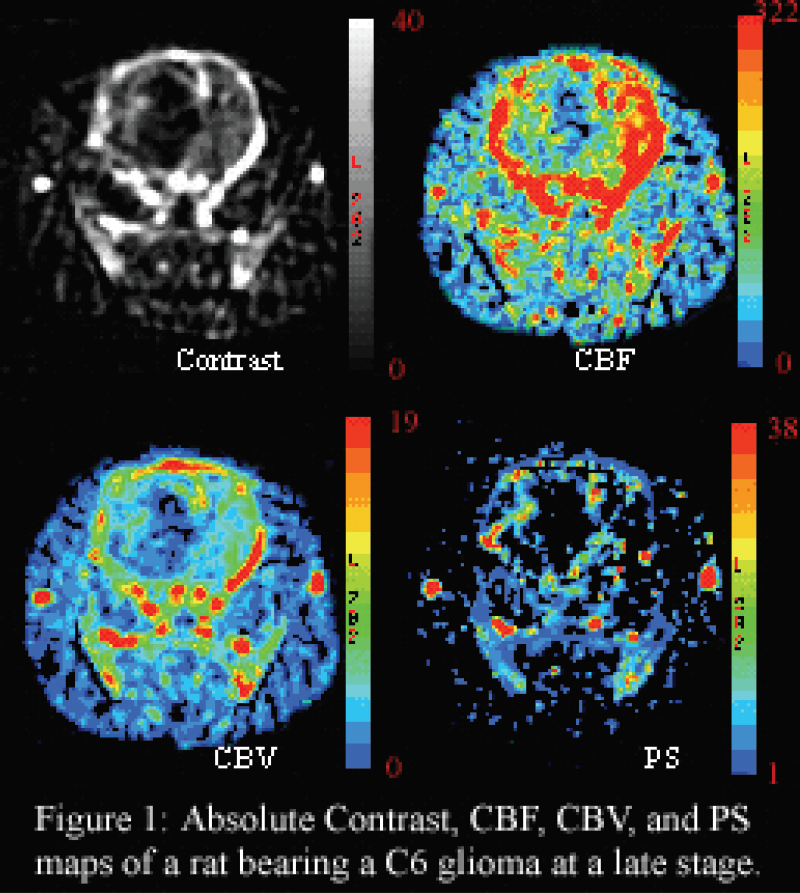
Absolute Contrast, CBF, CBV, and PS maps of a rat bearing a C6 glioma at a late stage.
Absolute brain perfusion values measured in various regions of interest of the rat brain.

Abstract ID: 566 Poster board space: 189
The chick embryo chorioallantoic membrane (CAM) is composed of highly vascularized tissue that develops early in chick development; this model has been used extensively in the study of angiogenesis (Brooks et al. Methods Mol Biol 1999, 129:257-69). Tumor cells can be applied along the chick CAM for rapid tumorigenesis over a period of days, in a living environment with multiple blood vessels suitable for intravascular administration of contrast agents. Ten-day chick embryos were maintained at approximately 37°C and 51% relative humidity. On day 11, eggs were illuminated using a 35W lamp at the air sac of the egg to identify prominent blood vessels. The CAM was separated and dropped. Subsequently, 2 × 10⁶ CCL-138 human pharyngeal squamous carcinoma cells were implanted along the CAM. Eggs were assessed for tumoral growth over a period of 2 days. A window was also generated over previously identified blood vessels suitable for intravascular contrast agent administration. On day 13, eggs were injected with gadopentate dimeglumine or experimental dendrimeric MR contrast agents for preliminary investigation of delivery of these experimental contrast agents. T1-weighted imaging has been consistently performed on multiple eggs on both 1.5T and 7T magnets for evaluation of pre- and post contrast signal intensities, demonstrating the efficacy of MRI in documenting signal enhancement in implanted tumors due to contrast administration. Chick CAM assays allow a cost-effective and rapid means of evaluating novel MR contrast imaging agents and their use in imaging tumors prior to their in-vivo evaluation in higher animal models.

Abstract ID: 567 Poster board space: 190
Joseph Nwankwo, Elizabeth Chen, Ben Durkee, Marc Longino, Sharon Weber,
Abstract ID: 568 Poster board space: 191
David Atkinson, Ann Mladek, Brett Carlson, Mark Schroeder, Dan McConnell, Mark Jacobs, Mark Jacobson, C. David James, Val Lowe,
Mammalian target of rapamycin (mTOR) signaling is important for tumor proliferation in glioblastoma multiforme (GBM), and mTOR inhibitors have promising efficacy in early clinical trials. Because only a minority of patients benefit from mTOR inhibitor therapy, developing non-invasive methods for demonstrating treatment efficacy could be used to select patients for therapy. In this study we evaluated whether FDG-PET could be used to monitor the efficacy of short-term treatment with RAD001, a rapamycin analogue. From a panel of human GBM xenografts, we selected one sensitive and one resistant line for this study. Treatment of intracranial GBM10 xenografts with RAD001 results in a 33% prolongation in median survival (p=0.02), while the same treatment has no significant effect on the survival of GBM12 xenografts. In mice with established GBM10 or GBM12 flank xenografts, FDG-PET imaging was conducted before and after four days of therapy with placebo or 10 mg/kg RAD001. Surprisingly, FDG uptake was suppressed in both lines following therapy: median percent change from pre- to post-treatment in tumor SUV for placebo versus RAD001 groups was +5% versus −23%, respectively, for GBM10 (p=.009) and +9% versus −45%, respectively, for GBM12 (p = 0.001). From these results, we concluded that monitoring changes in tumor FDG uptake following treatment will not be useful in distinguishing between sensitive and resistant tumors. However, we have demonstrated in mice that a 10-fold higher dose of RAD001 is required to inhibit mTOR signaling in intact brain versus peripheral tissues, and the appropriate dosing schedule for complete inhibition of mTOR in patients with brain tumors is unknown. Since tissue sampling of brain tumors before and after treatment is not practical, we hypothesize that suppression of FDG uptake within tumors could be incorporated into Phase I dose-escalation trials to define a dosing schedule for mTOR inhibitors that effectively suppresses mTOR function.
Abstract ID: 569 Poster board space: 192
Additionally, we observed two patients with GBM treated with adjuvant TMZ therapy up to 20 and 27 cycles, respectively. Due to a stable clinical status TMZ was discontinued and reduced, respectively. Three MET-PET follow-up scans in both patients were performed until the change of adjuvant therapy regimen, and two MET-PET scans were performed afterwards. Additionally, to measure proliferative activity of the GBM two [18F]fluoro-
After discontinuation and dose reduction of adjuvant TMZ therapy in two patients with GBM the metabolic activity of the tumor as measured by MET- and FLT-PET increased.
Abstract ID: 570 Poster board space: 193
Carcinoembryonic antigen (CEA) is a surface glycoprotein and a marker for colon cancer. Liver metastases are a common complication in patients with colon cancer. To simulate metastatic disease, CEA-expressing human colorectal LS174T cells, transfected with luciferase-expressing vector, were injected into the livers of athymic mice. Three sets of mice were produced by growing the cells for 5, 12 and 16 days. Optical imaging confirmed the presence of cancer cells in each animal. An anti-CEA antibody fragment, scFv-Fc H310A with optimized serum kinetics, was labeled with 124I and injected into the three sets of mice for serial microPET imaging. All tumors were visible by 24 hours and the signal persisted through 72 hours. The biodistribution showed tumor activity from 1.21% to 3.50% ID/g with tumor-to-blood ratios from 5.76 to 8.33 (average tumor weight −40–196 mg). This study demonstrates the ability to detect CEA-expressing liver metastases using a 124I-labeled antibody fragment at an early disease stage. In a second model, B or T cells were tagged with a recombinant form of CEA for detection by microPET imaging using the 124I-labeled scFv-Fc tracer. For the PET reporter, N-A3 (CEA epitope recognized by tracer) was fused to the FcγRIIb extracellular and transmembrane domains. Jurkat (T) and Daudi (B) cells were transfected with the N-A3-FcγRIIb construct and subcutaneous tumors were established in mice for serial imaging by microPET and microCT. Results confirmed that the 124I-labeled tracer localized at the N-A3-positive tumor with little (19 h) or no (48 h) background. The biodistribution showed 7.35% ID/g in the N-A3-positive tumor, with low activity in the control tumor and normal tissues. These results reveal the CEA PET reporter - PET tracer system feasibility for marking therapeutic T cells. Thus, radiolabeled anti-CEA antibody fragments, such as the scFv-Fc, are useful in variety of models of CEA-expressing cells.
Abstract ID: 571 Poster board space: 194
A commercially available dental microscope was altered to rapidly acquire quasi-monochromatic reflectance, polarized reflectance, and fluorescence images in vivo for the dual purpose of screening for oral neoplasia and real time margin determination during surgery. The system is designed to enhance visual inspection of the oral mucosa at wavelengths previously determined to improve contrast between clinically normal and dysplastic regions. Fluorescence and reflectance images are taken in the 350–650 nm range and wavelengths were chosen both to suit detection of topically applied contrast agents described below and to detect such endogenous fluorophores such as NADH and FAD. A 100 W mercury arc lamp is used as the excitation source which provides adequate illumination power for both reflectance and fluorescence imaging and allows for exposure time of sub-100ms for most fluorescence excitation/emission pairs. Two high-resolution, cooled, color CCD cameras are used simultaneously to acquire images and allow for accurate ratio imaging in polarized reflectance mode eliminating differential movement artifacts that may occur with sequential imaging. The device has a 15 micron spatial resolution at a field of view of 1 square cm.
The device is designed to be used in conjunction with nanoscale metallic contrast agents which bind to molecular targets overexpressed in neoplastic tissue. Monochromatic excitation filters in the device match the peak scattering wavelengths of gold and silver nanospheres and nanorods. The contrast agent is applied topically to oral mucosal excised tissue or in vivo. The nanoscale contrast agents are conjugated either directly or with a PEG linker to anti-EGFR antibodies. Because the scattering and absorption characteristics of nanoparticles are highly dependent on size and shape as well as dimerization and conglomeration effects, modeling efforts have been made using the Finite-Difference Time Domain (FDTD) method to find the most suitable nanoparticle parameters to be used with the device.
Abstract ID: 572 Poster board space: 195
Several strategies that allow conditional expression of oncogenes have been used for the generation of mouse models in which spontaneous tumours develop. One of the most prominent is the CRE/loxP system. In this system, a transcription blockage unit including a polyadenylation signal is flanked by loxP sites and located upstream of the oncogene. Using CRE recombinase, the transcription unit is excised from the genome thus allowing the expression of the oncogene. However, this recombination is irreversible. The widely used tetracycline dependent expression system allows a reversible expression of the gene of interests. Depending on the transactivator that binds the TRE promoter, the presence of tetracycline will either induce or suppress the gene expression. In this study we have investigated the outcome of combining the CRE/loxP and the tetracycline contolled gene expression systems in order to express the SV40 large T antigen linked to luciferase, which enables the monitoring of gene expression using bioluminescence imaging. Using this strategy allowed the regulation of gene expression after CRE-mediated recombination. Transfection of the constructs into primary murine embryonic fibroblasts demonstrated function of the regulatory elements and reversible transformation of illuminated cells.
Abstract ID: 573 Poster board space: 196
Confocal imaging systems have the ability to produce high-resolution images of a 2-dimensional plane within turbid media. A major limitation of most confocal systems is the depth of penetration into tissue of the excitation light required to generate an image. To extend and simplify this approach, we have developed a simple fiber-optic based system that has similar capabilities, but is not limited by penetration depth. We have observed that by placing a tissue in contact with an optically flat window, it is possible to magnify and transmit an in-focus, high-resolution image with the use of a small image guide. This image guide consists of thousands of individual coherent optical fibers and is 400 microns in diameter, small enough to pass through the lumen of an 18-gauge needle. When the distal end of the probe is placed in contact with a biological specimen such that the tissue is essentially flat against its surface, the proximal end of the fiber can be imaged onto a commercial CCD camera, yielding a high-resolution image. This approach allows the fiber bundle to be placed directly into deep tissues, where traditional confocal microscopy would be limited. To reduce the effect of specular reflection from the image guide and highlight cell surface tumor markers, the fiber bundle microscope will be used in conjunction with antibody targeted quantum dot conjugates. These conjugates fluoresce under the illumination of a simple LED and specifically bind to extracellular receptors, such as EGFR, which are overexpressed in tumor cells. Taking advantage of the Stokes shift of the quantum dots, excitation light can be effectively filtered out. This combination of the fiber bundle microscope and quantum dot conjugates would enable clinicians to rapidly determine whether a suspicious lesion is potentially cancerous, reducing any delay before a final diagnosis is made.
Abstract ID: 574 Poster board space: 197
Dynamic Contrast Enhanced MRI (DCEMRI) is gaining popularity for staging, detecting and characterizing prostate tumors in patients who present with elevated PSA, because most groups now agree that some form of DCEMRI adds diagnostic value to the conventional T2-weighted MRI examination. This conclusion is based on the observations that tumors have relatively faster wash-in and wash-out of the low-molecular weight gadolinium based contrast agents than the surrounding normal prostate tissue. There are some abnormal benign regions of the prostate that may sometimes mimic the behavior of tumors leading to false positives on the DCEMRI scans. Solvent suppressed MRS of the prostate has also been employed to improve the diagnostic accuracy of the overall MRI examination. In MRS, elevated choline and reduced citrate are indicative of cancer. We undertook the present study to determine whether the microvascular density (MVD), and/or the expression of angiogenic factors correlated with abnormal DCEMRI findings and whether elevated choline levels correlated with choline kinase or choline transporter expression. We employed both real time PCR maps (see Figure 1) and immunohistochemical staining of whole mount specimens obtained post radical prostatectomy sectioned in the same location as both DCEMRI and MRS.
Our preliminary results indicate that neither the expression of VEGF, nor MVD spatially correlate with patterns seen on DCEMRI. However there appear to be correlations between some novel angiogenic factors. The elevated choline levels do appear to correlate spatially with the expression of choline kinase. This approach to matching immunohistochemical maps with in vivo MRI offers the opportunity for interpreting MRI findings in molecular, rather than morphological terms.

Abstract ID: 575 Poster board space: 198
Cu-ATSM, a hypoxia PET imaging agent, is reduced and retained in hypoxic tissue but remains intact and rapidly diffuses out of normoxic tissue. As a defense mechanism in prostate cancer cells, the fatty acid synthesis (FAS) pathway harnesses its oxidizing power for improving the redox balance despite conditions of extreme hypoxia, potentially altering Cu-ATSM hypoxia selectivity. Researchers have shown that C75 inhibits FAS by 89–95%, and in this study it was administered to observe possible changes in Cu-ATSM uptake due to FAS inhibition.
PC-3, LNCaP, and 22Rv1 prostate tumor cells were plated in 6-well plates and grown to ≈75% confluence at 37°C and 5% CO2. Four hours prior to the study, the cells were supplemented with 50 μM C75. 64Cu-ATSM was added to the cells, and the plates were placed in an anoxic gas mixture (5% CO2 / 95% N2) to facilitate Cu-ATSM uptake. At various times (15, 32, 64, and 83 minutes) the cells were lysed by addition of 0.15% sodium dodecyl sulfate, after washing in triplicate. Lysis extracts were counted in a γ-counter. Uptake data for all experiments were normalized for the amount of protein present and calculated as the percentage uptake (cell-associated).
Uptake of 64Cu-ATSM increased over time in the C75 treated cells when compared to controls, with differences at 83 minutes of 15, 11, and 26% for PC-3, LNCaP, and 22Rv1 cell lines, respectively. This suggests that 64Cu-ATSM may be an ineffective marker of hypoxia in prostate tumor models due to the increased presence of FAS which alters the redox properties of the cell. Further in vitro studies of cells in suspension will enable more uniform hypoxic conditions. These findings will be validated in vivo using small animal PET imaging.
We are grateful for financial support from the DOD (PC040435) and NIH (F32CA110422-1).
Abstract ID: 576 Poster board space: 199
Frozen tissue redox scanning can provide high resolution 3-D images of metabolic states in tissue through the detection of the endogenous fluorophores NADH and FP. The redox ratio, defined as the NADH signal divided by the total fluorescence (Fp+NADH), is a direct indicator of steady-state oxidative metabolism. We have performed redox imaging of frozen tumors grown from LnCaP cells orthotopically in the prostates of scid mice, and are investigating the effect of androgen deprivation (AD) therapy on redox potential. Tumors were allowed to grow to a volume of 100 mm3, at which point animals were castrated, and tumors harvested after 72 hours. The histograms of the redox ratio from the tumors are shown below, with results from both an untreated animal and an animal receiving AD therapy. A total of 3 tumors from control animals have been scanned, along with 4 tumors from treated animals. Two of the three untreated tumors demonstrated high heterogeneity in the redox ratio, with a significant fraction of the tumor demonstrating high oxidative metabolism. This can be seen in the figure labelled ‘untreated’, where a large fraction of the tumor has a redox ratio lower than 0.5. In contrast, all four treated tumors demonstrated much less heterogeneity in the redox ratio, with all tumors demonstrating low levels of oxidative metabolism. We hypothesize that the effect of AD therapy is to suppress an aggressive sub-population of cells that exist in the rapidly growing untreated tumors.

Abstract ID: 577 Poster board space: 200
Understanding the molecular pathogenesis of androgen-independent prostate cancer has become exceedingly important due to the limited treatment options available once the disease has reached this advanced state. It has been proposed that posttranscriptional regulation of androgen receptor (AR) mRNA could be responsible for AR protein accumulation and uncontrolled cell growth in some cases of androgen-independent prostate cancer. HuR, an RNA binding protein that targets AR mRNA, is known to affect the localization, stabilization, and translational efficiency of its mRNA targets, therefore making it a likely candidate to force this androgen-inde-pendent growth mechanism. The visualization of AR mRNA localization and stability changes when HuR activity is impaired could elucidate the importance of this mRNA-protein interaction in promoting androgen-inde-pendent prostate cancer growth. To visualize AR mRNA in live prostate cancer cells, we used a dual molecular beacon approach where two molecular beacons, each targeting a different site on the AR mRNA transcript, were used in concert to enhance signal-to-noise amplification. The specificity of the molecular beacons was validated using LNCaP (AR+) and DU-145 (AR–) prostate cancer cell lines. The ability of the probes to detect changes in mRNA level was determined using siRNA knock-down of AR mRNA in LNCaP cells as well as by transfecting DU-145 cells with a full length AR plasmid. We found that the live-cell imaging data correlated well with our real-time PCR results for cellular AR mRNA levels. HuR protein level was decreased with siRNA treatment and the subsequent effects on AR mRNA localization and expression level were examined via confocal microscopy and real-time RT PCR. Using molecular beacons to image live cell mRNA in conjunction with modulating the function of posttranscriptional regulating proteins provides a powerful tool for elucidating the roles of these proteins, which has potential utility for a wide range of biological and medical applications.
Abstract ID: 578 Poster board space: 201
Angiogenesis, the formation of new blood vessels, is an important process in sustaining tumor growth. Inhibition of angiogenesis could therefore be of considerable therapeutic use. Traditionally, the efficacy of angio-static therapy is evaluated using methods like measuring tumor growth profiles and ex vivo histological determination of the microvessel density (MVD). Non-invasive imaging methods to establish the efficacy of anti-angiogenesis therapies are under development and becoming increasingly important.
In this study we used αvβ3 targeted bimodal RGD-liposomes to quantify angiogenesis in a mouse tumor model with molecular MRI and evaluated the therapeutic efficacy of the angiogenesis inhibitors anginex [1] and endostatin [2]. 30 6-wk old C57BL/6 mice were inoculated with B16F10 melanoma cells subcutaneously on the right flank. Mice were treated either 3 or 14 days with anginex or endostatin using subcutaneously placed osmotic minipumps. The percentage of the tumor area with significant signal enhancement, upon intravenous injection of the contrast agent, was quantified. After the MRI measurements the tumors were dissected and frozen. Microvessel density (MVD), which is as an ex vivo surrogate marker for angiogenic activity, was determined by counting vessels in 5 random tumor areas.
The in vivo MRI data of the 3 days treatment group and 14 days treatment group were compared to the ex vivo determined MVD (Fig. 1). Overall the trend was found that in vivo molecular MRI of tumor angiogenesis reflected closely the treatment effects as deduced from ex vivo MVD determinations.

Abstract ID: 579 Poster board space: 202
Molecular imaging of human disease models with multi-modality imaging holds great potential in pharmaceutical discovery. Our micro-SPECT unit is composed of NaI crystals coupled to nine flat panel multi-anode PMT's. Molecular GRP oncology imaging in pre-clinical models and micro-SPECT lesion quantification properties were assessed in different mediums (air and water) with dedicated phantoms and metastatic disease models. A tissue equivalent micro-hollow sphere phantom with internal diameters ranging from 3.95 to 7.86 mm with volumes ranging from 31 to 250 μL was employed. Tc-99m activity levels ranging from 124 to 968 μCi were placed inside the micro-spheres. The phantom was scanned without and with water to simulate hot micro-lesions. Metastatic models were injected with In-111-DOTA-8-AOC-BBN(7-14)NH2 and Tc-99m agents to assess the extent of SPECT lesion detectability. The phantoms were scanned for 360-degree, 60 projection views at 45 cm radius of rotation for 30 minutes with a 30% energy window. Projection data were reconstructed with an OSEM algorithm and ROI segmentation was performed. Pinhole phantom quantification results indicate a lesion gamma-ray detection of 1.17 cps/μCi and 0.95 cps/μCi for the scans performed without and with water, respectively. SPECT ROI segmentation analysis revealed that in the water phantom, 19% less counts were detected. Micro-SPECT was successful in detecting receptor mediated localization of In-111-DOTA-8-AOC-BBN(7-14)NH2 in prostate bone metastases and HDP uptake in tumor models. Micro-SPECT tibial lesions as small as 1.5 mm were detected. Micro-CT co-registration confirmed the location and lesion extent of the metastatic model analyzed. ROI tumor SPECT measurements were correlated with corresponding data obtained by pharmacokinetic analysis. The phantom data suggest that in the presence of water higher attenuation and absorption of photons are occurring than in air. Furthermore, these data support the fact that attenuation and scatter corrections are required for even small volumes such as the model investigated.
Abstract ID: 580 Poster board space: 203
Met and its ligand hepatocyte growth factor/scatter factor (HGF/SF) mediate tumorigenesis, and angiogenesis. Anthrax toxin (AnTx) inhibits HGF/SF-Met signaling by inhibiting its downstream signaling molecule, mitogen activated protein kinase kinase (MAPKK), resulting in inhibited HGF/SF-induced cell scattering and motility. We have previously shown that HGF/SF-induced Met activation increased tumoral blood volume. The specific increase was used to develop contrast media (CM) ultrasound-based functional molecular imaging (FMI). Here we demonstrate the use of Met FMI on mice bearing mouse mammary tumors for imaging tumor borders and Met-HGF/SF signaling inhibition in vivo.
HGF/SF-induced blood volume alteration maps demonstrated a dramatic increase in blood volume in the center and margins of the tumors. Fine needle biopsies taken from tumor margin areas with increased CM signal contained local metastasis, compared to normal areas with no increase in CM signal intensity.
AnTx inhibited HGF/SF effect on tumoral blood volume in time and dose dependent manners, in vivo. AnTx treated tumors also demonstrated decreased growth rate relative to untreated mice (2.4 and 2.6 folds after 7 and 14 days, respectively, n = 10, p = 0.02). Thus, HGF/SF-induced hemodynamic alteration and tumor growth were dependent on the MAPK signaling.
This unique ultrasound-based Met FMI technique can be utilized to screen tumor borders and invasion and be used for personalized anti Met therapy.
This work was supported in part by the NIH research grant (P50CA93990)
Abstract ID: 581 Poster board space: 204
Liposomes are a promising vehicle for the delivery of therapeutic, diagnostic and analytical agents to the developing vasculature in breast cancer. However, specific targeting remains a critical problem. Using an in vivo phage display approach we have recently identified a novel peptide that binds specifically to breast cancer vasculature. Here we describe our progress in using this peptide to deliver multifunctional liposomes with both therapeutic and analytical cargo. Cationic liposomes are a class of lipid vesicles with demonstrated potential in systemic gene delivery. The incorporation of magnetic resonance imaging contrast agents within these liposomes allows in vivo visualization to assess the dynamics of accumulation at the target. The approach is to load the liposomes with the cargo in question, and subsequently couple the targeting peptide to the surface using maleimide chemistry. Cationic liposomes with the base formulation DOTAP:DOPE:DOPE:MPB (1:0.95:0.05 molar ratio) were prepared by extrusion in the presence of Magnevist. A small amount (0.2 mole %) of Rhodamine B-DOPE was included to allow for high-resolution fluorescence microscopy to confirm localization. Alternately, Gd-DTPA-bis(oley-lamide) (Gd-BOA) was added to the base formulation in place of varying fractions of the DOTAP. Following coupling of the peptide via the DOPE-MBP, the liposomes were purified using size exclusion chromatography. The liposomes were then characterized by transmission electron microscopy (TEM), dynamic light scattering (DLS), and MR spectroscopy. The TEM and DLS revealed a normal liposomal morphology with an average radius of 70–80 nm. MR spectroscopy revealed a T1 relaxation of 200 ms for the Magnevist based liposomes and 600 ms for the optimal Gd-BOA compositions. Targeted liposomes were detected with fluorescence microscopy in MDA-MB-231 tumor sections within 60 minutes of intravenous administration. MRI studies are currently under way to image the targeted liposomes in tumors. This work was supported by DOD COE W82X-04-1-0595.
Abstract ID: 582 Poster board space: 205
Hypoxia is known to influence a series of biologic parameters that also affect the malignant potential of a neoplasm. Because of hypoxia intrinsic resistance to radiation and chemotherapy may be enhanced. Vasculature is an essential component of tumor microenvironment and a key component of tumor growth, invasion, and metastasis. The main aim of this work was to concurrently image tumor hypoxia and tumor vasculature during the tumor development and therapy.
MicroPET and contrast-enhanced (CE) microCT were used to monitor tumor hypoxia and tumor vasculature, respectively. 64Cu-diacetyl-bis(N4-methylthiosemicarbazone) (64Cu-ATSM) was used as a PET imaging surrogate of tissue hypoxia. A polyiodinated triglyceride lipid emulsion with high retention in the vasculature was used as a contrast agent for CT imaging. A Siemens/CTI microPET R4 scanner was used for PET imaging and a GE eXplore Locus microCT scanner for CT imaging. PET and CT images were coregistered using AMIRA software. A transgenic adenocarcinoma of the mouse prostate (TRAMP) model and a squamous cell carcinoma (SCC-1) xenograft model were investigated. The tumors were followed weekly during their growth and after therapy.
Vasculature delineation was dependent on the amount of injected contrast as well as CT image quality. PET imaging with CuATSM was stable for imaging points 3 hours post injection. Both of the investigated tumor types showed high heterogeneity of tumor hypoxia. The heterogeneity corresponded to the imaged vasculature, but not exclusively. Inverse correlation between large vasculature and hypoxia was observed, even though it was not perfect. A significant change in tumor hypoxia during the growth as well as therapy was observed.
Feasibility of concurrent vasculature and hypoxia imaging was demonstrated. Inter-relation between the two was found. Significant changes during the tumor growth and after the therapy were observed.
Abstract ID: 583 Poster board space: 205
Abstract ID: 584 Poster board space: 206
Activation of the apoptotic cascade plays an important role in the response of tumors to therapy. The ability to non-invasively image apoptosis would facilitate studies on the optimization of therapeutic protocols regarding dosing, schedule, and identification of efficacious combination therapies. Previously, a reporter construct was developed that fused estrogen receptor (ER) regulatory domains with luciferase to quench luciferase activity (ER-DEVD-Luc-DEVD-ER) [1]. When apoptosis occurs, caspase 3 cleaves the reporter construct, allowing ER domain dissociation and luciferase activation. Although this luciferase reporter has greatly improved apoptosis detection, its sensitivity is limited by high background activity despite silencing efforts. To enhance the signal to noise of this apoptosis reporter, we have adapted the split firefly reporter strategy [2] and fused N-Luc and C-Luc to strongly interacting peptides, Pep A and Pep B, respectively[3]. Construction of a polypeptide wherein PepA-NLuc and PepB-CLuc are in a head to head position with an intervening caspase 3 cleavage site significantly reduces bioluminescence activity. After caspase dependent cleavage, significant restoration of bioluminescence occurs. We have created stable glioblastoma cell line with this reporter and have imaged caspase 3 activation using chemotherapeutic drugs (Temozolomide and Perifosine) both in vitro and in vivo. We have observed a 6–18 fold increase in luciferase activity over pretreatment in vivo. These studies demonstrate that the reporter has an improved signal to noise compared to ER-Luc-ER and will significantly enhance studies in the evaluation of experimental therapeutics.

Abstract ID: 585 Poster board space: 207
Due to the excitation of localized plasmons, gold nanoparticles demonstrate extremely high absorption and scattering in visible/NIR depending on their size, shape and concentration. Our project is focused on the employment of gold nanoparticles as optical contrast agents in spectrally resolved endoscopy based early cancer detection tasks either by i) exploiting differences in extravasation between normal and neoplastic tissue or ii) conjugating the particles with molecular biomarkers specific to disease. As a first step, the absorption and scattering properties of gold nanostructures of various sizes and geometries (spheres and core-shells) were measured in biological tissue. In particular, we examined spectral characteristics of the diffuse reflection of visible light from the ex vivo pig's colon using a dark field contrast before and after the injection of the gold nanoparticles of various sizes (3-100 nm) into the connective tissue of the colon wall. In our experiments, the presence of small gold nanoparticles (3-30 nm) within the tissue was detected due to strong absorption of light near the peak of the plasmon excitation, whereas the presence of larger nanoparticles affected scattering rather than absorption of diffuse reflected light. These preliminary data demonstrated that the absorption and scattering properties of plasmonics nanostructures can potentially provide a necessary cancerous/non-cancerous contrast in detecting tumor/dysplasia. Compared to the other inorganic nanomaterials, the biocompatibility of gold nanoparticles offers a unique advantage for future clinical applications.
Abstract ID: 586 Poster board space: 208
Abstract ID: 587 Poster board space: 209
Effects of VEGF on Intra-tumor Physiological Heterogeneity and Anti-Angiogenic Therapy as Measured by Dynamic Contrast Enhanced CT.
Abstract ID: 588 Poster board space: 210
Fluorescent imaging technology has been developed for life science research and mainly used in in vitro study. This technology has many advantages of being able to label tissues readily by fluorescent protein or fluorescent reagent, detect multiple tissues simultaneously by different wavelength probes and get high resolution images. It also allows us to visualize cells with single-cell resolution in vivo. Fluorescent proteins can be used to visualize any type of cancer process including primary tumor growth and angiogenesis. In this study, we tried to trace, in real time, tumor growth and angiogenesis in living animals using fluorescent imaging technology. HT1080 human fibrosarcoma cells were transfected with a green fluorescent protein (GFP) gene and implanted into nude mice mammary gland subcutaneously. Blood vessels were labeled with a near infrared fluorescent (NIR) probe which is useful for in vivo imaging by reason of lower auto fluorescence and deeper penetration in vivo, compared to short wavelength probes. Using Olympus in vivo fluorescent imaging system OV100 and IV100, the interaction of the green tumor cells and red blood vessels can be visualized in living animals. Four days after implantation, we could detect the fluorescent probe signal in tumor region and trace angiogenesis from early stage. Micro blood vessel and single cell in tumor was also observed with high resolution image. This technique allows us to visualize, in real time, important aspects of cancer in living animals including primary tumor growth and angiogenesis.
Abstract ID: 589 Poster board space: 211
Abstract ID: 590 Poster board space: 212
Most tumors contain hypoxic regions. Hypoxia activates several hypoxia-inducible genes associated with tumor proliferation and metastasis. Hypoxia inducible factor (HIF)-1α is stabilized under hypoxia and binds to hypoxia response elements (HREs) in these hypoxia-inducible genes. Combined optical and magnetic resonance spectroscopic (MRS) imaging studies revealed a coarse colocalization between hypoxic regions and regions of elevated total choline (tCho) in human PC-3 prostate cancer xenografts stably expressing green fluorescent protein (GFP) under the control of an HRE (PC-3-HRE-GFP). These in vivo findings were confirmed in PC3-HRE-GFP prostate cancer cells in culture. Phosphocholine (PC) and tCho levels significantly increased following 24 hours of hypoxia in wild-type PC-3 and PC-3-HRE-GFP cells (Fig. 1a). This was accompanied by increased choline kinase expression (Fig. 1b). These data indicate that hypoxia drives up choline kinase expression and subsequent PC generation. To further test if choline kinase expression may be driven by HIF-1α-mediated hypoxia response, we cloned the full-length human choline kinase promoter and six different deletion promoter constructs in a luciferase reporter vector. The deletion promoter reporter construct pGL4 (−1068 to −338) containing three putative HREs exhibited a twofold induction following 24 hours of hypoxia in both wild-type PC-3 and PC-3 HRE-GFP cells (Fig. 1c). Our results suggest that the choline kinase promoter contains HREs that bind stabilized HIF-1α under hypoxic conditions, inducing choline kinase expression with an increase in PC and tCho levels as an adaptive response to hypoxia. These findings may also explain why tumor xenografts and clinical prostate tumors exhibit a distinctly heterogeneous distribution of tCho. This work was supported by NIH P50 CA103175.

(a) Representative ‘H spectra from normoxic and hypoxic PC3-HRE-GFP prostate cancer cells and the corresponding (b) western blots scored for choline kinase expression. (b) Twofold induction of pGL4 (−1068 to −338) was detected following 24 h hypoxic treatment in pC3 wild-type and PC-3-HRE-GFP prostate cancer cells (n=2).
Abstract ID: 591 Poster board space: 213
Somatic mutations in the epidermal growth factor receptor (EGFR) have been identified in NSCLC patients and are both prognostic for outcome and predictive for sensitivity to the tyrosine kinase domain inhibitor erlotinib (OSI-774, Tarceva). To evaluate the influence of EGFR mutations on erlotinib sensitivity we have generated three stable NR6 cell lines expressing equivalent levels of EGFR, or the clinically detected mutants EGFR-L858R or EGFR-Del(746-752). NR6 cells do not express EGFR or detectable levels of ErbB2, ErbB3, or ErbB4. Both mutations facilitate ligand-dependent anchorage-independent growth and are more sensitive to erlotinib than wild-type EGFR both in vivo and in vitro. In this study, we have used positron emission tomography (PET) of 2-fluoro-2-deoxy-
Abstract ID: 592 Poster board space: 214
Lysosomes play a major role in cancer invasion and metastasis by mediating protease routing, regulation, and secretion. They may therefore be an ideal molecular imaging target for assessing the metastatic potential of a given tumor. We are currently developing and characterizing optical probes such as 6′-O-lissamine-rhodamineB-glucosamine for noninvasive imaging of lysosomes [1]. In this proof of principle study, we observed significant differences in the overall lysosomal parameters between highly metastatic human breast tumors (MDA-MB-231) and poorly metastatic human breast tumors (MCF-7) inoculated in the mammary fat pad of severe combined immune suppressed mice. Tumors were frozen, sectioned at 15-μm thickness, fixed, and immunofluorescence staining of lysosomes was performed using anti-CD63 antibody, followed by confocal laser-scanning fluorescence microscopy. The mean pixel intensity ratios of lysosome fluorescence from the anti-CD63 antibody (red), divided by nuclei fluorescence (blue) was analyzed for several slices throughout the tumor from three independent experiments for each tumor model (n=20 for MCF-7, n=27 for MDA-MB-231). This mean pixel intensity ratio significantly increased in MDA-MB-231 tumors compared to MCF-7 tumors, demonstrating increased lysosomal burden in the more metastatic tumor model (see Fig). In addition, a less clustered lysosome pattern was observed in MDA-MB-231 tumors compared to MCF-7 tumors (Fig). The increase in lysosomal burden in the more metastatic tumor model may result in an increased capacity of cells within these tumors to degrade extracellular matrix components, thereby potentially facilitating invasion and metastasis. Noninvasive imaging probes to determine the lysosomal burden of a tumor may be useful for evaluating the ‘metastatic threat’ of a tumor. This work supported by NIH R21 CA112216 (awarded to K.G.).

Immunofluorescence staining of lysosomes (red) and nuclei counterstain (blue) in a representative (a) MDA-MB-231 tumor, and (b) MCF-7 tumor. Field of view in both images is 115 μm × 115 μm. (c) Mean pixel intensity ratio red/blue of MCF-7 (n=20 images) and MDA-MB-231 (n=27 images) tumors was significantly increased in MDA-MB-231 tumors, reflecting a higher lysosomal burden in MDA-MB-231 tumors.
Abstract ID: 593 Poster board space: 215
Abstract ID: 595 Poster board space: 217
In the present study, we report tumor perfusion and permeability using gadolinium based DCE-MRI and post-processing compartmental pharmacokinetic modeling. Human MCF-7 breast cancer cells (5times106 cells per mouse) were injected subcutaneously into the rear flank of ten female balb/c athymic nude mice. Tumor growth was assessed by proton density weighted MRI for three weeks. Gadolinium (gadodiamide) based DCE-MRI was performed on each animal to assess gadolinium uptake into the tumor tissue. Four axial T1-weighted images were taken with the time resolution of 15 seconds for total MRI time of 15 minutes. After 1 minute of the T1-weighted baseline scans, 0.1 mM Omniscan injection was injected through a tail vein catheter. T1-signal intensities were calculated for the tumor and adjunct muscle tissue from each image. A compartmental pharmacokinetic model in which the tissue clearance of gadolinium was related to its transcapillary exchange in tumor and adjacent muscle tissue was constructed, based on T1-signal intensities. Gadolinium transfer rate constants (tissue perfusion) were 0.37÷0.99 for the tumor and 0.10÷0.39 for the muscle tissue. Capillary surface factor (tissue permeability) was 10.12÷58.67 in the tumor and 1.25±15.05 in the muscle tissue. Gadolinium clearance into the middle and periphery of the tumors was 3.27+1.62 and 3.82+2.09 times greater than into adjacent muscle, respectively. Estimates from the transcapillary exchange analysis indicated that blood flow to the middle and periphery of the tumors were 2.12+1.29 and 2.80+1.61 times greater than blood flow to muscle, respectively. Our first DCE-MRI results, coupled with compartmental pharmacokinetic modeling, provide the quantitative evidence for increased tumor perfusion and tumor permeability when compared with adjunct muscle tissues. This technology can be successfully used in the future animal and clinical studies to assess anti-angiogenic properties of novel targeted therapies.
Abstract ID: 597 Poster board space: 219
Shan-Yun Cheng, Ya-Fang Chang, Ren-Shen Liu,
Abstract ID: 598 Poster board space: 220
Wei-tien Tai, Ya-Fang Chang, Ren-Shen Liu,
Recently, the progress in the development of multimodality molecular imaging techniques has made real-time monitoring of transgenic expression possible, opening a vista of potentially important in vivo animal models for the study of prostate diseases. Here we demonstrate the tumor progression and metastases in a human-prostate carcinoma (LNCaP) bearing mouse model, which constitutively expresses herpes simplex virus type-1 thymidine kinase (HSV1-tk) and luciferase (luc) genes. LNCaP-tk/luc human prostate carcinoma xenografts provide tumor growth kinetics, metastatic spreading, and permit in vivo detection over times using bioluminescent imaging (BLI).
LNCaP-tk/luc human prostate cancer cells were implanted subcutaneously into SCID mice and were monitored for the tumor progression. Both primary tumors and micrometastases were detected by BLI in vivo. Ex vivo bioluminescent imaging of excised lung lobes, bones and prostate further confirmed the in vivo signals and indicated a slightly higher frequency of metastasis in some mice. Intensity levels of bioluminescence from in vivo and ex vivo images were well correlated to the frequency and size of metastatic lesions in lungs and bones, and were subsequently confirmed by histopathology.
The longitudinal evaluation of bioluminescent tumors and metastatic development within the same cohorts of animals permitted sensitive and quantitative assessment of both primary and metastatic prostate tumor kinetics in vivo. These results demonstrate the potential application of a non-invasive optical imaging modality in preclinical diagnostic and therapeutic strategies to manage prostate cancer. This study was supported by a grant NSC95-3112-B010-004 from the National Science Council, Taipei, Taiwan.
Abstract ID: 599 Poster board space: 221
Absract ID: 600 Poster board space: 222
Abstract ID: 601 Poster board space: 223
Abstract ID: 602 Poster board space: 224
For higher resolution image in MR, macromolecular MRI contrast agents have been actively studied as tumor-selective contrast agents. However, their application has been largely limited mainly due to their failure in selective enhancement of images at solid tumor sites.
In the field of anti-cancer drug delivery to solid tumors, polymeric micelles emerged as a novel carrier system. The polymeric micelle carrier system showed highly tumor-selective delivery by a passive targeting mechanism (the ERP effect) with greatly enhanced in vivo anti-tumor activity.
We have studied a tumor-specific MRI contrast agent with polymeric micelles that can change signal intensity in MRI. By the use of polymeric micelles as a carrier, Gd ions can be selectively delivered to solid tumor sites. In addition to this targeting effect, the polymeric micelle system is considered to possess another advantage for high contrast at the tumor tissues. In the micelle form in the blood stream, the Gd ions incorporated in the inner cores of the micelles show lower relaxivity because of their separation from the outer aqueous environment. After dissociation of the micelles at the tumor tissues of the Gd ions can easily access the outer aqueous environment, and consequently show higher relaxivity. In this paper, we report the molecular design of the polymeric micelle MRI contrast agents.
We have synthesized block copolymers constituted of PEG and P(Asp) chains with different chain length. Micelles were formed by complexation between polyanionic block copolymers and polycations. The Gd encapsulating block copolymer showed 4 to 5 folds higher relaxivity than the micelles. The effect of polymer length and composition for relaxivity will be discussed.

Abstract ID: 603 Poster board space: 225
Abstract ID: 604 Poster board space: 226
At present there is considerable interest in the in vivo fluorescence and bioluminescence imaging of oncogenic processes in small animals. In vivo fluorescence and bioluminescence imaging offers the ability to characterize whole-body tumor burden, detect small isolated metastases, visualize metastases spontaneously generated from primary tumors, and follow therapeutic treatments non-invasively via longitudinal study. Yet despite such attention and potential, in vivo imaging of fluorescence and bioluminescence in small animals remains at best a qualitative endeavor; unable to determine in situ labeled-cell numbers and locations in any but the most superficial (e.g., subQ) cases. The most sensitive in vivo imaging systems to date can only detect ≈103-104 bioluminescent cells or ≈106-107 fluorescent cells that have been placed subQ. This handicap arises largely from the inherently high degree of inhomogeneous scattering and absorption of light by mammalian tissue and the deleterious effects of skin autofluorescence.
To improve fluorescence/bioluminescence imaging sensitivity and quantization, we evaluated in vitro the optical and cell adhesion properties of a variety of transparent flexible plastics, for use as surgically-placed windows. Such flexible transparent windows permit visualization of much larger areas than afforded by glass windows, with fewer impediments to animal locomotion. Materials demonstrating the lowest cell adhesion and highest light transmission were tested in vivo by their surgical placement in the abdomen of nu/nu mice bearing fluorophore/lumiphore-labeled SKOV3ip.1 ovarian cancer micro-metastases. Following post-op convalescence, mice were periodically imaged over 3 weeks using a Xenogen IVIS 200 imaging system and Leica MZ16F microscope. Figure attached shows (a) surgical and (b) through-window views of individual, fluorescent cancer cells on the omentum that were not detectable through intact skin.

Abstract ID: 605 Poster board space: 227
A non-invasive technique to identify and quantify metastatic tumor burden would be a valuable clinical tool and would benefit both preclinical and clinical oncology drug development. Although viable tumor tissue exhibits a low apparent diffusion coefficient (ADC), it cannot be distinguished from other low diffusion tissues by ADC alone. In this study, whole-body MRI diffusion-weighted imaging (DWI), combined with multispectral analysis, was evaluated as a method of detecting metastases in a clinically relevant murine model of metastatic breast cancer (4T1 cell line).
4T1 cells were injected into the mammary fat pad of 20 BalbC mice. Whole-body MRI was performed six weeks post cell implantation. ADC maps and T2 and proton density (M0) maps were constructed from DWI and T2-weighted images, respectively. K-means clustering of ADC, T2, and M0 was employed to classify tissue populations hierarchically in three steps: All tissue was separated from background based on M0, and then classified to low and high diffusion tissue based on ADC. Finally, low diffusion tissue was classified into four classes based on ADC, T2, and M0. Brain and spinal cord were removed from the viable tumor tissue class by morphological operations.
The viable tumor tissue in primary tumors and lymph node metastases were easily identified from the tissue class maps (Figure 1). To our knowledge, this is the first reported attempt to apply whole-body DWI with multispectral analysis as a means to identify metastases. This study has shown that this technique can be employed preclinically to assess metastatic tumor burden in a murine model of breast cancer.

Axial and coronal slices of a mouse with a lymph node metastasis.
Abstract ID: 606 Poster board space: 228
Abstract ID: 607 Poster board space: 229
Dynamic contrast enhanced (DCE) MRI using macromolecular or nanoparticle-based media has been previously used to assess vascular permeability in animal models of cancer. Vascular permeability is of interest because it reports upon the angiogenic status of tumors and thus it can potentially serve as a biomarker in preclinical studies of antiangiogenic drugs. The apparent permeability that is measured by MR depends not only on the vessel permeability to the contrast agent but also on variable factors such as perfusion and blood pressure, which is the driving force for convective mass transport of the contrast agent across the vessel wall into the tumor interstitium. In this study, we correlate the physiologic effects of common anesthetics, including inhaled isoflurane and injected ketamine/xylazine, ketamine/diazepam and alpha-chloralose, with the apparent permeability assessed by macromolecular DCE MRI in a rat tumor model. We found that ketamine/diazepam, but not the more commonly used anesthetics isoflurane and ketamine/xylazine, maintained near-normal respiratory, cardiovascular, and thermoregulatory function. Furthermore, uniquely with the ketamine/diazepam anesthetic we observed dynamic tumor uptake of the macromolecular contrast agent that was comparable to unanesthetized animals. In contrast, isoflurane and ketamine/xylazine yielded distinctly reduced measures of apparent permeability in the same tumor model. Dynamic MR studies of animal tumor models using macromolecular media can be extremely useful for assessing the efficacy of new drugs or for the development of new contrast agents themselves, however we conclude that the physiologic effect of anesthesia on the permeability measure should not be overlooked in implementing these studies.
Abstract ID: 610 Poster board space: 73
To develop more sensitive and selective radiotracers for PET imaging reporter gene HSV1-tk. 2-amino-6- [18F] fuoro-9-(4-hydroxy-3-hydroxymethylbutyl) purine (6-[18F]Fluoropenciclovir) is potential PET probe for HSV1-tk imaging. The reference compound 6-Fluoropenciclovir was synthesis as shown in scheme 1. 2-Amino-6-chloro-9- (4-acetyl-3-acetyl-methylbutyl) purine
Abstract ID: 611 Poster board space: 74
Qi-Huang Zheng, Bruce Mock, Toshihiko Hara, Mingzhang Gao, Shuyan Wang, Aditya Bansal, Rachid Nazih,
The choline transporter represents a novel potential target for oncologic PET probes. The goal of this work was to develop 11C- and 18F-labeled probes based on the well-known high-affinity choline transport inhibitor, hemicholinium-3 (HC-3). The bis-tertiary amine precursor 4,4′-bis-(1-methyl-3-hydroxy-morpholinyl-(3))-biphenyl was synthesized from 4,4′-bis-bromoacetyl-biphenyl and 2-(methylamino)ethanol. The precursor was labeled by [11C]methyl triflate in acetonitrile through the primary N-[11C]methylation and trapped on a cation-exchange CM Sep-Pak cartridge to release the non-reacted bis-tertiary amine precursor with ethanol and to retain the pure 11C-methylated single-side quaternary amine intermediate on the same CM Sep-Pak. The labeled intermediate underwent the secondary 12C-methylation by addition of methyl iodide in ethanol to the same cartridge. After 2 min, unreacted methyl iodide was removed from the cartridge by rinsing with ethanol. The final bis-quaternary amine carbon-11 labeled product [11C]HC-3 was then eluted from the cartridge with saline. The synthesis was performed in an automated multi-purpose 11C-radiosynthesis module, allowing measurement of specific activity during synthesis. The radiochemical yields were 50–60% based on 11CO2 decay corrected to end of bombardment (EOB), and specific activity was in a range of 4.0–6.0 Ci/μmol at EOB. Using similar methodology, 18F-labeled target tracer [18F]HC-3 was prepared by N-[18F]fluoromethylation of the precursor using [18F]fluoromethyl triflate followed by N-methylation using methyl iodide and purified by the CM Sep-Pak method in an automated multi-purpose 18F-radiosynthesis (FBM) module with 5–10% radiochemical yields. Preliminary biodistribution studies in a subcutaneous 9L-glioma rat model showed highest uptake in kidneys and liver. Tumor uptake was moderate for both 11C- and 18F-labeled HC-3 probes (0.46–0.95 % dose/g at 5–30 min post-injection). Tumor:muscle ratios of 3.0–5.1 were significantly higher than for positron-emitter labeled choline analogs. The data encourage further evaluation of the new HC-3 analogs as PET tracers for the choline transporter.
Abstract ID: 612 Poster board space: 75
In vitro investigations have suggested that the nicotinic acetylcholine receptor (nAChR) α7 subtype is implicated in Alzheimer's disease, schizophrenia and others. However, there is no suitable imaging agent for α7 nAChR for in vivo use at present. We developed a new ligand for α7 nAChR, 2-amino-5-bromo-benzoic acid 1-aza-bicyclo[2.2.2]oct-3-yl ester (Br-QAA). The iodine-125 labeled derivative, 2-amino-5-[125I]iodo-ben-zoic acid 1-aza-bicyclo[2.2.2]oct-3-yl ester ([125I]I-QAA), was synthesized and its potential as an imaging agent for α7 nAChR in brain was evaluated. In vitro binding affinity of Br-QAA was measured in rat brain homogenates using [125I]α-bungarotoxin and the resulted Ki value was 0.50 μM. [125I]I-QAA was successfully obtained by Br-I exchange reaction from Br-QAA with [125I]NaI and radiochemical yield was 83%. The uptake in the mouse brain was high and the radioactivity was gradually decreased (5.59 and 0.67 %ID/g at 5 and 60 min, respectively). The hippocampus to cerebellum uptake ratio was increased with time and reached to plateau at 60 min post-injection (2.04 at 60 min). Since α7 nAChR density is high in the hippocampus and low in the cerebellum, this observation should indicate the receptor-specific binding of [125I]I-QAA in vivo. However, in competitive drug treatment studies, pretreatment with neither non-radioactive I-QAA nor MLA demonstrated a significant decrease in the accumulation of radioactivity. This implies that [125I]I-QAA accumulation in the brain includes a high non-specific binding in vivo and α7 nAChR imaging with [125I]I-QAA is difficult at the present time. Further structural optimization studies are underway to reduce non-specific binding fraction.
Abstract ID: 613 Poster board space: 76

Abstract ID: 614 Poster board space: 77
Fluorescent antisense DNA oligomers are being used in in vitro assays for bacterial identification. We hypothesize that antisense oligomer directed against bacterial ribosomal RNA may be used as imaging agents and would be specific for bacterial infections. Furthermore, different antisense oligomers could eventually be used to differentiate among bacterial strains. In this proof-of-concept study, two labeled phosphorodiamidate morpholino (MORF) were studied, one against the universal bacterial ribosomal sequence Eub338 (5'GCTGCCTCCCGT) and a partial sense control (5'AGGGCATCCTCA). Each MORF was radiolabeled with 99mTc via NHS-MAG3 and incubated in bacterial cultures. Serial dilutions (1 nM to 1 μM) of the labeled anti-Eub338 and control MORF were incubated at 37°C with fixed concentrations of four gram negative (G–) and two gram positive (G+) bacteria strains. At 1 h, accumulations in all bacteria at all concentrations were nonspecific since accumulations were similar for both MORFs. However, at 4 h, accumulations were significantly higher for the anti-Eub338 over the control MORF in Staphylococcus (G+), Enterococcus (G+) and Pseudomonas (G–). For Staphylococcus and Enterococcus this difference was significant only to 10 nM while for Pseudomonas significance extended up to 1 μM but was most pronounced at the lowest concentration. Thereafter both MORFs were incubated at a fixed 2 nM concentration over 6 serial dilutions of in Enterococcus and Pseudomonas bacteria. The highest percent accumulations occurred at the lower cell densities for both MORFs, but accumulation of the 99mTc-anti-Eub338 MORF was 2-fold greater than the control for both bacteria. These results show that MORF oligomers crossed both the cell wall and membrane of these bacteria and suggest that specific binding to ribosomal RNA occurred and became evident at 4 h. We conclude that radiolabeled antisense MORFs may have potential as infection and possible bacterial specific imaging agents.
Abstract ID: 615 Poster board space: 78
Abstract ID: 616 Poster board space: 79
Norman Koglin1, Martina Anton2, Andrea Hauser1, Maria F. Montoya-Bravo1, Katja Ahrens2, Dieter Saur3, Hana Algul3, Luciana Marinelli4, Oliver Demmer5, Burkhardt Laufer5, Horst Kessler5, Roland M. Schmid3, Bernd Gansbacher2, Markus Schwaiger1,
A key role in metastasis and organ specific homing of tumor cells is attributed to the chemokine receptor CXCR4 and its endogenous ligand SDF-1a. For targeting of CXCR4 expression in vivo, we recently developed a radiolabeled cyclic peptide, 124I-CPCR4. This PET imaging probe was the first that showed high affinity binding to CXCR4 (KD=0.4nM), high accumulation in a CXCR4 tumor model (5.5%ID/g,1h p.i.), and thus allows a clear delineation of CXCR4 positive tumors in vivo.
To correlate tumor development with receptor expression and to monitor potential therapeutic interventions using the non-radiolabeled probe by multimodal imaging, tumor cells were transduced for CXCR4 and luciferase (luc) expression. Therefore lentiviral vectors were constructed either with CXCR4 and luc genes or only luc or eGFP as controls and were successfully used for stable transduction of murine CMS5 fibrosarcoma cells. Transduced CMS5/CXCR4/luc cells were investigated in FACS studies and radioligand binding assays and showed surface expression of CXCR4 and high affinity and specificity for CPCR4-binding. Functional expression of luc was ascertained in cell assays.
For in vivo studies transduced cells were injected subcutaneously into nude mice. Tumor bearing mice were analyzed with μ-PET using radiolabeled CPCR4 and optical imaging. Ex vivo tumor analysis included autoradiography, bioluminescence measurements and immunohistochemistry. For a better understanding of CPCR4-binding and to design ligands with improved pharmacokinetics, a newly proposed CXCR4 receptor model has been developed and is currently validated by investigating CXCR4 receptor mutants. Furthermore structure-activity relationship studies of CPCR4-derivatives are performed for tracer optimization and investigation of other labeling options.
In conclusion, this approach allows in vivo imaging of CXCR4 expression and provides valuable tools for the development of enhanced imaging probes for non-invasive investigation of the metastatic potential of tumors and determination of CXCR4 expression for individualized therapy.
Abstract ID: 617 Poster board space: 80
Bacteriophage (phage) display is a valuable combinatorial tool for the selection of peptides that bind with high affinity to a desired target. However, the potentially large number of resulting phage and corresponding peptides can be difficult and time consuming to validate and characterize. We propose the use of multivalent, bifunctional phage for quick and efficient in vivo screening of peptides and their tumor targeting propensity in mouse models of cancer. Phage displaying multiple PC-3 prostate carcinoma tumor homing peptides and multiple copies of an alpha-melanocyte stimulating hormone (α-MSH) peptide analog were employed to image prostate and melanoma tumors, respectively. Phage were modified by covalent addition of biotin and shown to retain avidity for the appropriate cancer cell line. Indium-111 radiolabeled DTPA-conjugated streptavidin was prepared for biodistribution and imaging studies. Biodistribution studies indicated that the biotinylated phage extravasated into the implanted tumor. Tumor uptake (for both the prostate and melanoma tumor mouse models) was on average 2.3, 0.5 and 1.0%ID/g at 0.5, 6, and 24 hours post injection of 111In-streptavidin. Radioactivity in the melanoma tumor was selectively reduced by greater than 50% via competition with a non-radiolabeled α-MSH peptide analog, demonstrating specific targeting. Radioactivity was primarily excreted through urinary and hepatobiliary systems. B16-F1 melanoma bearing mice were imaged using a two-step MSH-phage pretar-get approach with 111In-streptavidin. SPECT/CT image analysis showed that the intravenously injected biotinylated MSH phage were retained within the melanoma tumor four hours post injection of 111In-labeled streptavidin. Successful imaging of solid tumors demonstrated two-step targeting with bifunctional phage as a promising strategy for peptide validation and tumor visualization.

Abstract ID: 618 Poster board space: 81
Xiaoxia Wen, Zhi Yang, Wei Wang, Nathaniel Albert, Suren Soghomonyan, Sherita Daniel, Juri Gelovani, Dimitrios Kontoyiannis,
Invasive aspergillosis is the most common opportunistic mycosis in immunosuppressed patients with leukemia and transplant recipients and a frequent cause of morbidity and mortality in this patient population. Early detection along with early institution of antifungal therapy to the site of infection when the tissue fungal burden is relatively low is of critical importance in improving the poor outcome of invasive aspergillosis. For the diagnosis of invasive pulmonary aspergillosis, chest X-ray and high-resolution chest computed tomography (CT) are often performed to obtain anatomic information. However, early stages of pulmonary infection are difficult to diagnose by these methods. Nuclear imaging techniques, particularly positron emission tomography (PET), offer additional values in that they provide physiologic and molecular information that are not accessible with CT. We have tagged a cyclic peptide c(CGGRLGPFC) with either a gamma emitter (111In) or a positron emitter (68Ga) for noninvasive imaging of invasive aspergillosis in the lung. The peptide was previously identified to target to Aspergillus fumigatus hyphae using phage display technology [1]. Significantly higher uptake (8-fold) at 24 hr after intravenous injection of 111In-DTPA-c(CGGRLGPFC) was seen in the lungs of mice infected with Aspergillus than that of uninfected mice. Increased radioactivity in the lung of infected mice was clearly visualized at 5 min and 2 hr after radiotracer injection. Moreover, μPET images were acquired at 30 and 90 min after intravenous injection of 68Ga-DOTA-c(CGGRLGPFC). Uptake of the radiotracer in infected lung, but not in the lungs of control mice, was clearly visualized. These results indicate that radiolabeled cyclic peptides targeted to Aspergillus hyphae may have the potential for the detection of invasive aspergillosis. Studies addressing the comparative sensitivity of this approach with computed tomography and its specificity with other pathogenic molds are planned. Supported by John S. Dunn Foundation.
Abstract ID: 619 Poster board space: 82
PEPT2 is a transporter found predominantly in the kidney, but is also present at the brain choroid plexus epithelial cells within the cerebral ventricles. This transporter facilitates the transmembrane movement of small di- and tripeptides from ventricle to blood. We have recently developed [11C]GlySar (glycylsarcosine) as an in vivo imaging agent for PEPT2[1]. In this study, the brain and kidney pharmacokinetics following ICV injection of [11C]GlySar into mice were followed using microPET imaging. Wild type and PEPT2 knockout mice were anesthetized (isoflurane) and [11C]GlySar (5-30 microcuries, 1 microliter) injected into a lateral ventricle. Animals were placed into microPET scanners (either Concorde R4 or P4 microPET) and the entire animal imaged for one hour. At one hour, [15O]water (to localize entire brain) or [11C]GlySar (to localize kidneys) was injected via the lateral tail vein, and imaging continued for 5–30 minutes. In wild-type mice, loss of radioactivity from the whole brain, movement down the spinal column, and continual accumulation of radioactivity in the kidney were observed during the first 60 minutes. In contrast, knockout animals showed slower brain clearance, and no evidence of any kidney uptake of radioactivity was seen. Comparison of wild and PEPT2 (–/–) rat imaging studies support transporter-mediated movement of [11C]GlySar from the ventricles into the bloodstream, via the choroid plexus, with subsequent uptake by transporters into the renal tissue. These studies demonstrate the ability of microPET to determine the kinetics of disposition of drugs and other radiolabeled substrates directly injected into the ventricles of the mouse brain.
Abstract ID: 620 Poster board space: 83
Lipids are one of the main components of many drug delivery vehicles, such as liposomes and microbubbles. We therefore believe that the use of radiolabeled lipids can improve our understanding of the biodistribution of such vehicles. Here we describe a process to synthesize 18F−radiolabeled 1,2-dipalmitoylglycerol (DP) and its application in molecular imaging using liposomes. As shown in Figure 1, DP was converted to a precursor (DPTs) by reaction with p-toluenesufonyl chloride (TsCl). The precursor was reacted with 18F-fluoride to form 18F-DP in 50% radiochemical yield and with radiochemical purity of 90–95%. Liposomes were prepared by sonication of a mixture of 18F-DP, 1,2-dipalmitoyl-sn-glycero-3-phosphocholine (DPPC), 1,2-distearoyl-sn-glycero-3-phosphoethanolamine-N-[methoxy(polyethyleneglycol)-2000] (ammonium salt) (DSPE-PEG2000), and cholesterol. Liposomes were sized by extrusion through 200 and 100 nm polycarbonate filters and purified using Sephadex G75 gel filtration. After purification, 70% of the radioactivity remained in the 100 nm vehicles. One mCi of the radiolabeled liposomes was injected into a rat and the biodistribution assessed using MicroPET. Early imaging data suggest that the radiolabeled liposomes remained within the circulatory system for more than 90 minutes as shown in Figure 2, producing a high resolution image of the vasculature. This new probe could be useful in the assessment of drug delivery vehicles, functional vascular morphology, and cell trafficking.

The synthesis of 18F-DP.

Maximum intensity projection (MIP) showing the biodistribution of F-18 DP lipsomes.
Abstract ID: 621 Poster board space: 84
Molecular modeling is well established in the design and optimization of novel therapeutics; however, it is equally applicable to the design of substrates for imaging. We have initiated the design of novel substrates that will bind to thymidine kinase 1 (TK1) and not to thymidine phosphorylase (HTP), with the plan to use them in the context of PET imaging. The designs were based on a thymidine core with multiple sites of variation. A virtual combinatorial library of 34,122 compounds was built and evaluated through a series of filters that combined the best aspects of docking, QSAR models, and post-filtering. More specifically, the docking results were passed through a binding mode filter to ensure compounds met the minimal criteria for the possibility of phosphorylation (Fig. 1). This was followed by a selectivity filter against HTP and an existing QSAR model that predicted phosphorylation rates. The best candidate molecules passing this aggressive combination of filters were synthesized and assessed for substrate affinity to TK1 enzyme. The best substrates were radiofluorinated for further in vitro and in vivo studies.

Abstract ID: 622 Poster board space: 85
Abstract ID: 623 Poster board space: 86
Ryuichi Nishii, Ioseb Mushkudiani, Seok Lim, Peggy Tinkey, Agatha Borne, Osama Mawlawi, Richard Wendt, Mian Alauddin, Carlos Gonzales, William Tong,
Abstract ID: 624 Poster board space: 87
Abstract ID: 625 Poster board space: 88
Uday Mukhopadhyay, Mian Alauddin, William Tong,
Abstract ID: 626 Poster board space: 89
Pardep Ghosh, Z. Peng, Mian Alauddin, William Bornmann,
Abstract ID: 627 Poster board space: 90
Yunming Ying, Uday Mukhopadhyay, Seok Tae Lim, Aleksander Shavrin, Dawid Maxwell, Mian Alauddin, William Bornmann,

Abstract ID: 628 Poster board space: 91
Uday Mukhopadhyay, Yin Ying, Mian Alauddin, William Bornmann,
Abstract ID: 629 Poster board space: 92
F-18 labeled fluoroacetate ([18F]FA) has been developed as an oxidative metabolism imaging agent for positron emission tomography instead of [11C]acetate. However, in the case of whole-body screening, an anionic form like FA causes a fundamental problem in such as brain because of the low membrane permeability. In this study, we selected ethyl [18F]fluoroacetate ([18F]EFA) and estimated the potentiality of [18F]EFA for brain imaging as a candidate for acetate metabolism imaging agent in whole body. Blood brain barrier permeability was measured by brain uptake index (BUI) method using rat. The BUI of [14C]EFA was 125.9 ± 4.1% (n=4) and this value was much higher than that of [14C]FA and also higher than that of [3H]H2O as a reference of blood flow. In vitro studies, [14C]EFA was instantly hydrolyzed to the water-soluble compounds in rat brain homogenate. The stability of EFA in plasma among different species was examined using [14C]EFA. In primate plasma, such as human and monkey, over 80% of the radioactivity existed as intact EFA after 30 min, although immediately hydrolyzed in rat and rabbit plasma. Thus, primate model was considered to be essential for further studies. The PET study using common marmoset was revealed that [18F]FA was low uptake in the brain; however, [18F]EFA was accumulated in the brain rapidly and the radioactivity was retained at 90 min after injection. These results indicated that [18F]EFA has a potential for PET imaging of acetate metabolism in whole body.
Abstract ID: 630 Poster board space: 93
Pardip Ghosh, Uday Mukhopadhyay, Mian Alauddin,
Abstract ID: 631 Poster board space: 94
Development of small molecular probes specific binding to β-amyloid (Aβ) plaque is useful for diagnosis and monitoring the progression of Alzheimer's disease (AD). Radioligand [123I]IBOX, 2-(4-dimethy-laminophenyl)-6-iodobenzoxazole, a thioflavin derivative, has been developed by Kung et al to image Aβ plaques in the brain. For characterization of this radioligand, ex vivo autoradiography of transgenic Tg2576 mouse brain was studied and compared to the in vitro thioflavin-S and antibody staining results.
Labeling yield of [123I]IBOX was over 40% and radiochemical purity of the product was higher than 90%. The partition coefficient (PC) of the synthesized [123I]IBOX is determined as 1.90. Eighteen-month old transgenic mice Tg2576 and age-matched controls were used for imaging, biodistribution and staining studies. A commercial microSPECT (X-SPECT scanner, Gamma Medica Inc.) was applied for dynamic scan.
SPECT Images showed 123I-IBOX were accumulated both in the brain of AD mouse and control mouse. We can differentiate AD mice in subregion area has higher uptake than control mice. From the biodistribution data, uptake of [123I]IBOX in brain for both transgenic mouse and control mouse was found about 0.01%ID/g at 30 min after radioligand injection.
Autoradiography of the sagittal brain section showed the radioligand was concentrated in hippocampus and cortex areas. Specific binding in hippocampus and frontal cortex of Tg2576 mouse was 15% and 12% higher than those of control mouse. In the in vitro assay, both of thioflavin-S and immunocytostain (by anti-1-17 β-amyloid, 6E-10) clearly demonstrated the plaques deposited in the hippocampus and frontal cortex of the transgenic mouse.
In conclusion, an Aβ plaque binding radiotracer [123I]IBOX showed higher specific binding ratio in the hippocampus and frontal cortex by ex vivo autoradiography and in vivo microSPECT imaging. Maybe it could be one of the candidates for AD radioligand.
Abstract ID: 632 Poster board space: 95
Visualization of peripheral benzodiazepine receptor (PBR) in activated microglia cells with positron-emitting a radioligand have shown to be useful tools for in vivo imaging of neurodegenerative diseases such as Alzheimer's disease. We previously presented promising imidazopyridine (IP) derivatives for this purpose. Radiosynthesis and evaluation of novel high affinity IPs with O-methyl structure are described here. Two tracer candidates were successfully labeled by 11C-methyl triflates from corresponding desmethyl precursors. Both IP compounds showed higher initial uptake in cerebellum and olfactory bulb comparing to other brain regions in ddY mice. This initial uptake decreases gradually up to 60 min post injection with slower clearance rate than 11C-PK11195, an established lig- and for PBR. Co-administration of unlabeled PK11195 reduced this high uptake. These findings suggest IPs are potent candidates for PBR imaging.
Abstract ID: 633 Poster board space: 96
Multipinhole collimator techniques were developed from the single pinhole in order to have improved sensitivity without decreasing the resolution of the image.

Prism 2000 was used for this work, which was equipped with two multipinhole collimators consisting of 12 holes each, made of lead consisting of NaI (TI) crystal.
The mice studies was done by injecting 111In-lipo intravenously through the tail veins of the mice and data acquisition was obtained using the SPECT gamma camera.
Abstract ID: 634 Poster board space: 97
[18F]fluoroacetate (FA) has been proposed as an analog of [11C]acetate offering the possibility for wider distribution. Several synthesis and purification methods have been proposed for this tracer. Given the small size of the anionic molecule, purification from the reaction mixture also containing other anions such as fluoride is difficult.
Abstract ID: 635 Poster board space: 98
Abstract ID: 636 Poster board space: 99
We have designed and synthesized a collection of activatable SPECT imaging agents to characterize MMP-14 activities in breast cancer. Each probe contains a poly-
Abstract ID: 637 Poster board space: 100
Fluorine-18 labeled peptides are a rapidly emerging field of probes for targeted PET imaging. It is therefore important to have simple, reproducible and automatable synthetic radiolabeling strategies. Several approaches to incorporate an 18F bearing prosthetic group have been developed in the past decade. The prosthetic groups are first labeled with F-18 and subsequently attached to the peptide.
The most common reaction for peptide labeling is acylation of an amino group using activated esters of 4-[18F]fluorobenzoic acid or 2-[18F]fluoropropionic acid. These reactions are not chemoselective. However, the solid phase labeling approach developed and extensively used in our laboratory allows us to selectively incorporate 18F into complex peptides. We have labeled a wide variety of peptides, including 22mers and cyclic peptides containing a disulfide bridge using a solid phase approach in moderate yields. This method was automated and the labeled peptides were used for tumor imaging with microPET.
We also developed a method for highly chemoselective conjugation of ω-[18F]fluoroalkynes to N-azidopropionyl peptides using Cu(I) catalyzed 1,3-dipolar cycloaddition “click chemistry”. The reaction is orthogonal to any functional group found in peptides and it is performed in aqueous solution. The ω-[18F]fluoroalkynes were obtained by nucleophilic substitution in 36–81% yields in 10 minutes. Subsequently, the ω-[18F]fluoroalkynes were conjugated to N-azidopropionyl peptides in 10 minutes at room temperature and the products were obtained in 60–99% decay corrected yield and 81–99% radiochemical purity. The 18F-labeled peptides were obtained in two steps in 30 minutes. We therefore now have both solution and solid phase approaches for selective radiolabeling of peptides with fluorine-18.

Abstract ID: 638 Poster board space: 101
Abstract ID: 639 Poster board space: 102
Protease-specific probes with activated transmembrane transport targeting prostate cancer have been developed and tested by our laboratory. Here we demonstrate continued optimization of a prostate-specific antigen (PSA) activatable probe for imaging applications. These PSA probes are comprised of: i) a cleavable PSA peptide substrate; ii) a radiolabeled membrane translocation peptide and iii) an attenuation sequence that complements the translocation peptide to create an “on-off” switch for radioactivity uptake by the targeted cells. When these imaging probes are exposed to PSA over-expressed cells, selective cleavage of the PSA peptide substrate will release the radiolabeled fragment for successful membrane translocation, thereby delivering radioactivity from the extracellular environment to the cytoplasm of the targeted cells. This PSA activatable probe not only offers an in vitro quantification of PSA activities, but may provide high sensitivity for in vivo molecular imaging. Here we present the rational design optimization of the probe construct, the optimization of the PSA cleavable peptide sequence and the in vitro PSA cleavage efficiency and selectivity.
Abstract ID: 640 Poster board space: 103
The integrin alpha(v)beta(6) is a heterodimeric transmembrane cell surface receptor. It is expressed exclusively on epithelial cells. Expression is low or at undetectable levels in normal adult tissues. However, expression is increased dramatically following injury or inflammation and in many epithelial cancers such as oral squamous cell carcinoma. We have demonstrated the feasibility of 4-[18F]-fluorobenzoyl labeled A20FMDV2, a linear, twenty amino acid containing peptide comprised of the key residues of the foot-and-mouth-disease virus VP1 GH loop (FMDV loop) binding site, to selectively image alpha(v)beta(6) in vivo using MicroPET. Images revealed rapid uptake (< 30 min) and good retention (>5 h) of radioactivity in the alpha(v)beta(6)-positive tumor following i.v. injection, together with fast renal elimination of nonspecifically bound activity. Blood and urine analysis (10, 30, 60 min p.i.) in conjunction with biodistribution studies (1, 2, 4 h p.i.; 3 mice/ time point) revealed the need to improve the biological half-life of this promising first-generation small peptide imaging agent. In addition, we synthesized and evaluated small fragments of A20FMDV2 and assessed their binding in vitro. ELISA experiments revealed that over half of the N- and C-terminal residues could be deleted without negatively influencing the affinity towards alpha(v)beta(6). Thus, nanomolar and sub-nanomolar IC50-values for alpha(v)beta(6)-binding were retained for unmodified and 4-[19F]-fluorobenzoyl labeled fragments. Despite the fact that these peptides contain a RGD motif they preserved good selectivity for alpha(v)beta(6), with IC50-values for alpha(v)beta(3)-binding in the micromolar range. Modifications to improve the biological half-life of these imaging agents are under development.
Abstract ID: 641 Poster board space: 104
The macrocyclic chelating agent 1,4,7,10-tetraazacyclododecane-N,N′,N“,N′”-tetraacetic acid (DOTA) serves to conjugate of radioactive metal elements. Labeling with DOTA for bioactive macromolecules, such as peptides, proteins, and oligonucleotides, has been proposed as potential tools for molecular imaging. We here developed a simple chemical method for labeling with DOTA for macromolecules using commercially available reagents. The preferential activation of one carboxyl group of DOTA (4.2 mg, 10 °mol) undergoes with N-hydroxysuccinimide (1.2 mg, 10 °mol) using 1-ethyl-3-[3-(dimethylamino)propyl]carbodiimide hydrochloride (2.0 mg, 10 °mol) in dimethylsulfoxide (1 mL). The suspension was stirred at room temperature for 3 hours. The supernatant obtained was used as DOTA active ester solution. Somatostatin containing three amino groups was selected as a model peptide for labeling. DOTA active ester solution (15 °L) was added to somatostatin solution in Dulbecco's PBS (100 °M, 85 °L). The resulting mixture was injected to RP-HPLC column and the isolated peaks were freeze-dried to yield the three kinds of mono-DOTA labeled somatostatin. Mono-DOTA-somatostatins obtained were radiolabeled with 68Ga and used for animal PET studies. Then, albumin was selected as the labeling protein. DOTA active ester solution (5 °L) was added to albumin solution in Dulbecco's PBS (25 °M, 95 °L). The resulting mixture was purified on PD-10 column. DOTA -albumin was radiolabeled with 68Ga and used for PET studies with ischemic model animals. Anti-GFP antibody was selected as a model antibody for labeling. DOTA active ester solution (8 °L) was added to anti-GFP in Dulbecco's PBS (25 °M, 192 °L). The resulting anti-GFP-DOTA was estimated for the binding activity to GFP by fluorescence intensity. After the labeling, the binding potential of anti-GFP-DOTA was equal to that of unlabeled anti-GFP. Thus, the method opens the possibility of simple and efficient bioactive macromolecular imaging. We will show the data from some other application PET studies.
Abstract ID: 642 Poster board space: 105
Chromen-4-one derivatives have been identified which possess high affinity for mitochondrial complex 1 (MC-1). The abundance of mitochondria in the myocardium facilitates accumulation of avid MC-1 inhibitors in the heart. This study was to examine if chromen-4-one derivatives can be used for development of a myocardial perfusion imaging (MPI) agent. Two chromone derivatives, 2-[4-(4-fluoro-butyl)-benzylsulfanyl]-3-methyl-chromen-4-one [
Abstract ID: 643 Poster board space: 106

Left panel: overview in three directions of a rat that suffered from ischemia/reperfusion injury. The upper row shows the PETimages, bladder liver and heart are clearly visible. The second row shows the CT images and details of anatomical information about the rat are visible. The third row shows the combined PET/CT images and allows us to link biological information to an exact anatomical localization.Right panel: closeup in three directions of the CT and PET/CT images from the left panel, allowing us even to determine with part of the heart infarcted.
Abstract ID: 644 Poster board space: 107
Overexpression of the integrin alpha(v)beta(6) has been linked to several diseases such as oral squamous cell carcinoma, breast, ovarian and pancreatic cancers. Our goal is to develop molecular imaging agents that are specific to the alpha(v)beta(6) integrin thus allowing for important early detection of these diseases in vivo using imaging. For this purpose, two peptide libraries containing the previously described binding motif DLXXL were prepared using the one bead one compound combinatorial chemistry approach (OBOC). The libraries were screened in a stringent cell based on-bead binding assay. Forty-three peptide sequences were identified in the preliminary screen. Interestingly, only one peptide contained the familiar RGD motif. These peptides were then screened in vitro by ELISA using immobilized recombinant alpha(v)beta(6), as well as alpha(v)beta(3), alpha(v)beta(5), alpha(5)beta(1) and alpha(iib)beta(3), to evaluate their affinity and selectivity. In general, the peptide sequences that were identified from the library showed greater affinity and selectivity for the desired alpha(v)beta(6) integrin than for the other integrins evaluated. To allow for these peptides to be utilized as molecular imaging probes in vivo, we developed a solid phase approach for the incorporation of 4-[18F]fluorobenzoic acid (4-[18F]FBA) onto the N-terminus of the peptide sequence. The effect of this prosthetic group on the binding properties of the peptides was also assessed using ELISA. The results indicated that, in most cases, the addition of the radiolabel did not have a deleterious effect on the peptide properties. Sequences that showed significant binding to alpha(v)beta(6) were subjected to systematic modifications (alanine walk) in order to identify the essential amino acid residues involved in binding to the integrin. To date several peptides have been labeled with 4-[18F]FBA and are currently under evaluation in vivo using MicroPET.
Abstract ID: 645 Poster board space: 108
The Bombesin family of receptors has been shown to overexpress on a variety of cancers. Of particular interest to our group is the high degree of overexpression of the BB2 receptor subtype in prostate cancer. Bombesin is an amphibian peptide that has been shown to target the BB2 receptor subtype with high affinity. Our group and others have been involved in the development and evaluation of Bombesin analogs for diagnostic imaging and radionuclide therapy. The work presented here deals with recent Indium-111 Bombesin analogs developed in our laboratory. Specifically, we synthesized DOTA-8-AOC-[(D)Tyr6,Thi13,Nle14]BN(7-14)NH2, DOTA-PEG1-BN(7-14)NH2 and DOTA-PEG2-BN(7-14)NH2 (where PEG1 and PEG2 are linkers derived from one and two units of polyethylene glycol, respectively). The Indium-111 radioconjugates were synthesized by incubation of 111InCl3 for 30 minutes at 60°C in the presence of the peptide. The resulting radioconjugate was then purified by RP-HPLC. In vitro studies of the Indium-111 Bombesin analogs using the PC-3 cell line demonstrated nanomolar binding affinity of the radioconjugates for Bombesin receptors. Expanding on the work of Jensen and Maecke, 111In-DOTA-8-AOC-[(D)Tyr6,Thi13,Nle14]BN(7-14)NH2 demonstrated comparable pharmacokinetic properties in CF-1 mice with pancreatic tissue uptake (33.63±8.02% ID/g at 1h p.i.) compared to 111In-DOTA-8-AOC-BN(7-14)NH2 (27.0±4.0 %ID/g at 1h p.i.). In vivo pharmacokinetic investigations of 111In-DOTA-PEG1-BN(7-14)NH2 and 111In-DOTA-PEG2-BN(7-14)NH2 in CF-1 mice have shown excellent retention (10.01±2.84 and 11.52±1.03% ID/g at 24h p.i. for PEG1 and PEG2, respectively) in the mouse pancreas, a tissue that is known to express the BB2 receptor subtype. This is consistent with prolonged retention time of bombesin receptor targeting compounds in pancreatic tissue, as demonstrated by Maecke and co-workers. Further evaluation of the tumor uptake and retention characteristics as demonstrated using microSPECT imaging of these compounds are currently in progress.
We acknowledge support from DHHS-2RO1-CA72942 (TJH) and DHHS-1P50-CA13013 (WAV).
Abstract ID: 646 Poster board space: 109
Pradip Ghosh, Yunming Hing, Liwei Guo, Mian Alauddin, William Bornmann,
Abstract ID: 647 Poster board space: 110
Abstract ID: 648 Poster board space: 111
Uroguanylin is an endogenous peptide hormone that binds the guanylate cyclase C receptor (GC-C). GC-C is over-expressed on human colorectal cancers, and provides a specific target for molecular imaging vectors. Human uroguanylin is a 16 amino acid peptide with 2 disulfide bonds, while the E. coli heat-stable enterotoxin (STh), a bacterial uroguanylin mimic, presents a third disulfide bond which is implicated in affording STh the highest affinity for GC-C of any known ligand. We investigated the effect of affinity differences between uroguanylin and STh for the GC-C receptor on tumor localization in vivo in SCID mice bearing T84 human colon cancer tumor xenografts. An analog of uroguanylin was N-terminally labeled with the DOTA moiety, purified by RP-HPLC and analyzed by MALDI-TOF MS and in vitro receptor binding assay. Competitive binding analysis using T84 human colon cancer cells and 125I-F19-STh(1-19) demonstrated an IC50 of 2.0 ± 0.4 for uroguanylin, significantly lower than the previously reported value of 0.5 ± 0.1 for F19-STh (1-19). In vivo, 111In-DOTA-uroguanylin demonstrated tumor uptake of 1.04 ± 0.07 %ID/g at 1 hr pi. The specificity of tumor localization was demonstrated by co-injection of 4 mg/kg unlabeled uroguanylin, which reduced tumor uptake by 70%. Tumor/blood, tumor/muscle, and tumor/liver ratios were 4.7, 20.8, and 6.1 respectively at this time point. Previously reported in vivo tumor localization studies utilizing 111In-DOTA-F19-STh(1-19) demonstrated tumor uptake of 1.81 ± 0.63 %ID/g, with tumor/blood, tumor/muscle, and tumor/liver ratios of 5.7, 18.1, and 7.2, respectively. Use of the structurally less complex uroguanylin targeting vector may provide an alternative to the use of the E. coli heat-stable enterotoxin for imaging of colorectal cancers expressing GC-C in vivo.
Abstract ID: 649 Poster board space: 112
Liwei Guo, Dongmei Han, David Yang, William Bornmann,
Wortmannin is a fungal metabolite that specifically inhibits phosphatidylinositol 3-kinases (PI-3), mitogen-activated protein kinase (MAPK) and myosin light-chain kinase (MLCK), which are important components of intracellular signal transduction systems that have been implicated in cell growth and oncogenesis. We intended to synthesise a radiolabeled derivative of wortmannin, 17β-hydroxy-16,[124I]iodowortmannin. With the availability of this radiolabeled derivative, these enzymes could be easily detected on PET, and novel wortmannin-binding component(s) might be discovered, which would lead to a clearer understanding of the roles of these kinases in cells and of the details of signal transduction pathway. Although 17β-hydroxy-16,[125I]iodowortmannin has been reported previously, the synthesis was carried out in a extremely low total yield (< 5%) [1]. Here we report a novel and more effective synthesis of 17β-hydroxy-16,[124I]iodowortmannin, as shown in the following scheme. The exploration of its use as a PET probe is under way.

Abstract ID: 650 Poster board space: 113
Dongmei Han, Liwei Guo, David Maxwell, Ismael Samudio, Michael Andreeff, David Yang, William Bornmann,
Proteins in the Bcl-2 family are central regulators of programmed cell death, and members that inhibit apoptosis, are overexpressed in many cancers and contribute to tumor initiation, progression and resistance to therapy. ABT-737 (

Abstract ID: 651 Poster board space: 114
Asutosh Pal, Zhehong Peng, Mian Alauddin, William Bornmann,
18F- and 11C- labeled 2′-fluoro-5-methyl-arabinofuranosyluracil (FMAU) have previously been used as effective radiotracers for be imaging tumor proliferative activity with PET. Several procedures have been reported for the synthesis of 18FMAU, all of which require multiple steps (i.e., coupling of radiolabeled sugar and base, separation of anomers, etc.) after radiolabeling with 18F in the 2′ position of sugar moiety (i.e., coupling of radiolabeled sugar and base, separation of isomers, etc.). The latter affects radiochemical yields as well as radioactivity decay during these lengthy synthesis and purification procedures. Therefore, we developed an effective method of synthesis of a novel precursor for a more simplified radiofluorination of [18F]FMAU, shown below:

Abstract ID: 652 Poster board space: 115
Asutosh Pal, Zhehong Peng, Mian Alauddin, David Yang, William Bornmann,
A series of structurally simple substituted N-hydroxy-N′-phenyloctanemide has been synthesized. We have developed an efficient synthesis of N-hydroxy-N-[3-tert-butylstannyl]phenyloctanediamide from the monoester of suberoyl chloride, which would be the precursor of the radioiodo(I-124)labeled SAHA. The starting material, suberoyl chloride, was converted to a monoester of suberoyl chloride. The monoester was condensed with 3-stannylbutylaniline in the presence of triethyl amine. On further treatment with methanolic hydroxylamine hydrochloride and sodium methoxide, this compound gave [3-SnBu3]SAHA. The schematics of synthesis of the precursor for radioiodination and various newly synthesiszed SAHA analogues is shown below:

Abstract ID: 653 Poster board space: 116
Seok Tae Lim, Zhehong Peng, Pardep Ghosh, Suren Soghomonyan, Andrei Volgin, Aleksander Shavrin, Mian Alauddin, William Bornmann,

PET imaging demonstrated that [18F]-STI571 accumulated in K562 tumors over-expressing Bcr-Abl with tumor-to-muscle ratios (T/M) of 1.16, 2.23, 2.04, and 1.8, at 30, 60, 90, and 120 min, respectively. Autoradiography confirmed accumulation of [18F]-STI571 in viable tumor regions, but not in necrosis.
Abstract ID: 654 Poster board space: 117
Abstract ID: 655 Poster board space: 118
Ryuchi Nishii, Seok Tae Lim, Uday Mukophadhay, Ioseb Mushkudiani, William Tong, Julius Balatoni, Osama Mawlawi, Peggy Tinkey, Agatha Borne, Richard Wendt, Carlos Gonzales, Mian Alauddin,
Abstract ID: 656 Poster board space: 119
Abstract ID: 657 Poster board space: 120
Abstract ID: 658 Poster board space: 121
Mihaela Ginj1, Hanwen Zhang1, Damian Wild1, Jean Claude Reubi2, Hans Rink3,
Somatostatin is a tetradecapeptide with potent inhibitory actions on several tissues. The inhibition is mediated by at least 5 somatostatin receptor subtypes (sst1-5). Interestingly, the majority of human sst-positive tumors simultaneously overexpress multiple sst subtypes, the sst2 being the most frequently and most densely expressed. Therefore we developed pansomatostatin radiopeptides for imaging (PET, SPECT) and targeted therapy and studied the suitability for the imaging of tumors expressing multiple somatostatin receptor subtypes.
From a small library a DOTA-coupled cyclic octapeptide, DOTA-cyclo (dDab-RFFdWKTF) (= KE88; DOTA=1,4,7,10-tetraazacyclododecane-1,4,7,10-tetraacetic acid; dDab=diaminobutyric acid), was selected which could be labeled with the SPECT, PET and therapy nuclides 67,68Ga, 111In, 90Y, 177Lu. It showed high affinity to all somatostatin receptor subtypes, either approaching (sst1, sst2) or even excelling (sst3-5) the natural peptide in binding affinity. The radioligand was shown to have agonistic properties on sst1-5. An important property of radioligands is to rely on an active uptake mechanism; therefore internalization is of major importance. Interestingly, this radiopansomatostatin ligand internalized efficiently only into a HEK cell line expressing sst3. The internalization into sst2 and sst5 was negligible. A nude mouse model carrying two tumors in their flanks (HEKsst2 and HEKsst3) showed specific uptake of 111In-KE88 in both tumors at early time points. The tracer is quickly washed out from the sst2 tumor whereas high and persistent uptake in the sst3 tumor is seen in the SPECT images. We conclude that this pansomatostatin radioligand shows no internalization in sst2 but efficient internalization in sst3 also in vivo. The KE88-based radioligands may be of interest to image sst2-expressing tumors and tissues at early time points (e.g. with 68Ga) and sst3-expressing tumors at later time points using longer-lived positron-emitters (64Cu, 55Co) or γ-emitters (111In).
Abstract ID: 659 Poster board space: 122
Auger electron-emitting radionuclides like 111In, 67Ga, and 125I have potential for the therapy of small size cancers due to their high level of cytotoxicity, high LET and short-range biological effectiveness. As these radionuclides either emit also γ-rays (67Ga, 111In) or have congeners emitting γ-rays for SPECT (123I) or positrons for PET (124I, 68Ga, 110mIn), radiopeptides with improved pharmacology may be used in a multimodality approach (imaging and therapy). We have previously reported an approach to specifically target the Auger-emitting isotopes into the nuclei of tumor cells in a controlled manner to ensure rapid nuclear localization of high levels of intact radiolabeled peptide [1]. The addition of an NLS moiety to [111In-DOTA, Tyr3]octreotide (DOTATOC) increased the internalization rate, prolonged the cellular retention and dramatically augmented the nuclear uptake in comparison to the parent compound.
In order to ascertain the viability of this concept, in this study we investigated: 1) the in vitro cytotoxic capability of the Auger electron-emitter in the trifunctional derivatives and in DOTATOC, respectively; 2) biodistribution and imaging in Lewis rats bearing an sst2-positive tumor; 3) the influence of an additional DNA pyrene intercalator on the biological properties of the NLS-DOTATOC conjugate.
The cytotoxicity data on HEK-sst2 cells were collected with In-DOTA-Lys(NLS)-TOC, 111In-DOTATOC and 111In-DOTA-Ala(pyrenyl)-Lys(NLS)-TOC, respectively. Both the clonogenic and the MTT-based assays show increased cytotoxicity of the NLS-conjugated derivatives. In Lewis rats the uptake of 111In-DOTA-Lys(NLS)-TOC in the sst2-positive tumor is almost three times as high as the one of the parent 111In-DOTA-TOC. Imaging studies (SPECT/CT) showed that the higher tumor uptake and the prolonged retention resulted in improved images.
These findings confirm the previous in vitro data reported for this type of trifunctional conjugates, showing that the principle of two-step-targeting might work in practice and potentially opening new radiothera-peutical windows.
Abstract ID: 670 Poster board space: 123
Radiolabeled peptides that specifically target malignant tissues can be employed in non-invasive molecular imaging to study tumor targeting and therapy. Overexpression of the ErbB-2 receptor is important in the pathogenesis and progression of many cancers including breast cancer. In this study, we investigated the properties of an ErbB-2 receptor-targeting small linear peptide KCCYSL that was selected through bacteriophage (phage) display, for breast carcinoma cell targeting as well as tumor imaging. In vitro binding to ErbB-2 overexpressing breast carcinoma cells (MDA-MB-435) was examined both with biotinylated and radiolabeled peptide constructs. Biotinylated KCCYSL bound to cultured MDA-MB-435 carcinoma cells with low micromolar affinity. Studies with 111InCl3-labeled DOTA-KCCYSL peptide confirmed this binding. In vivo biodistribution studies demonstrated radiolabeled-KCCYSL peptide accumulation in MDA-MB-435 tumor-bearing SCID mice (≈1%ID/g in tumor, 4%ID/g in kidneys) at 2 hrs. Little accumulation occurred in other organs. MicroSPECT/CT studies were performed with the 111InCl3-labeled peptide (135 μCi) and showed good tumor uptake after 2 hours post-injection. Thus, the phage-display derived radiolabeled peptide conjugate can be employed for the non-invasive imaging of ErbB-2 expressing tumors in a mouse model of human breast cancer.
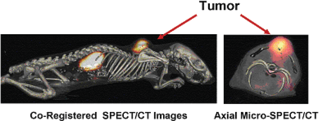
Abstract ID: 671 Poster board space: 124
We have developed a hypothesis-driven, systems biology approach to identify a small subset of proteins that are induced at the tissue-blood interface and are inherently accessible to antibodies injected intravenously. Using this new, integrated analytical approach we have found new tissue-specific endothelial cell surface proteins that can act as vascular targets to bring us one step closer to achieving the elusive goal of targeting single organs or solid tumors in vivo. Intravital microscopy and SPECT imaging show rapid, specific targeting and transport of antibodies targeted to the caveolae trans-cellular pathway in both normal and tumor-bearing tissues. In normal tissues, the transport occurs within seconds to minutes after injection, while in tumor-bearing tissues, the transport occurs within minutes to hours after injection. This unprecedented speed in caveolar trafficking in vivo may underscore a key physiological mechanism for selective transvascular exchange and elucidate a strategy to enhance tissue-specific functional imaging as well as drug, viral and nanomedicine delivery in vivo.
Abstract ID: 672 Poster board space: 125
Subtractive proteomic mapping has revealed specific expression of AnnexinA1 on the neovascular endothelium of human and rat solid tumors. We have used intravital microscopy and SPECT imaging of intravenously injected antibodies to AnnexinA1 (mAnnA1) to assess specific tumor targeting. Using fluorescently labeled mAnnA1 we show rapid targeting and extravasation of the labeled antibody to gain access to and accumulate inside solid tumors within minutes to hours after injection. 125I- mAnnA1 also showed rapid targeting within solid tumors using SPECT imaging in orthotopic models and spontaneous solid tumors as well as exhibiting dose-dependent hemorrhage in the periphery of implanted tumor spheroids 24 hours after injection with complete tumor eradication by 5 days. This is consistent with the histopathology of treated in vivo rat lung tumors. We are now using other radionucleotides, 177Lu, 131I, 90Y and 225Ac, to see if they are advantageous over the effects caused by 125I-labeled mAnnA1. We have seen some similarities, but there are also differences which we are further investigating.
Abstract ID: 673 Poster board space: 126
Progressive deposition of amyloid plaques and neurofibrillary tangles is a critical event for the pathogenesis of Alzheimer's disease (AD). Extensive deposition of amyloid plaques and neurofibrillary tangles in the brain is present even in very mild AD and precedes the presentation of cognitive decline. These evidences indicate the existence of a wide gap between clinical and neuropathological findings of AD. In vivo detection of these pathological lesions using positron emission tomography (PET) would thus prove useful for preclinical diagnosis of AD and tracking disease progression. To develop a PET probe for imaging amyloid in the brain, we have screened over 2600 compounds and found benzoxazole derivatives as candidate agents for in vivo imaging amyloid. One of these agents, 2-[2-(2-dimethylaminothiazol-5-yl)ethenyl]-6-[2-(fluoro)ethoxy]benzoxazole (BF-227), displays high binding affinity to Aβ fibrils. BF-227 specifically binds amyloid plaques in AD brain sections. Intravenous administration of BF-227 displayed excellent brain uptake, rapid clearance and specific in vivo labeling of amyloid deposits in APP transgenic mice. In acute toxicity study using mice, no mortality was observed on intravenous administration of BF-227 up to 10 mg/kg dose. Clinical PET study using [11C]BF-227 demonstrated the retention of [11C]BF-227 in the predilection site for amyloid deposition in AD patients. Quantitative analysis of PET images distinctly differentiated AD patients from normal individuals. These findings suggest that [11C]BF-227 is a promising PET probe for in vivo imaging amyloid in AD patients.
Poster Session III: P11: Imaging Cell Signaling Pathways
Abstract ID: 675 Poster board space: 1
The ability to specifically label proteins with a wide range of optical properties and functionalities can help reveal information about protein functions and dynamics in living cells. Here we describe a novel protein labeling technology developed for cell imaging, protein capture and immobilization applications. The technology is based on the efficient formation of a covalent bond between a specially designed reporting protein and its specific ligand, either in living cells, in solution, or on a solid support. The reporter protein is a genetically engineered catalytically inactive derivative of hydrolase. The ligands are small chemical tags capable of carrying a variety of functionalities, such as fluorescent labels, environmental sensors, affinity handles, or attachments to a solid phase. The covalent bond forms rapidly under general physiological conditions, is highly specific, and essentially irreversible. The stability of the bond allows imaging of live cells during long periods of time, imaging of fixed cells, and multiplexing with different cell/protein analysis techniques. The flexible nature of the technology allows labeling of different protein pools with a variety of fluorescent ligands and separating these pools by imaging cells at different wavelengths without requiring changes to the underlying genetic construct. The technology provides new options for analysis of dynamic cellular events, and can be used for direct capture, isolation and surface immobilization of protein fusions and/or protein complexes expressed in vivo or in vitro.
Abstract ID: 676 Poster board space: 2

Abstract ID: 677 Poster board space: 3
Rapid time-domain in vivo small animal EPR imaging strategies have been developed using water-soluble, non-toxic paramagnetic spin probes derived from triarylmethyl radical. By using Single Point Imaging, which employs pure phase-encoding, and using gradient setting modalities that allow rapid two and three dimensional EPR imaging, we are able to perform in vivo high resolution 2D and 3D spin as well as quantitative oxygen mapping based on the broadening effect of oxygen molecule on the local spin probe line width. Two dimensional images which take less than 10 sec. provide dynamic spin perfusion and oxygen distribution maps. The data collection mode allows several hundred T2* weighted single point image data to be collected on-the-fly, allowing accurate oxymetry. By generating multiple images with a number of gradients that have different T2* weighting, but almost identical resolution, we are able to generate oxygen and spin distribution maps and that are reliable, and can be accomplished at times an order of magnitude faster than conventional CW spectral-spatial imaging. Rapid time-domain EPR imaging allows monitoring of dynamic changes in spin and tissue oxygen distribution in mice, breathing different gas mixtures. Dynamic oxygen images from mouse SCC tumor models subject to Carbogen® (95% O2, 5% CO2) breathing will be presented and discussed. Three dimensional EPR images with high resolution obtained by this method clearly demonstrate the heterogeneity in spin and oxygen distribution in these SCC tumors, the presence of hypoxic core, and the improved oxygenation upon Carbogen breathing. It is known that oxygen is essential in the killing of tumor cells by radiation, and a non-invasive monitoring of in vivo pO2 can play a key role in radiation staging to improve the overall efficiency of radiation treatment of tumors.
Abstract ID: 678 Poster board space: 4
We assess image quality by measuring the ability of an observer to perform a scientifically relevant task using images produced by the system. This approach to image-quality assessment accounts for patient variability, differing probe characteristics, and can be applied to any imaging modality. Traditionally, image quality in molecular imaging has been measured using contrast-to-noise ratio, sensitivity limits, or resolution as the figure of merit. While these measures are descriptive and quantitative, they do not correlate with any known measure of task performance. The purpose of this presentation is to demonstrate this disconnect between these traditional figures of merit used in molecular imaging with those defined using task-based measures of image quality. We will also present computationally feasible measures of image quality that can be applied to the field of molecular imaging. Emphasis will be placed on comparisons across imaging modalities such as SPECT imaging, PET imaging, and optical imaging, as well as data-presentation methods such as presenting 2D projections compared to 3D reconstructions.
Abstract ID: 679 Poster board space: 5
Cyclooxygenases (COX) are rate-limiting enzymes involved in the conversion of PLA2-mobilized arachidonic acid into prostaglandins (PG), leukotrienes and thromboxanes. The major product of the COX-2-catalyzed reaction is PGE2, an inflammatory mediator participating in many biological processes including development and tissue specificity, pain, immunity and angiogenesis. Aberrant COX-2 function has been linked with many human diseases such as arthritis, gastric inflammation, atheromatous plaque formation and cancer. Its overexpression has been detected in different cancers, but with higher frequency in cancers of the colon and the breast. Using RNA interference we have reduced the expression of COX-2 in the highly malignant breast cancer cell line MDA-MB-231 below detectable limits, even in response to its natural stimulus IL-1β, or the mitogen TPA. Microarray analysis of the cells lacking COX-2 implicates the enzyme in many processes necessary for tumor formation, establishment and metastasis. A group of the transcripts that appeared down-regulated in the absence of COX-2 were related to breast cancer invasive phenotype. Cells lacking COX-2 were found to be less able to invade reconstituted extracellular matrix than parental cells in vitro. Cells lacking COX-2 were injected in the mammary fat pad of SCID mice to assess their tumorigenic potential. While parental and empty vector cells gave rise to large tumors within 4–7 weeks of injection, only one of the injected mice injected with COX-2 knock-down cells developed measurable tumors within the first 9 weeks post-injection. Additionally, cells lacking COX-2 were unable to metas-tasize to lungs of SCID mice when they were injected in the tail vein, in contrast to parental and empty vector cells that showed multiple metastatic foci within 5 weeks from injection. Taken together, these data show that COX-2 plays a crucial role in the tumorigenic and metastatic profile of the poorly differentiated, ER-/PR- breast cancer cell line MDA-MB-231 in vivo.