




Abstract
The endothelin (ET) receptor system has been shown to play a role in a number of vascular diseases. We have synthesized 18F-and 11C-labeled radioligands to enable in vivo imaging of the fundamental processes involved in ET receptor pharmacology in normal and diseased tissue using positron emission tomography (PET). One aim is to elucidate the proposed role of the ETB subtype as clearing receptor, removing ET-1 from the circulation, and whether this is an important mechanism to limit the detrimental effects caused by upregulated ET-1 in disease. To image ETB receptors we have labeled the selective agonist BQ3020 with 18F. In vitro characterization verified that [18F]-BQ3020 bound with a single subnanomolar affinity (KD == 0.34 ± 0.10 nM, Bmax == 9.23 ± 3.70 fmol/mg protein) to human left ventricle. Binding of [18F]-BQ3020 to human kidney was inhibited by ET-1 and unlabeled BQ3020 but not by the ETA selective antagonist FR139317, confirming that selectivity for the ETB receptor was retained. In vitro autoradiography revealed, as expected, high levels of ETB receptor densities in lung and kidney medulla, whereas kidney cortex and heart showed lower levels of ETB receptor densities. Furthermore, a high level of [18F]-BQ3020 binding was found to colocalize to macrophages in atherosclerotic coronary arteries. MicroPET studies demonstrated high uptake of [18F]-BQ3020 in ETB receptor–rich tissue, including lung, liver and kidney. The in vivo biodistribution of [18F]-BQ3020 was comparable to that previously obtained for [18F]-ET-1, supporting our hypothesis that the ETB receptor plays a significant role in the uptake of ET-1. In conclusion, [18F]-BQ3020 has retained high affinity and selectivity, allowing imaging of ETB receptor distributions in vitro and in vivo in human and animal tissue. Furthermore, in vitro data suggest that [18F]-BQ3020 potentially can be used to image atherosclerotic lesions in vivo using PET.
Introduction
Positron emission tomography (PET) is the most sensitive technique for imaging and quantifying receptor-bound radioligands in vivo. With the recent introduction of dedicated PET scanners for small animals, such as the microPET, it is now possible to perform functional imaging in rodents at high resolution (<2 mm), enabling delineation of receptor distributions within discrete organs and their larger substructures.
Vascular receptors for peptides such as endothelin (ET) are a diverse group of potential drug targets that have not been studied extensively using PET. We have recently demonstrated that binding to ET receptors in rat can be imaged dynamically using the microPET (1). Our aim is to use microPET to image the ET receptor system in vivo to elucidate the fundamental processes involved in ET receptor pharmacology in normal and diseased tissue, and particularly the role of the ETB receptor subtype in tissues including lung, kidney, and liver, where high densities of this subtype are expressed. We hypothesize that tissue-specific removal of circulating ET-1 is mediated by ETB receptors and that this will be an important mechanism to limit the detrimental effects caused by upregulated ET-1 in disease.
To enable PET imaging of both receptor subtypes (ETA and ETB), we have labeled the endogenous ligand ET-1 and the mixed antagonist SB209670 with 18F (2, 3). In addition, the ETA selective antagonist PD156707 and the precursor peptide big ET-1 have been labeled with 11C and 18F, respectively (4, 5), to allow imaging of ETA receptors and enzyme conversion of big ET-1 to ET-1 in vivo. To image the ETB subtype, the selective agonist BQ3020 was labeled with 18F (4). The purpose of the present study was to validate that the label was tolerated using in vitro receptor binding characterization, as well as to perform initial microPET imaging in the rabbit to verify that the in vivo biodistribution of [18F]-BQ3020 reflected binding to tissue expressing high densities of ETB receptors.
Materials and Methods
Peptides and Radiolabeled Compounds.
ET-1 and BQ3020 ([Ala11,15]Ac-ET-1(6–21)) were obtained from Peptide Institute Inc. (Osaka, Japan) and Neosystem (Strasbourg, France), respectively. FR139317 was synthesized by Dr. A. M. Doherty (Parke-Davis Pharmaceuticals Research Division, Ann Arbor, MI). [18F]-BQ3020 was labeled in the ε-amino group of Lys9 by conjugation with the Bolton-Hunter type reagent N-succinimidyl 4-[18F]-fluorobenzoate (4).
In Vitro Characterization.
Cryostat sections (30 μm) were cut from the left ventricular wall and from atherosclerotic coronary arteries from explanted hearts of recipient patients undergoing cardiac transplantation, from normal renal tissue (containing cortex and medulla) obtained from the opposite pole to the nonobstructing tumors following nephrectomy, and from normal lung tissue from patients undergoing lobectomy for carcinoma. All tissues were obtained with local ethical approval.
[18F]-BQ3020 was characterized using ligand binding assays as described previously (6). For saturation experiments, sections of human left ventricle were incubated with increasing concentrations of [18F]-BQ3020 (5 pM–2 nM) for 60 mins. In inhibition experiments, sections of kidney were incubated with a fixed concentration of [18F]-BQ3020 (0.5 nM). Inhibition of binding was tested by co-incubation with 1 μM FR139317 (ETA selective antagonist) and 1 μM unlabeled BQ3020 (ETB selective agonist), respectively. For autoradiographic visualization of ETB receptor densities in normal tissue (heart, lung, and kidney) and diseased tissue (atherosclerotic coronary arteries), sections were incubated with a fixed concentration of [18F]-BQ3020 (0.4 and 0.5 nM, respectively). Nonspecific binding in all assays was defined by co-incubating adjacent sections with ET-1 (1 μM). Specifically-bound ligand was measured by gamma counting or by apposing the tissue to a storage phosphor imaging screen for autoradiographic analysis. Binding was quantified using 18F standard curves for gamma counting or by coexposing the standards with the tissue sections.
To enable histologic examination of atherosclerotic coronary arteries, adjacent sections to those used in autoradiography were stained with hematoxylin-eosin and to visualize the presence of macrophages, a monoclonal mouse anti-human antibody (CD68) and methods described previously were used (7).
Data are expressed as mean ± SEM. Data from the saturation experiments were fitted to a one- or two-site model using nonlinear iterative curve fitting (8). A two-site model was accepted only if it resulted in a significantly better fit as judged by an F test (P < 0.05).
MicroPET Imaging.
PET imaging was performed in two New Zealand White rabbits using a microPET P4 scanner (Concorde Microsystems, Knoxville, TN). All experiments were performed in accordance with the United Kingdom Animal Scientific Procedures Act, 1986, and complied with guidelines of the local animal ethics committee.
General anesthesia was used during surgery and PET imaging. Induction was achieved using intravenous alphaxalone/alphadalone (0.2 ml/kg; Schering-Plough, Welwyn Garden City, UK) diluted in saline to a volume of 5 ml, and anesthesia was maintained with a mixture of isofluorane (2.5–4%), nitrous oxide (0.8–1 liters/min), and oxygen (0.5 liters/min), delivered by secured face mask. There was no need for endotracheal intubation during any of the surgical or imaging sessions. Rectal temperature was monitored and the animal placed on a padded warming blanket. An ear vein was cannulated for administration of [18F]-BQ3020 and the rabbits were then placed prone on the scanning bed. [18F]-BQ3020 (10 and 20 MBq, respectively) was administered as a bolus injection and data were acquired by the microPET in 3 × 5 min time frames for each bed position starting 60 mins postinjection. To allow for all organs of interest (lung, liver, and kidney) to be scanned, 3–4 bed positions were used. The obtained imaging data were reconstructed into color-coded images describing the biodistribution of radioactivity in the rabbit. Regions of interest were delineated for lung, liver, and kidney to enable quantification of radiotracer concentration in these organs as described previously (9).
Results
In Vitro Characterization.
Binding of [18F]-BQ3020 to human left ventricle (n = 4) was concentration-dependent and saturable (Table 1). A one-site model was preferred to a two-site model and the Hill slope (nH) was close to unity. Binding of [18F]-BQ3020 to human kidney (n = 3) was inhibited by ET-1 (1 μM) and unlabeled BQ3020 (1 μM), but not by the ETA selective antagonist FR139317 (1 μM). Quantitative autoradiography using [18F]-BQ3020 revealed high levels of binding to ETB receptors in lung and kidney medulla, whereas kidney cortex and heart showed lower levels of ETB receptor densities (Table 2). In atherosclerotic coronary arteries, high levels of ETB receptor densities were found to colocalize with macrophages (Fig. 1).
MicroPET Imaging.
Following [18F]-BQ3020 infusion, there was a rapid redistribution of radioligand from the circulation to ETB receptor–rich tissue. High levels of [18F]-BQ3020 binding were found in lung, liver, and kidney (Figs. 2 and 3). In kidney, a heterogeneous distribution of radioactivity was observed, with lower levels of uptake in the kidney cortex compared with the medulla (Figs. 2B and C, and 3), demonstrating that the microPET scanner has sufficient resolution to differentiate uptake in subrenal structures. As expected, ETA-rich tissue in the heart could not be visualized in the microPET images.
Discussion
We have demonstrated in vitro that [18F]-BQ3020 has retained the expected subnanomolar affinity and selectivity to the ETB receptor. Furthermore, we have shown using microPET that following infusion of [18F]-BQ3020 the expected biodistribution of radioligand was obtained, with high levels of uptake in ETB receptor–rich tissues in the rabbit.
We have previously shown that 18F labeling of ET-1 in Lys9 did not affect affinity and specificity for ET receptors (Table 1) and that [18F]-ET-1 had the required pharmacokinetic properties to permit binding and imaging of ET receptors in vivo using microPET (1). We therefore hypothesized that using this approach for 18F labeling of BQ3020 would not affect the affinity and selectivity to the ETB receptor. In vitro characterization confirmed that [18F]-BQ3020 had retained high-affinity binding with the expected single subnanomolar affinity comparable to values observed in vitro for [125I]-BQ3020 (Table 1; Ref. 10). Binding was inhibited with ET-1 (mixed ETA/ETB) and BQ3020 (ETB-selective) but not with FR139317 (ETA-selective), demonstrating that [18F]-BQ3020 had retained its selectivity for the ETB receptor. High levels of binding were detected in lung and kidney medulla, tissue expressing high densities of ETB receptors, whereas, as expected, heart and kidney cortex showed low levels of ETB receptor densities (11). In atherosclerotic coronary arteries, [18F]-BQ3020 binding was colocalized to macrophages, in agreement with the observation by Bacon et al. using [125I]-BQ3020 (12), suggesting that [18F]-BQ3020 potentially can be used for the visualization of atherosclerotic plaques in vivo with PET.
We have previously demonstrated, using dynamic microPET imaging, that [18F]-ET-1 is rapidly cleared from the circulation by binding to ET receptors in lung, liver and kidney. This uptake was significantly blocked when the animal was pretreated with the ETB selective antagonist BQ788, confirming that this fast clearance from the circulation was ETB receptor–mediated (1). In support of these findings, we obtained a comparable biodistribution with high levels of uptake in lung, liver, and kidney when [18F]-BQ3020 was infused in the rabbit, clearly demonstrating that the ETB receptor plays an important role in the uptake of ET-1. In images reconstructed from the micro-PET, it was possible to delineate the expected heterogeneous distribution of ETB receptor densities in the kidney, with high levels of [18F]-BQ3020 binding in the medulla and low levels of binding in the cortex as demonstrated in vitro in this and previous studies (13), as well as in vivo using microPET and [18F]-ET-1 (1).
In conclusion, this study shows that [18F]-BQ3020 has retained high affinity and selectivity for the ETB receptor and has the potential to image ETB receptor distributions in vivo in normal and diseased tissue using positron emission tomography. In addition, the in vivo biodistribution of [18F]-BQ3020 was comparable to that of [18F]-ET-1, supporting our hypothesis that the ETB receptor plays a significant role in the uptake of ET-1.
Dissociation Constant (KD), Maximal Density of Receptors (Bmax), and Hill Coefficient (nH) for [18F]-BQ3020 in Human Left Ventricle a
In Vitro Quantification of ETB Receptor Densities in Various Human Tissues Using [18F]-BQ3020 a
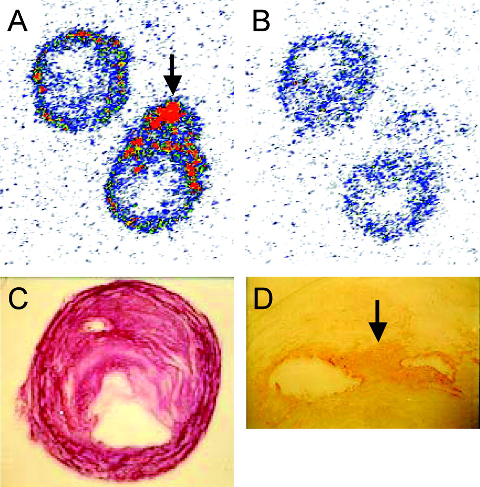
In vitro autoradiography showing (A) total and (B) nonspecific binding of [18F]-BQ3020 in transverse sections of atherosclerotic coronary arteries. Images are color-coded to express level of radioactivity, with red indicating high levels and blue indicating low levels. (C) Hematoxylin and eosin–stained adjacent section showing the atherosclerotic plaque. (D) High densities of ETB receptors were found to colocalize to areas with high densities of macrophages (arrows), as seen by macrophage staining using CD68 and the peroxidase-antiperoxidase technique yielding a brown reaction product.

MicroPET images showing in vivo biodistribution of [18F]-BQ3020 after intravenous infusion in New Zealand White rabbit (coronal planes). Images are color-coded to express level of radioactivity, with red indicating high levels and black/blue indicating low levels. (A) High levels of uptake were found in lung and liver. (B and C) In kidney, suborgan distribution of radioligand could be visualized with high levels of uptake in medulla and lower levels of uptake in cortex.
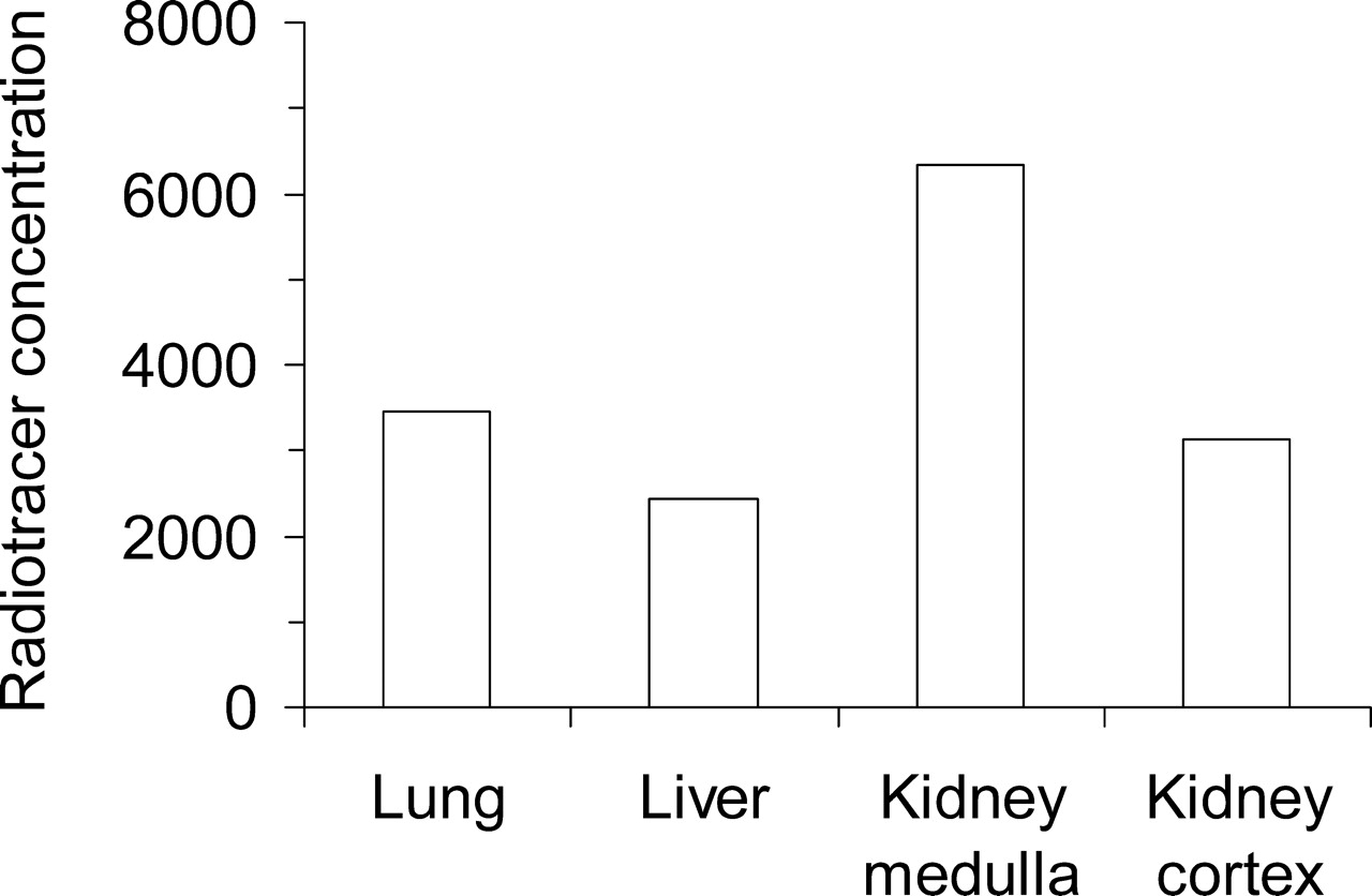
In vivo concentrations of [18F]-BQ3020 in lung, liver, and substructures of kidney. Regions of interest were delineated in the microPET images to enable quantification of radiotracer concentration as described previously (9).
Footnotes
This work was supported by grants from the British Heart Foundation, the Medical Research Council Technology Foresight (U.K. Government), and, for the microPET, a JREI grant from HEFCE and Merck Sharp & Dohme, Ltd.
Acknowledgements
We thank Mrs. Rhoda Kuc for technical support.