



Abstract
Trabecular meshwork (TM) cells are now considered to play an active role in the aqueous outflow mechanism because they exhibit smooth muscle–like contractile properties. Endothelin-1 (ET-1), a potent vasoconstrictor peptide, has been proposed to play a role in the local regulation of aqueous outflow and intraocular pressure (IOP) control. We propose an in vitro culture model as a method for the study of ET-1–induced human TM (HTM) cell contractility and for the study of whether pre-incubation with flunarizine, a calcium-channel blocker, can inhibit the action of ET-1. Experiments were performed on semiconfluent HTM cells (primary cultures established from normotensive human donor eyes) at the second passage, with phosphate-buffered saline (PBS) as a control. The contractile status of the cells was evaluated by a morphometric analysis of cell area, assuming that HTM cells in culture are able to reduce their area as a consequence of cytoskeletal contraction, rather than regulatory volume decrease. After incubation with 10 μM ET-1 for 5 mins, we observed a reduction of HTM cell area with respect to PBS-treated cells: 2425 ± 876 μm2 versus 3125 ± 987 μm2 (P < 0.001); and cells exhibited a retraction in shape and a reduction in number of indented profiles. Administration of ET-1 at progressively lower doses produced a corresponding lower reduction of HTM cell area, suggesting a dose-response effect of ET-1. Pre-incubation with 10 μM flunarizine strongly inhibited the ET-1 effect on HTM cell contraction: 2806 ± 865 μm2 versus 2910 ± 846 μm2 (P = not significant). Our data indicate that ET-1 induced a statistically significant reduction in the area of HTM cells versus controls, and that ET-1 can directly influence the aqueous outflow. Moreover, we observed that flunarizine inhibited the effect of ET-1 on the HTM cells.
Introduction
A number of endothelium-derived vasoactive substances play key roles in the maintenance of basal vascular tone in the body and in ocular circulation (1). In particular, endothelin-1 (ET-1) is a potent vasoconstrictor peptide that is mainly released by vascular endothelial cells, but also by epithelial and glial cells (2, 3).
In chronic openangle glaucoma, the ET-1 levels are particularly high in the aqueous humor
(4, 5); this might be one of the causes of
high intraocular pressure (IOP), because ET-1 causes contraction of the trabecular meshwork
(TM; Ref. 6) with a consequent reduction in aqueous outflow (7, 8). ET-1–induced contractions are partly
dependent on extracellular calcium and
Materials and Methods
HTM Cell Culture Model System.
HTM cell cultures were established from normotensive donor eyes. The globes were soaked
in Dulbecco’s modified Eagle’s medium (DMEM) supplemented with 10% fetal bovine serum
(FBS) plus 5% penicillin/streptomycin solution and 1%
The HTM cells were cultured in 6-well plates at 37°C in a 5% CO2 atmosphere in the same medium; confluence was reached after 3 weeks. HTM cells from the established three primary lines were characterized with respect to their growth characteristics, morphology, and cytoskeletal proteins: the immunocytochemical procedures for detection of smooth-muscle actin (antisera purchased from DakoCytomation, Glostrup, Denmark) were performed as described elsewhere (10). HTM cell viability was checked with trypan blue staining.
Low-confluence HTM cell cultures at the first or second passage were used. All experiments were performed in triplicate wells and repeated at least two independent times using cells from the three established cell lines.
For each set of experiments, a concentration of 2000 cells/ml culture medium was used, and HTM cells were set in 24-well plates.
Effect of ET - 1 on HTM Cells.
ET - 1 (C109H159N25O32S5, batch No. L02049/e) was purchased from Alexis Biochemicals, Lausen, Switzerland, and stored at −20°C until used.
A 20 μl aliquot of 10 μM endothelin dissolved in 0.01 M phosphate-buffered saline (PBS), pH 7.2, was added to the medium and administered to the cells, wells were carefully shaken, and the plates were set in the CO2 incubator. As a control, 20 μl PBS was added to the medium of control wells. Cells at baseline, 5 mins, and 10 mins after the addition of ET-1 or PBS were observed with a Zeiss Axiovert inverted microscope, and digitized micrographs of the cells were acquired with the Nikon Coolpix 990 camera.
To characterize the mechanisms by which ET-1 stimulated contractile force generation, we
added 10 μM ET-1 to cells treated with 10 μM flunarizine
(a calcium channel blocker that affects vessel calcium
Cell Contraction and Image Analysis.
The contractile status of the cells was evaluated by a morphometric analysis of cell area, assuming that, as previously demonstrated (12, 13), HTM cells in tissue culture are able to reduce their area as a consequence of cytoskeletal contraction rather than regulatory volume decrease.
Image analysis was performed on the computer-acquired digitized micrographs taken in all of the experiments using image-processing software (Image Pro Plus, version 3.0.2; Media Cybernetics, Silver Spring, MD).
The morphometric evaluation of the cell area was semi-automatically performed on a cell-to-cell basis by overlying the cell periphery with the digital cursor. A total of 250 cells were measured for each experiment. Statistical evaluations were performed with the Statistical Package for Social Science (SPSS) program package (SPSS Inc., Chicago, IL) using an unpaired Student’s t test; values of P < 0.05 were regarded as statistically significant.
Results
Cells at baseline (Fig. 1) showed a regular and elongated shape, with smooth profiles. Cells treated with PBS did not change in shape and morphology with respect to the baseline cells. After incubation with 10 μM ET-1 for 5 mins, we observed a reduction of HTM cell area with respect to PBS-treated cells: 2425 ± 876 μm2 versus 3125 ± 987 μm2 (P < 0.001); and cells exhibited a retraction in shape and a reduced number of indented profiles (Table 1 and Fig. 2). Pretreatment of the HTM cells with 10 μM flunarizine prevented the contraction of the cells after stimulation with 10 μM ET-1, with respect to baseline: 2806 ± 865 μm2 versus 2910 ± 846 μm2 (P = not significant) and inhibited the reduction in cell area (Table 2 and Fig. 3).
Discussion
ET-1 belongs to a family of vasoactive peptides (ET-1, ET-2, and ET-3) isolated from vascular endothelial cells. ET-1 (and its ET-3 but not ET-2 isoform) is also abundantly distributed in the iris, ciliary body, retina, choroid, TM (14), and aqueous humor, where ET-1 levels are normally two to three times greater than in plasma (15). There is much evidence suggesting a role of ET-1 in the local regulation of IOP, but it remains unclear whether elevated ET-1 levels occur in response to, or are themselves a cause of, glaucoma.
The purpose of this study was to evaluate the effect of ET-1 on HTM cell contractility; contraction and relaxation of HTM cells may influence IOP in the sense that their relaxation makes a trabecular space enlargement and facilitates outflow, whereas contraction produces the opposite effect. This study also investigated whether calcium channel blockers could inhibit the effect of ET-1 on HTM cells.
TM contractility was usually studied in TM strips from bovine eyes because of the anatomic difficulty in isolating these strips from human eyes (6). In bovine eyes, an ET-1–induced TM contraction was observed (9), and it was also suggested that ET-1 may be involved in the regulation of aqueous humor dynamics by changing the calcium concentration and the intracellular pH in TM cells (16, 17). Moreover, calcium-channel blockers can inhibit the ET-1 effect in the TM strips from bovine eyes (18).
In the present work, we proposed a cultured in vitro HTM cell model that offers the ability to analyze ET-1–induced contractility on human TM cells and offers interesting opportunities for pharmacologic evaluation of drugs that could control the condition of HTM cells.
In this study, we demonstrated that ET-1 induced a statistically significant reduction in HTM cell area versus control. The dose-response study confirmed that the effect depends on the concentration of ET-1 applied, as demonstrated previously by others (9) using the same ET-1 doses. Moreover, we demonstrated that the calcium-channel blocker, flunarizine, strongly inhibited the effect of ET-1 on HTM cells, and thus, it can perhaps lower the in vivo IOP, increasing the TM aqueous outflow.
Effect of 10 μM ET-1 Treatment Compared with PBS Treatment on HTM Cells a
Effect of 10 μM ET-1 Treatment on HTM Cells After Pretreatment with 10 μM Flunarizine a
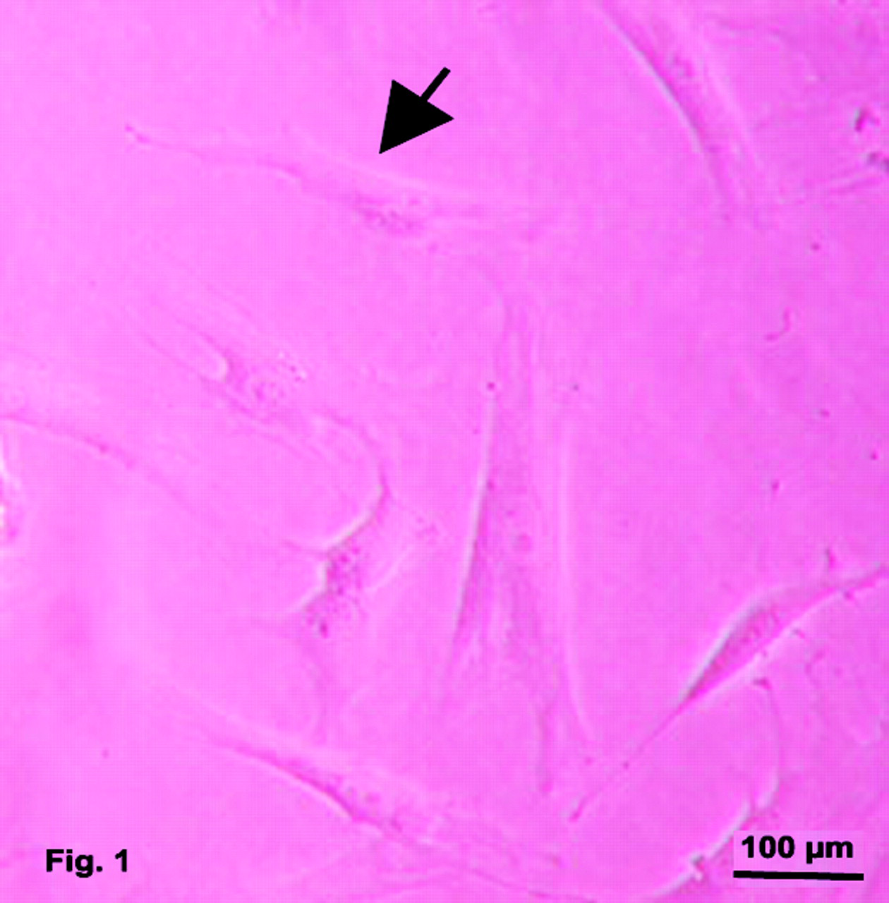
HTM cells from eyeglobe n.2 (female, age 48 years) at the second passage, in a state of subconfluent culture. Cells display an elongated shape with narrow profiles and a central bulge containing the nucleus.

HTM cells treated with 10 μM ET-1 for 5 mins. Cells rearrange their shape and show indented cell profiles (arrows) at the edges of the cells where the contractile forces exert their action.
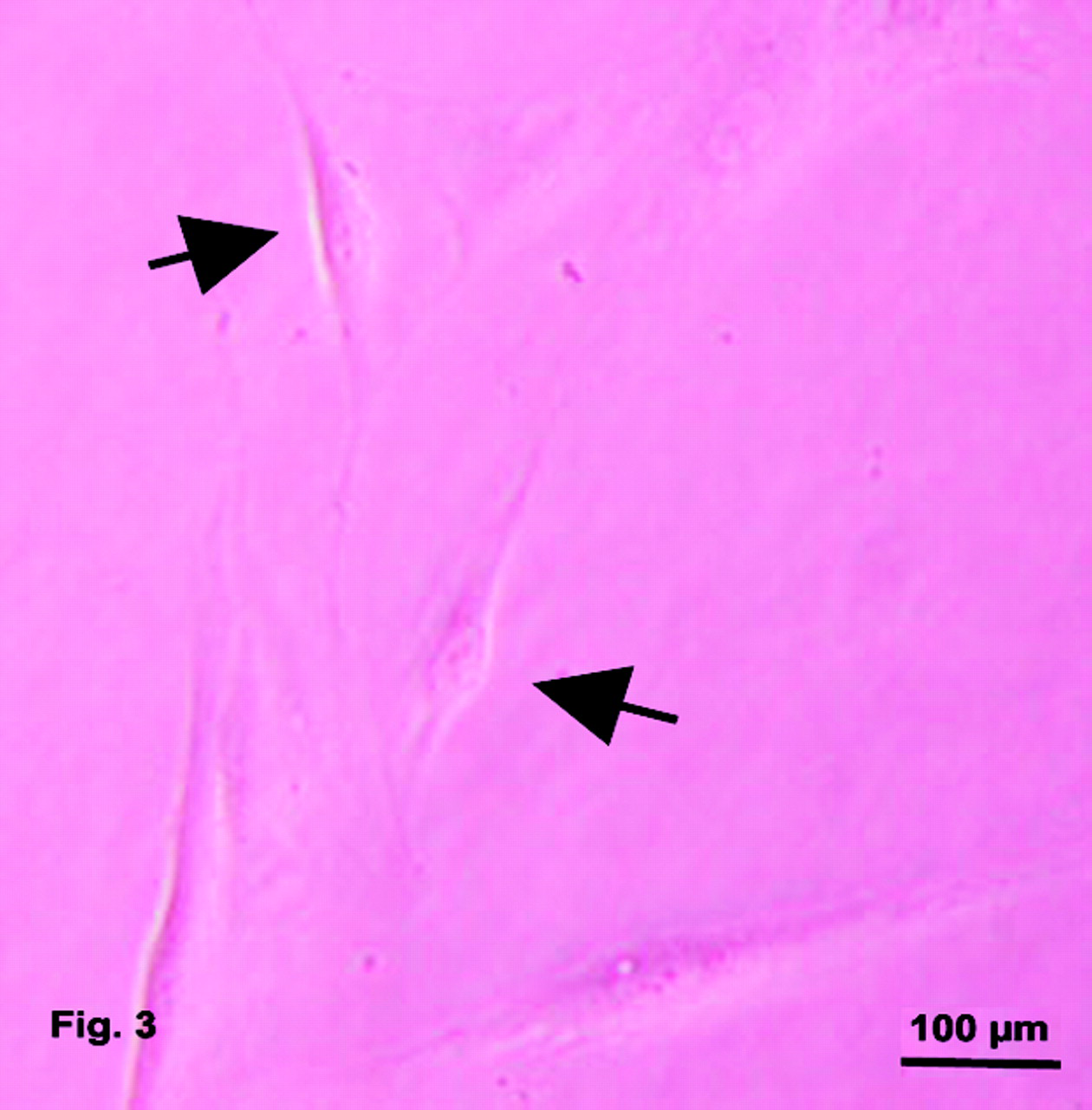
HTM cells treated with 10 μM ET-1 for 5 mins after pretreatment with 10 μM flunarizine. The inhibitory contraction effect produced by flunarizine on ET-1–stimulated HTM cells is shown by cells that show an elongated shape and seem to have the same baseline cell morphology.
Footnotes
Acknowledgements
We are indebted to Ms. Chiara Coslovi for her skillful and experienced technical assistance.