
Abstract
DA-9102 isolated from Actinidia arguta is a candidate of natural medicine currently under Phase II clinical trial for atopic dermatitis in Korea. In this study, spontaneous dermatitis was induced by magnesium deficiency in hairless rats and this system was applied to assess the suppressive effects of DA-9102 on atopic dermatitis-like skin disease. Oral administration of DA-9102 at a dose of 100 mg/kg for 16 days substantially suppressed the occurrence of spontaneous dermatitis. Eczematous skin lesions, water loss and scratching behavior were significantly decreased by DA-9102 in a dose-dependent manner. Infiltration of inflammatory cells into the skin and pathologic remodeling of the epidermis and dermis were much less than the Mg-def. group. Results from flow cytometry analysis of peripheral blood mononuclear cells indicated that DA-9102 suppressed activation of leukocytes. The decrease in the number of CD45RA+ cells was accompanied by a lower level of IgE in DA-9102 treated rats, and the reduction in the number of CD11b+ cells by DA-9102 in both periphery and skin was significant. Further, DA-9102 not only suppressed the mRNA expression of TH2 cytokines including IL-4 and IL-10 in the lymph node but it also decreased the levels of inflammatory mediators such as nitric oxide and leukotriene B4 (LTB4) in the serum. Taken together, these results suggest that DA-9102 is an orally applicable potent immune modulator capable of controlling the occurrence of atopic dermatitis-like skin disease.
Introduction
Considering the limitations of current systemic treatments for atopic dermatitis, developing safe and effective pharmacological agents is greatly needed (1, 2). Orally applicable antihistamines seem to be ineffective except for lessening sleep disturbance by pruritus (3). Systemic corticoids are effective against atopic dermatitis in adults, however, rebound flare-ups and diminishing effectiveness severely limit their use (4). While cyclosporin, an immunosuppressive agent, is permitted for use by patients with severe clinical manifestations, it has been associated with toxicity and adverse effects such as risk of hypertension and nephropathy (5, 6). Azathioprine is widely used for atopic dermatitis in the UK, but it has been reported to produce various side-effects including gastrointestinal disturbances, myelotoxicity and hepatotoxicity (7).
In recent years, natural bioactive products have attracted considerable attention as a source of medicinal substances. In Asian countries, extracts from the fruits, leaves, and stems of Actinidia arguta (hardy kiwi) are used as traditional medicines for the treatment of inflammatory diseases, and various constituents including quercetin, kaempferol, catechins, and volatile compounds have been identified (8–10). In addition, its safety is well established through a long history of human utilization. It was previously demonstrated that several extracts isolated from Actinidia arguta not only exert regulatory activities on immune systems by controlling the production of selective cytokines and IgE in the OVA-sensitized allergic model (11), but that they also have suppressive effects on the development of spontaneous dermatitis in the NC/Nga mouse model of human atopic dermatitis (12).
As an animal model of spontaneous dermatitis, hairless rats, the dominant mutants of the Sprague-Dawley (SD) rats, develop erythromatous skin lesions when placed on magnesium deficient diets (13, 14). Although the pathogenic mechanisms of dermatitis in this model still remain unclear, the clinical manifestations are very similar to the dermatitis shown in many human skin diseases including atopic dermatitis (15, 16), and the development of dermatitis has been known to be associated with several systemic immunological abnormalities including lymphocyte hyper-reactivity (17), hyper proliferation of monocyte and granulocytes (17, 18), deregulation of mast cells and histaminemia (19, 20), increased levels of IgE and high levels of pathologic cytokines (21, 22), as well as the overproduction of inflammatory mediators such as nitric oxide (23) and leukotrien B4 (24). The purpose of this study was to assess whether oral administration with DA-9102, a candidate of natural medicine isolated from Actinidia arguta and standardized for human clinical trial, has beneficial effects on clinical manifestations for acute forms of dermatitis and to further examine possible mechanisms of the pharmacological effects at the cellular and molecular levels in the immune system.
Materials and Methods
Preparation and Standardization of DA-9102.
Dried Actinidia arguta from Shaan-xi (China) were purchased at a specialized market for traditional herbal medicine (Kyungdong herb market, Seoul) in Korea and their identity was kindly confirmed by Dr. Changsoo Yuk (specialist in plant classification). Dried fruits (water contents were less than 10%) were extracted with a mixed solution containing water/BuOH/EtOH at 60°C for 2 days and filtered. The soluble fraction was dissolved in the same solution used in the previous extraction (1:1), and the solution was resolved in a mixture of water and BuOH. The BuOH layer was collected and concentrated by rotary evaporation (EYELA, Japan) at 50°C. The concentrates, named DA-9102, were determined by means of HPLC. Typically, more than 0.06–0.18% of esculetin as a surrogate marker is usually contained in DA-9102. Endotoxin was assayed under endotoxin-free experimental conditions using a Limulus Amebocytes Lysate (LAL) pyrogen kit (Bio-Whittaker, Walkersville, MD). Experiments were performed according to the manufacturer’s protocol. Briefly, 100 μl standard reagent of DA-9102 or phosphate buffered saline (PBS) was mixed with 100 μl of LAL reagent and was incubated for 1 hr at 37°C. Each tube was then examined for gelation. The quantity of endotoxin in DA-9102 was negligible.
Animal Experimental Design.
Male hairless rats (Ico:OFA hr/hr), 4 weeks of age, were purchased from Iffa Credo (Les Oncins, France). The rats were housed in a temperature and humidity-controlled room with a 12:12 hr light:dark cycle. All rats were allowed to freely access deionized water. Following the adaptation period, rats were divided into 3 groups (n = 6–7/group). The naive group was provided with a standard diet, and two groups were provided with a standard diet low in magnesium (Altromin, Lage, Germany, C1045:0.012% Mg 2+/Kgts) described by Ponvert et al. (16). This diet rendered the rats to be magnesium deficient. From the first day of the magnesium deficient diet, the experimental group was orally administrated with 100 μl of DA-9102 at the concentration of 100 mg/kg everyday for 16 days, and the Mg-def. group was administrated with 100 μl of vehicle (3% HPMC). Research was conducted according to the principles enunciated in the Guide for the Care and Use of Laboratory Animals, prepared by the Institute of Laboratory Animal Resources, National Research Council (http://www.nsa.edu/nrc).
Macroscopic Analysis of Atopic Dermatitis-like Skin Disease.
The severity of dermatitis was assessed every other day according to the modified SCORAD (Severity Scoring of Atopic Dermatitis) index described by Leung et al. (25). Clinical signs of dermatitis were evaluated by five categories; erythema, edema, excoriation/erosion, hemorrhage/oozing, and scaling/lichenification. The total score was taken from the grades of each symptom; 0 (no symptoms), 1 (mild), 2 (moderate), to 3 (severe). Without prior knowledge of the experimental groups, at least three veterinarians performed the scoring. Scratching incidence was evaluated by measuring the duration of itching time for 15 min per rat on day 15 when dermatitis was substantially present. Transepidermal water loss (TEWL) was assessed on the dorsal skin of the rat on days 0 and 16 using TM 310 (Courage+Khazaka Electronic GmbH, Cologne, Germany) according to the protocol as previously described (26). The temperature ranged from 23°C to 24°C and the relative humidity was maintained at 40–45%. Measurements recorded by TEWL reading were stabilized at approximately 45 sec after the probe was placed on the skin. The values were automatically calculated as g/m2h, and displayed digitally.
Histological Analysis and Immunohistochemistry.
For histological examination, rats were sacrificed and small biopsies were obtained from the dorsal skin on day 16. Skin sections were fixed in 10% phosphate-buffered formalin (pH 7.2), embedded in paraffin, and cut at 4 μm. Slides of paraffinized sections of skin were dewaxed by xylene and hydrated with 50% ethanol, and then stained with hematoxylin and eosin or 0.05% toluidine blue for 30 min, respectively. Histological features of the skin were observed under a microscope (Nikon, Japan) at a magnification of ×100. Mast cells were easily identified by metachromatic staining of their granules. For immunohistochemistry, dewaxed and hydrated slides were incubated with mouse anti-rat CD11b antibody (BD Pharmingen, CA, USA) diluted in 1% BSA at room temperature for 1 hr, and followed by incubation with FITC-conjugated anti-mouse IgG (Molecular Probe, CA, USA). The sections were mounted with Gel/mount (Biomeda, CA, USA), and observed using Fluorescence Microscopy (Nikon, Japan). CD11b+ cell densities were quantified by counting the number of fluorescence positive cells in 50 separated areas.
Isolation of Peripheral Blood Mononuclear Cells and Flow Cytometry.
Peripheral blood mononuclear cells (PBMCs) were isolated by the Ficoll-Hypaque (Sigma, St. Louis, USA) density gradient centrifugation according to the manufacturer’s recommendations with some modification (27). Cells recovered from the interface were washed with HBSS and suspended in RPMI 1640 medium. Isolated PBMCs were fixed and stained 30 min at 4°C with fluorescence isothiocyanate (FITC) conjugated antibodies, OX-33 (28), WT.5 (29) (BD Pharmingen, CA, USA) specifically reacting CD45RA, and CD11b, which were B and monocyte/granulocyte markers, respectively, at the concentration of 5 μg/ml. Flow cytometry was performed on a FACSort (Beckon Dickinson, CA, USA) with CellQuest (Beckon Dickinson, CA, USA) data acquisition and analysis software. In all cases, isotype-matched antibodies controlled nonspecific staining.
Quantitative Real Time PCR.
Total RNA was isolated from lymph nodes using Trizol Reagent (Gibco BRL, Carlsbad, CA). Isolated total RNA was used in reverse transcription (RT)-PCR by the AMV RT System (Roche, Mannheim, Germany) followed by quantitative real time PCR using the RG-6000 (Corbett Research, Australia). Forward and reverse primer sets for rat genes were designed as follows:
Determination of the Levels of IgE, Nitric Oxide, and Leukotrien B4.
The plasma levels of IgE were measured by the commercially available IgE detection kit (Shibayagi, Gunma, Japan) according to the manufacturer’s instruction. The levels of NO were determined by the commercially available total NO detection kit (R&D Systems, MN, USA). The detection limit was 3.12 μmol/L. The plasma levels of LTB4 were measured by the LTB4 ELISA kit (Assay Designs, MI, USA) according to the manufacturer’s instruction. The detection limit was 11.7 pg/ml.
Statistics
Data were expressed as mean ± SEM, and the difference between mean values were analyzed by unpaired Student’s t test. P values less than 0.05 or 0.01, which were calculated as one-tail P values, were considered statistically significant.
Results
Blockade of Atopic Dermatitis-Like Skin Disease by DA-9102.
To assess the suppressive effects of DA-9102 on spontaneous dermatitis, we harbored a hairless rat model presenting clinical manifestations similar to human atopic dermatitis. While rats fed with a standard diet (naive group) showed no signs at all during the experimental periods, rats fed with the vehicle (3% HPMC) under a hypomagnesic diet (Mg-def. group) presented redness and erosion in the trunk within 7–9 days, after which the symptom expanded to eyelids and perioral regions. These signs subsequently progressed into erythematous pruritic rash and hemorrhage to the abdominal region at day 16, indicating intensive inflammation (Fig. 1A). Oral administration with DA-9102 at a dose of 100 mg/kg almost completely suppressed the onset of the erythematous pruritic rash, erosion and hemorrhage, which was easily determined by the naked eye during the experiments (Fig. 1A). The increase in the dermatitis severity score was determined by an evaluation system using SCORAD. The Mg-def. group treated with the vehicle showed that dermatitis significantly progressed from day 10 and reached the maximum on day 16. However, DA-9102 substantially inhibited a dramatic increase in the dermatitis severity score in a dose-dependent manner during this period (Fig. 1B), and on day 16, oral administration of DA-9102 at the concentrations of 50 and 100 mg/kg resulted in approximately 25% and 50% inhibition, respectively, while 5 mg/kg indicated no significant difference from that of the Mg-def. Next, transepidermal water loss (TEWL) was measured to examine the effects of DA-9102 on epidermal barrier function because cutaneous dryness is a pathologic factor for epidermis impairment. There was a two-fold increase from day 0 to day 16 (13.4 ± 1.2 g/m2h vs 31.2 ± 3.8 g/m2h) in the values of the Mg-def. group, resulting in keratinous dry skin. In contrast, there was no significant change in the TEWL values of the DA-9102 treated group (both at the concentration of 50 mg/kg and 100 mg/kg) during the same period. There was a 59% difference in the values of TEWL between the Mg-def. and the DA-9102 treated group (Fig. 1C) on day 16. DA-9102 also lessened the scratching time at the 100 mg/kg concentration by approximately 65% (P < 0.08) compared with the Mg-def. group (Fig. 1D). These results indicated that oral administration of DA-9102 might suppress various clinical manifestations including erythema and hemorrhage as well as dryness and itching in atopic dermatitis-like skin diseases.
Effects of DA-9102 on Dermis and Epidermis.
The histological examination using H&E and toluidine blue staining confirmed suppression of skin lesions by DA-9102. The dorsal skin from the Mg-def. group exhibited a marked thickening in both the epidermis and dermis with infiltration of inflammatory cells and hemorrhage (Fig. 2b and e). However, treatment with DA-9102 substantially inhibited the thickening of the epidermis and dermis, as well as the infiltration of eosinophil and mast cells (Fig. 2c and f), resulting in a histological environment very similar to that of the naive group (Fig. 2a and d). These results indicated that oral administration of DA-9102 could effectively suppress the infiltration of inflammatory cells, and the pathologic remodeling of the epidermis and dermis in atopic dermatitis-like skin disease.
Effects of DA-9102 on IgE and CD45RA+ Cells.
In addition to clinical manifestations mimicking human AD, rats experiencing magnesium deficiency were reported to show immunological abnormalities such as elevated levels of IgE in plasma. Therefore, it was examined whether DA-9102 affected the levels of IgE and the activation of B cells. After the onset of dermatitis, the Mg-def. group produced approximately 80 ng/ml of total IgE. Administration of DA-9102 significantly lessened the levels of IgE, resulting in 80% lower level of IgE on day 16. Additionally, OX-33, a monoclonal antibody, was used to examine the effects of DA-9102 on B cells as it only binds CD45RA expression on B cells (28). As shown in Figure 3B, there was a significant increase in the CD45RA+ cells in the PBMCs of the Mg-def. groups compared with the naive group, indicating activation of B cells. DA-9102 maintained the percentage of CD45RA+ cells similar to that of the naive group. These results suggested that DA-9102 might suppress the onset of dermatitis by magnesium deficiency through the inhibition of B cell activation and IgE production.
Effects of DA-9102 on CD11b+ Cells.
Not only is dermal infiltration of inflammatory cells including eosinophils an important feature of atopic dermatitis, but also the presence of inflammatory cells in skin lesions results from the mobilization of bone marrow cells to the blood (30). Therefore, CD11b, a well known marker for monocytes/granulocytes, was detected by a monoclonal antibody, WT.5 (29) both in PBMCs and skin lesions. As summarized in Figure 4A, magnesium deficiency induced a significant increase of about 11.5% in CD11b+ cells in PBMCs compared with the naive group. Oral administration with DA-9102 maintained the percentage of CD11b+ cells similar to that of the naive group. The CD11b+ cells in skin suffering from dermatitis were also increased compared with that of the naive group, but DA-9102 greatly decreased the positively stained cell number by similar to that of the naive group (Fig. 4B), indicating reduced infiltrations of inflammatory cells. These data implied that DA-9102 might suppress the onset of atopic dermatitis-like skin lesions through the inhibition of activation of CD11b+ cells induced by magnesium deficiency.
Effects of DA-9102 on Pathologic Factors.
Because aberrantly expressed cytokines and overly produced inflammatory mediators are involved in the pathogenesis of various skin diseases (30), we examined whether DA-9102 affects their expressions. When the gene expressions of pathologic cytokines were examined using quantitative real time PCR, mRNA expressions of TH2 cytokines including IL-4 and IL-10 in lymph nodes were significantly increased in the Mg-def. group compared with those of the naive group, and that of IL-10 were prominent (Table 1). DA-9102 considerably reduced the expression of IL-4 and IL-10 by up to 41% and 63%, respectively. On the contrary, the expression of TH1 cytokine IFN-γ was decreased in the Mg-def. group suffering from dermatitis compared with the naive group. DA-9102 amplified the expression of IFN-γ although the values were variable. The levels of nitric oxide (NO) and LTB4 were also greatly enlarged in the Mg-def. group, however, DA-9102 significantly lowered the levels of NO and LTB4 by approximately 65% and 68%, respectively (Table 1). These results suggested that DA-9102 might control the overexpression of pathologic factors induced by magnesium deficiency, resulting in suppression of atopic dermatitis-like skin disease in hairless rats.
Discussion
DA-9102 has potential as an alternative systemic therapeutic for the treatment of atopic dermatitis-like skin disease with safety. In this study, oral administration with DA-9102 substantially blocked the development of an acute form of atopic dermatitis-like skin lesion in magnesium deficient hairless rats, and showed its protective effects against functional impairments in the skin. The inhibition of water loss from the skin by DA-9102 was demonstrated (Fig. 1C), and histological examinations indicated that DA-9102 suppressed the infiltration of eosinophils and mast cells in the epidermis and dermis, and also inhibited tissue destruction (Fig. 2), which was confirmed by the immunohistochemistry of CD11b in the skin and the flow cytometry (Fig. 4). Although the pathologic mechanisms have not been fully understood in this model, we could perceive that significant immunological abnormalities are developed in the rats suffering from dermatitis, ensuing in an increase in CD11b+ (Fig. 3A) and CD45RA+ cells and a decrease in the CD3+ cells (approximately 14% reduction compared with naive group). Up-regulation of CD11b in monocyte/granulocytes has been reported to play an important role in cell migration into inflammatory lesions and development of atopic dermatitis (31, 32) as well as spontaneous dermatitis responding to various stimuli, for example, such as radiation (27). The percentage of CD11b+ cells was highly increased in rats suffering from dermatitis, and the increased expressions of CD11b in both periphery and skin were significantly reduced by DA-9102, which implied that DA-9102 might suppress the mobilization of CD11b+ cells from the bone marrow and blood into skin lesion and the activation of CD11b+ cells including eosinophils under magnesium deficiency. Since certain antigenic stimuli are capable of rendering CD11b+ cells to secrete cytokines such as IL-6 and IL-10 that lead to promoting B cell proliferation and antibody secretion (33), the increase in the number of CD45RA+ cells (B cells) and IgE might therefore be mediated by the activation of CD11b. Actually, we could detect down regulatory effects both on the plasma levels of IL-6 in the early stage of dermatitis development (approximately 25% reduction compared with Mg-def. group) and the CD11b expression by DA-9102. Therefore, the lowering of serum IgE, which is in good agreement with our previous studies (11, 12), might be a reflection of suppressive effects of DA-9102 on CD45RA+ B cells mediated by the down-regulation of CD11b+ cells. Additionally, we found that magnesium deficiency induced an increase in the expression of TH2 cytokines including IL-4 and IL-10 rather than TH1 cytokines such as IFN-γ. DA-9102 significantly reduced TH2 cytokines, which is also consistent with the results obtained from our previous studies (11, 12). The levels of nitric oxide and LTB4, which are thought to play key roles in various inflammatory reactions in atopic dermatitis (34, 35), were also suppressed by DA-9102. Taken together, DA-9102 might inhibit the development of atopic dermatitis-like skin lesions through the suppression of several immunological pathways including CD11b+ cells, IgE, and TH2 cytokines. The data obtained in this study using DA-9120 are consistent with our previous results from PG102T and PG102E (11, 12), a total water soluble extract isolated from Actinidia arguta and an ethylacetate soluble fraction of PG102T, respectively. Based on this fact we assume that all three preparations might have common active constituents for the pharmacological effects and immune modulating activities. Currently researches for searching active constituents are underway. Furthermore, our safety study for DA-9102 discovered no toxic effects at all for 6-month administration at the dose of 1000 mg/kg. There were no significant changes in body weight and biochemical parameters. Considering the increasing prevalence of atopic dermatitis worldwide combined with the absence of suitable systematic therapeutics (36), further studies including human clinical trials and identification of active constituents for DA-9102 are warranted.
Effects of DA-9102 on the Cytokine Gene Expressions and Inflammatory Mediators in Hairless Rats Under Magnesium Deficiency a
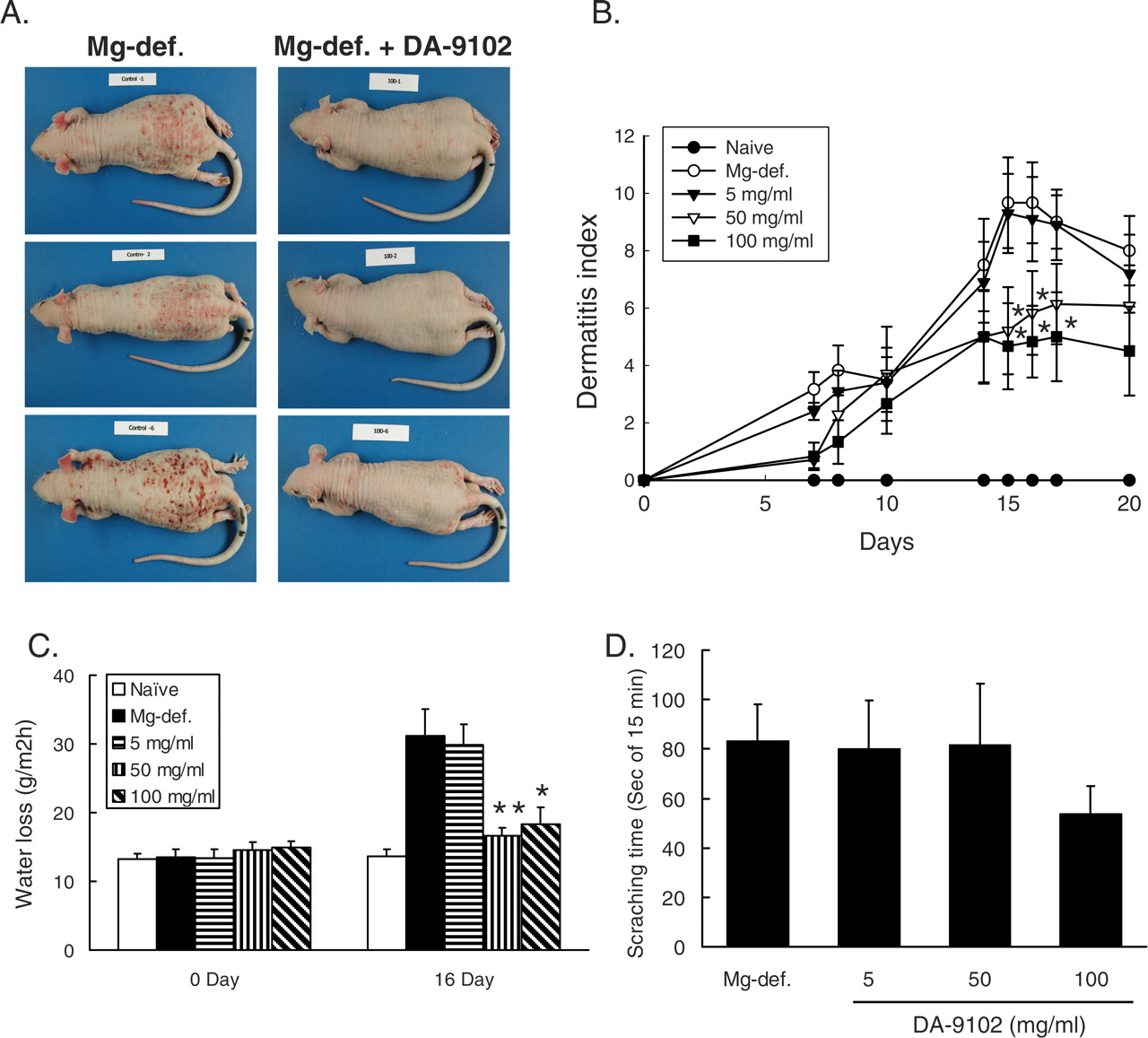
Effects of DA-9102 on spontaneous dermatitis in hairless rats under magnesium deficiency. (A) Clinical features were presented with three representative animals of the Mg-def. group and DA-9102 treated group. (B) Dermatitis index, (C) transepidermal water loss, and (D) scratching incidence were assessed by criteria described in Materials and Methods. Values are expressed as means ± SEM of 6 animals. * P < 0.05 vs. Mg-def. group (Student’s t test). A color figure is available in the online journal.

Effect of DA-9102 on the skin lesions in hairless rats under magnesium deficiency. The dorsal skin sections were stained with hematoxylin and eosine (H&E, a–c), and toluidine blue (d–e). Strong staining with H&E indicates leukocytes including eosinophils (arrows). Strong staining with toluidine blue indicates mast cells (arrowheads). The microscope original magnification is ×100. A color version of this figure is available in the online journal.

Effect of DA-9102 on IgE and CD45RA+ cells. (A) Serum was isolated from blood on day 16 and used for determination of the level of IgE with a commercially available ELISA kit. (B) PBMCs were isolated from the blood on day 16, and CD45RA+ cells were analyzed by flow cytometry. Values are expressed as means ± SEM for 6 animals. * P < 0.05 vs. Mg-def. group (Student’s t test). N.D., not detected.

Effect of DA-9102 on CD11b+ cells in hairless rats under magnesium deficiency. (A) PBMCs were isolated from the blood on day 16, and CD11b+ cells were analyzed by flow cytometry. (B) Skin biopsies were stained with FITC conjugated anti-CD11b antibody. Arrowheads indicate CD11b+ cells. The sections were examined under a fluorescence microscope (×400). (C) The CD11b+ cells were counted in 50 separated areas in each sample (n = 6). The relative number of CD11b+ cells was calculated by comparison with the cell number of the Mg-def. group. Values are expressed as means ± SEM for 6 animals. * P < 0.05 vs. Mg-def. group (Student’s t test). A color version of this figure is available in the online journal.
Footnotes
This study was supported by a grant from the Korea Health 21 R&D Project (A050440), and RIC grant (04700, 2006) (Traditional and Bio-Medical Research Center, Daejeon University) by ITEP.
Acknowledgements
We thank Kyung In Kim and Jiae Jun for their excellent technical support in the animal experiments.